BUBALIS), REVEALS MORE HYDROGENOTROPHIC METHANOGENS PHYLOTYPES
Singh K.M.1*, Pandya P.R.2, Parnerkar S.2, Tripathi A.K.¹, Rank D.N.3, Kothari R.K.4, Joshi C.G.1
¹Department of Animal Biotechnology, College of Veterinary Science and A.H., Anand Agricultural University, Anand (388 001),
Gujarat, India; ²Animal Nutrition Research Station, College of Veterinary Science and A.H., Anand Agricultural University,
Anand (388 001), Gujarat, India; ³Department of Animal Genetics & Breeding, College of Veterinary Science and A.H., Anand
Agricultural University, Anand (388 001), Gujarat, India; 4Department of Microbiology, Christ College, Rajkot, Gujarat, India.
Submitted: April 10, 2010; Returned to authors for corrections: May 12, 2010; Approved: August 26, 2010.
ABSTRACT
Methane emissions from ruminant livestock are considered to be one of the more potent forms of
greenhouses gases contributing to global warming. Many strategies to reduce emissions are targeting the
methanogens that inhabit the rumen, but such an approach can only be successful if it targets all the major
groups of ruminant methanogens. Therefore, a thorough knowledge of the diversity of these microbes in
breeds of buffaloes, as well as in response to geographical location and different diets, is required.
Therefore, molecular diversity of rumen methanogens in Surti buffaloes was investigated using 16S rRNA
gene libraries prepared from pooled rumen contents from three Surti buffaloes. A total of 171 clones were
identified revealing 23 different sequences (phylotypes). Of these 23 sequences, twelve sequences (12
OTUs, 83 clones) and 10 sequences (10 OTUs, 83 clones) were similar to methanogens belonging to the
orders Methanomicrobiales and Methanobacteriales, and the remaining 1 phylotype (5 clones) were similar
to Methanosarcina barkeri. These unique sequences clustered within a distinct and strongly supported
phylogenetic group. Further studies and effective strategies can be made to inhibit the growth of
Methanomicrobiales and Methanobacteriales phylotypes to reduce the methane emission from rumen and
thus help in preventing global warming.
Key word: Methanomicrobiales, Methanobacteriales, Surti buffaloes, phylotypes, 16s DNA.
INTRODUCTION
Methanogens are members of the domain Archaea, and fall
within the kingdom Euryarchaeota (48) Methanogens are
integral to carbon cycling, catalyzing the production of
methane and carbon dioxide, both potent greenhouse gases,
during organic matter degradation in anaerobic soils and
sediment (8). Methanogens are widespread in anaerobic
environments, including tundra (30), freshwater lake and
wetland sediments (7, 13), estuarine and marine sediments (2),
acidic peatlands (3, 17), rice field soil (8, 16), animal guts (33),
landfills (22), and anaerobic digesters treating animal manure
(1,34), food processing wastewater (23), and municipal
wastewater and solid waste (15, 53). Interest in methanogens
from ruminants has resulted from the role of methane in global
warming and from the fact that enteric methane emission is a
major source of greenhouse gas in agriculture
(http://www.indiastat.com). Currently, India possesses the
world’s largest livestock population of 485 million, which
accounts for 13% of the global livestock population
(Intergovernmental Panel on Climate Change. 2001). It has
57% of the world’s buffalo and 16% of the cattle population.
Contribution of methane emission in India by buffalo is 42%
(11). Reducing enteric methane emissions has been identified
as one way of lowering global methane emissions. However,
the effectiveness of any strategy that will reduce greenhouse
gas emissions and also increase production or nutritional
efficiency will likely depend upon having an understanding of
the numbers and/or distribution of methanogen species among
ruminant livestock. Several species of methanogens have been
isolated from ruminants, but few have been consistently found
in high numbers (35) and it is likely that major species of
rumen methanogens are yet to be identified (31, 50). The most
common species of methanogens isolated from the rumen are
strains of Methanobrevibacter, Methanomicrobium,
Methanobacterium, and Methanosarcina (50, 18).
Methanogens are difficult to study through culture-based
methods, and therefore many researchers have instead used
culture-independent techniques to study methanogen
populations. The 16S rRNA gene is the most widely used
target for gene surveys, and a number of primers and probes
have been developed to target methanogen groups (16, 26, 29,
28, 32, 39, 37, 45, 15, 49). Methanogens are frequently found
in association with protozoa (41, 21, 42, 43, 38). To date,
relatively little is known of the dominant methanogens in
ruminants, particularly Surti buffaloes in western India. This
paper uses comparative sequence analysis of cloned 16S rRNA
genes (rDNA) amplified from total DNA extracted from rumen
fluid to analyse the dominant methanogens present in the
rumen of Surti buffalo.
MATERIALS AND METHODS
Sampling and DNA extraction
The experiments were carried out on 3 adult Surti
buffaloes, approximately three years of age and with a mean
live weight of 201±18kg. They were reared at the Department
of Animal Nutrition, College of Veterinary Science and A.H.,
Anand. The permission of the Committee for the Purpose of
Control and Supervision of Experiments on Animals
(CPCSEA) was obtained prior to initiation of the study. All the
animals were maintained under uniform feeding regime (Indian
Council of Agricultural Research. 1998) for minimum 21 days.
The diet comprised of green fodder Napier bajra 21
(Pennisetum purpureum), mature pasture grass (Dicanthium
annulatum) and compound concentrate mixture (20% CP, 65%
TDN). The animals were offered 10 kg green, ad-lib dry grass
and 2.5 kg of concentrate mixture daily. Approximately 500 ml
of rumen fluid was collected via a stomach tube located in the
mid part of the rumen and connected to a vacuum pump at
0,2,4,6hrs post feeding (52,19). About 100 ml rumen fluid was
passed through four layers of cheese cloth to remove
particulate matter. Remaining rumen fluid was stored at -80°C
for further study. Total DNA (0, 2, 4, 6 hrs x 3 animals) was
extracted separately by using a commercially available kit
according to the manufacturer’s instructions (QIAGEN Stool
kit; QIAGEN, CA) and finally pooled the all DNA samples.
The total DNA mixture (pooled) was used as a template in
PCR to amplify 16S r DNA.
PCR primers and amplification
The primers used were 1Af (5'- TCYGKTTGATCCY
GSCRGAG-3') and 1100Ar (5'- TGGGTCTCGCTCGTTG-3').
(12).Subsequently 16S rDNA were amplified (1100bp) by PCR
using the metagenomic DNA and Master mix (Fermentas, UK).
A total of 25 l of reaction mixture consisted of 10 pmol of
each primer, 30 ng of template DNA, 12.5 l of Master mix
(Fermentas, UK). The PCR amplification was performed by
Thermal Cycler (ABI, USA) and PCR conditions were adjusted
in laboratory. The anticipated product of approximately1.1 kb
was purified using Qiagen DNA Gel Extraction Kits
(QIAGEN, CA) in accordance with the directions of the
Cloning and sequencing
The purified PCR products were cloned in PTZ57R/T
vector (Fermentas, UK) as per the instructions of the
manufacturer and transformed into E. coli DH alpha competent
cells. Ampicillin- and X-Gal
(5-bromo-4-chloro-3-indolyl-beta-D-galactopyranoside)-amended LB agar was used for
blue-white screening of transformants. The recombinant
plasmids then were extracted by the Qiagen mini-prep plasmids
extraction kit (QIAGEN, CA). Plasmid inserts were amplified
with primers M13F (5’-GTAAAACGAC GGCCAG-3’) and
M13R (5’-CAGGAAACAGCTATGAC-3’) and nucleotide
sequences of cloned genes were determined by sequencing
with M13F/ M13R primer in ABI Prism 310 Genetic analyser
(Applied Biosystems Inc., CA) using BigDye Terminator
(version 3.1) at Animal Biotechnology laboratory, AAU,
Anand, Gujarat, India.
Sequence analyses and phylogenetic tree constructing
All reference sequences were obtained from the
Genbank/EMBL/DDBJ/RDP (4). Sequences (~600 bp) from
the current study were trimmed (remove low-quality base calls
from the start and end of DNA sequence) manually and
analysed by the CHECK_CHIMERA program (27) to remove
any chimeric rDNA clone. The similarity searches for
sequences were carried out by BLAST
(http://www.ncbi.nlm.nih.gov/ BLAST/Blast.cgi (25) and
alignment was done using CLUSTAL W
(http://www.ebi.ac.uk/Tools/clustalw2/index.html (47).
Ambiguously and incorrectly aligned positions were aligned
manually. The distance matrix was calculated using the
DNADIST program included in PHYLIP (14) and used to
assign sequences in various operational taxonomic units
(OTUs) or phylotypes by DOTUR (40) with 95% confidence
intervals to quantify the diversity of phylotypes and total of 23
OTUs were generated based on furthest-neighbor algorithm at
cut offs of 10 % difference. The sampling effort of library was
evaluated by calculating the percentage of coverage (C)
according to the equation C= 1 - (n/N)X 100, where n is the
number of sequences represented by a single clone (Tables 1)
and N is the total number of clones analyzed in the library.
Phylogenetic tree was constructed by the neighbour joining
method using MEGA 4.0 (46). Bootstrap re-sampling analysis
for 1000 replicates was performed to estimate the confidence
of tree topologies (46).The prefix meth was used to denote
OTUs identified and nucleotide sequences have been deposited
in the Genbank database under the accession numbers are
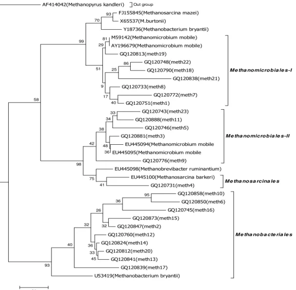
depicted in Fig 1.
RESULTS AND DISCUSSION
One hundred seventy one 16S rDNA sequences were
analyzed. On the basis of sequence similarity, all of the
sequences were related to methanogens. The sample
preparation technique, centrifugation before DNA extraction,
allowed us to preferentially examine methanogens isolated
from the fluid fraction of rumen contents. Two distinct clusters
were generated by Maximum Parsimony method analysis of
sequences (Fig. 1). The first largest cluster contained 12 OTUs
(83 clones) grouped with order Methanomicrobials forming
two distinct subclusters that were supported by high bootstrap
values (Fig. 1). Subcluster Methanomicrobials I consisted of 51
clones (7 OTUs) identical or nearly identical sequences
(similarity values ranged from 85 to 96 %) that were similar to
Methanomicrobium mobile-like clones. The second subcluster,
Methanomicrobials II, formed a deeper branch consisting of 32
clones (15 OTUs), similarity value of 16Ssequences that were
ranged from 85% to 90% and members of this group are
belonged Methanomicrobium mobile-like clones (Table 1).
Boone et al. (6) considered that a sequence similarity of 98 %
or less was evidence for separate species within the
methanogens. Based on this, Methanomicrobials would be
considered as Methanomicrobium mobile-like strains, however,
as pointed out by Martinez-Murcia et al. (24), 16S sequence
similarity values recommended to define a species provide a
working definition that has been empirically derived, and
values should not be treated as absolute or fixed. For example,
16S rRNA sequences from strains of Methanobacterium
study were greater than 98 % similar, yet these organisms are
considered distinct species. Shin et al. (51) reported that 85%
(89 of 104 clones) of the total clones from the bovine rumen
belonged to the order Methanomicrobiales, with 61 clones
resembling Methanomicrobium mobile. Similar results are also
reported by (53) in Feedlot Cattle from Ontario and Prince
Edward Island. Interestingly, Methanomicrobium mobile was
not detected in sheep from Western Australia (51). Phylotypes
within the Methanobacteriales represented 48.5% (83 clones)
of total clones and spanned ten OTUs. Within this cluster, the
cloned sequences formed two subclusters. It should be noted
that the significance of the subclusters is not supported by high
bootstrap values (Fig. 1).Although, the rDNA sequences may
represent species of Methanobacterium bryantii. A total of 1
OTUs representing 2.9% of total clones were closely related to
cultured species belonging to order Methanosarcinales.
Out group
! "# $%
& #"! % %
' % %
& '!!#' % %
() " '
() #
() #'
() "
() #""
() ## #
() #
M e tha no micro b ia le s-I
() # " "
()
() # !
() "
*+ ' % %
*+ ' % %
() ##! '
M e tha no micro b ia le s-II
*+ ' % , %
*+ %
() #"
M e tha no sa rcina le s ()
() !
() # !
() #"
() #
() #!
()
()
() "
() "' #
+ " ' % %
M e tha no b a cte ria le s
93
86 81 70
40 29
25
17 9 51 99
41 75
36 48
33 34 38
42
98
95
45 33 36
36
32 26
32
40
93 58
20
Figure 1. Phylogenetic relationships of partial 16S r DNA sequences of clones recovered from Surti rumen samples. The rooted
tree was constructed as a maximum parsimony tree using close-neighbor interchange level 1 and bootstrapped with 1,000 trials,
using the MEGA 4 tree building program. All positions containing gaps and missing data were eliminated from the data set .The
Methanopyrus kanderi (AF414042) are used as the out-group for rooting the tree. The scale bar represents the number of
Zinder (54) showed that in a typical cattle rumen the
approximate steady-state amounts of volatile fatty acids (VFA)
are 63% acetate, 21% propionate, and 16% butyrate and other
higher fatty acids. VFA are generally absorbed by the rumen
epithelium and subsequently converted to animal proteins, and
therefore not available for utilization as a carbon source by
acetoclastic methanogens (Methanosarcinales) residing in
rumen. Therefore, acetoclastic methanogens make up only a
small percentage of total methanogen community in cattle
rumen. The only carbon source available in plenty for
methanogens is H2/CO2, thus hydrogenotrophic methanogens
(Methanomicrobiales and Methanobacteriales) that are capable
of using H2/CO2 can multiply easily and are observed in high
abundance in rumen [54]. Earlier phylogenetic studies based on
the 16S rRNA and mcrA genes also revealed that majority of
the sequences retrieved from bovine rumens and cattle dung
were affiliated to hydrogenotrophic methanogens belonging to
Methanomicrobiales and Methanobacteriales (44, 14). The
methanogen community of buffaloes rumen should reflect the
same trend as observed in a typical cattle rumen such as the
greater abundance of hydrogenotrophic methanogens than the
acetoclastic methanogens. The results of the present study
corroborate with earlier observations where in the Surti rumen
16S r RNA library 97% clones belonged to the
hydrogenotrophic methanogens, while the acetoclastic
methanogens represented merely 3% of the total clone diversity
(Table 1). Previous culture based studies have isolated
methanogens of the genus Methanomicrobium and
Methanobacterium from the bovine rumen (18), although
Methanobrevibacter and Methanosarcina tend to be isolated at
higher population levels (29). While, Methanobrevibacter-like
clone could not detect in the present study. This may be due to
differences in sample preparation, animal diet or geographic
region. The methanogen community of Surti buffalo should
reflect the same trend as observed in a typical cattle rumen
such as the greater abundance of hydrogenotrophic
methanogens than the acetoclastic methanogens. The results of
the present study corroborate with earlier observations (33)
where in the fresh cow dung mcrA library 93.5% clones
belonged to the hydrogenotrophic methanogens, while the
acetoclastic methanogens represented merely 6.5% of the total
clone diversity, in the 8-month-old dung, which included 80%
of clones belonging to hydrogenotrophic methanogens, while
acetoclastic methanogens constituted only 20% of the total
clone diversity.
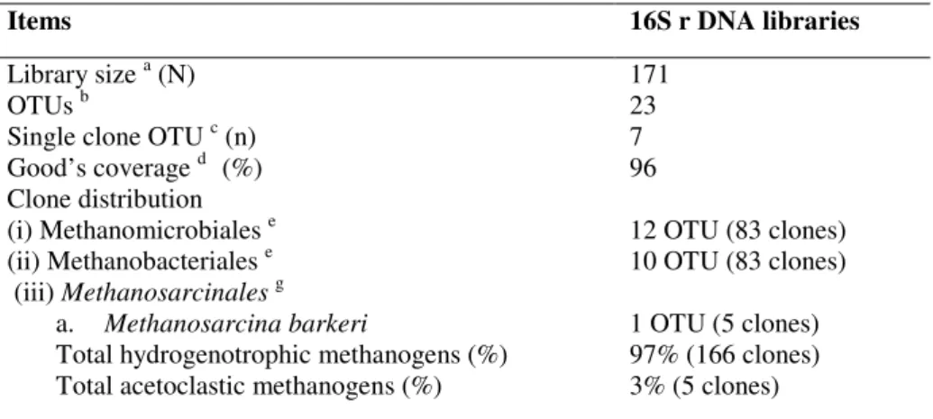
Table 1. Analysis of 16S rDNA phylotypes diversity retrieved from the rumen fluid of Surti buffaloes
Items 16S r DNA libraries
Library size a (N) OTUs b
Single clone OTU c (n) Good’s coverage d (%) Clone distribution (i) Methanomicrobiales e (ii) Methanobacteriales e (iii) Methanosarcinalesg
a. Methanosarcina barkeri
Total hydrogenotrophic methanogens (%) Total acetoclastic methanogens (%)
171 23 7 96
12 OTU (83 clones) 10 OTU (83 clones)
1 OTU (5 clones) 97% (166 clones) 3% (5 clones)
a Number of clones analyzed from library b OTUs based on 16S r DNA sequences c OTUs containing only single clone
d The higher percentage coverage means more diversity is captured eHydrogenotrophic methanogens
Although some feeding strategies reduce ruminal methane
emissions, the amount of CH4 produced during ruminal
fermentation is dependent upon the nature of the substrate
being fermented. In general, methanogenic potential of the
ruminal microflora is greatest for the fermentation of structural
carbohydrates compared to that of non-structural carbohydrates
(5).The addition of fat or individual fatty acids to ruminal
cultures decrease CH4 production (34). Ruminal methane is
formed by the action of methanogenic archaea typified by
hydrogenotrophic methanogens, which is present in ruminants
fed upon a wide variety of diets worldwide. Genome sequences
would provide new insights into the lifestyle and cellular
processes of this important rumen hydrogenotrophic
methanogens (Methanomicrobiales and Methanobacteriales)
under control feeding regime. It would also define vaccine and
chemogenomic targets for broad inhibition of rumen
methanogens and represents a significant contribution to
worldwide efforts to mitigate ruminant methane emissions and
reduce production of anthropogenic greenhouse gases.
Over all more studies are needed on the effects of diets
composition and animal species on the diversity of
methanogens and enteric methane emission in the rumen. This
study has revealed the largest assortment of hydrogenotrophic
methanogens phylotypes ever identified from rumen of Surti
buffaloes and the need to better understand the factors
influencing methanogen diversity with methane emission.
Further studies are needed to examine methanogen diversity in
goat, sheep and dairy cattle located in the Gujarat state. Such
studies would significantly enhance our knowledge and ability
to use novel methods to manipulate the rumen methanogen
populations to reduce methane production from ruminant
animals. Reducing enteric methane emissions is likely to be
one of the key mitigation strategies for the reduction of
greenhouse gas emissions in the agricultural sector.
ACKNOWLEDGEMENTS
Financial support provided by the Department of
Biotechnology Govt. of India, New Delhi to conduct the study
reported here is acknowledged with respect and gratitude.
REFERENCES
1. Angenent, L.T.; Sung, S.W.; Raskin, L. (2002). Methanogenic population dynamics during startup of a full-scale anaerobic sequencing batch reactor treating swine waste. Water Res. 36:4648–4654.
2. Banning, N.; Brock, F.; Fry, J.C.; Parkes, R.J.; Hornibrook, E.R.C.; Weightman, A.J. (2005). Investigation of the methanogen population structure and activity in a brackish lake sediment. Environ. Microbiol. 7:947–960.
3. Basiliko, N.; Yavitt, J.B.; Dees, P.M.; Merkel, S.M. (2003). Methane biogeochemistry and methanogen communities in two northern peatland ecosystems, New York state. Geomicrobiol. J. 20:563–577.
4. Benson, D.A.; Karsch-Mizrachi, I.; Lipman, D.J.; Ostelland, J. (2007). GenBank. Nucleic Acids Res. (35), D1–D25.
5. Boadi, D.; Benchaar, C.; Chiquette, J.; Massé, D. (2004). Mitigation strategies to reduce enteric methane emissions from dairy cows: Update review. Can. J. Anim. Sci.84:319-335.
6. Boone, D.R.; Whitman, W.B.; Rouviere, P. (1993). Diversity and taxonomy of methanogens. In: Methanogenesis Edited by Ferry JG. pp. 35-80. New York: Chapman and Hall, Inc.
7. Cakir, F.Y.; Stenstrom, M. K.; (2005). Greenhouse gas production: a comparison between aerobic and anaerobic wastewater treatment technology. Water Res. 39:4197–4203.
8. Castro, H.; Ogram, A.; Reddy, K.R. (2004). Phylogenetic characterization of methanogenic assemblages in eutrophic and oligotrophic areas of the Florida everglades. Appl. Environ. Microbiol. 70:6559–6568.
9. Chabra, A.; Manjunath, K.R.; Panigrahy, S.; Parihar, J.S. (2009). Spatial pattern of methane emissions from Indian livestock. Current Science.96: 5-10.
10. Chin, K.J.; Lueders, T.M.; Friedrich, W.; Klose, M.; Conrad, R. (2004). Archaeal community structure and pathway of methane formation on rice roots. Microb. Ecol. 47:59–67.
11. Cocetti, G.; Murto, M.; Bjornsson, L. (2006). An update and optimisation of oligonucleotide probes targeting methanogenic archaea for use in fluorescence in situ hybridisation (FISH). J. Microbiol. Methods 65:194–201.
12. Earl, J.; Hall, G.; Pickup, R. W.; Ritchie, D.A.; Edwards, C. (2003). Analysis of methanogen diversity in a hypereutrophic lake using PCR-RFLP analysis of mcr sequences. Microb. Ecol. 46:270–278.
13. Embley, T.M.; Finlay, B.J.; Thomas, R.H.; Dyal, P.L. (1992). The use of rRNA sequences and fluorescent probes to investigate the phylogenetic positions of the anaerobic ciliate Metopus palaeformis and its archaebacterial endosymbiont. J. Gen. Microbiol. 138:1479-1487. 14. Felsenstein, J.; 1985. Confidence limits on phylogenies: an approach
using the bootstrap. Evolution. 39:783-791.
Hir, R.P. (1994). Some rumen ciliates have endosymbiotic methanogens. FEMS Microbiol. Lett. 117:157-162.
16. Galand, P.E.; Saarnio, S.; Fritze, H.; Yrjal, K. (2002). Depth related diversity of methanogen archaea in finnish oligotrophic fen. FEMS Microbiol. Ecol. 42:441–449
17. Großkopf, R.; Stubner, S.; Liesack, W. (1998). Novel euryarchaeotal lineages detected on rice roots and in the anoxic bulk soil of flooded rice. 18. Jarvis, G.N.; Strompl, C.; Burgess, D.M.; Skillman, L.C.; Moore, E.R.;
Joblin, K.N. (2000). Isolation and identification of ruminal methanogens from grazing cattle. Curr. Microbiol. 40:327-332.
19. Khampa, S.; Wanapat, M.; Wachirapakorn, C.; Nontaso, N. (2006). Effects of urea level and sodium di-malate in concentrate containing high cassava chip on ruminal fermentation efficiency, microbial protein synthesis in lactating dairy cows raised under tropical condition. Asian- Aust. J. Anim. Sci. 19:837-844.
20. Liu, W.T.; Chan, O.C.; Fang, H.H.P. (2002). Characterization of microbial community in granular sludge treating brewery wastewater. Water Res. 36:1767–1775.
21. Lloyd, D.; Ralphs, J.; Durrant, L.; Williams, A.G.; Amann, R. (1994). Studies of the bacterial endosymbionts of "anaerobic protozoa" using fluorescently- labelled rRNA-targeted oligonucleotide probes. Biochem. Soc. Trans. 22:S323
22. Lloyd, D.; Williams, A.G.; Amann, R.; Hayes, A.J.; Durrant, L.; Ralphs, J.R. (1996). Intracellular prokaryotes in rumen ciliate protozoa – detection by confocal laser-scanning microscopy after in-situ hybridization with fluorescent 16S ribosomal-RNA probes. Eur. J. Protist. 32:523-531.
23. Luton, P.E.; Wayne, J.M.; Sharp, R.J.; Riley, P.W. (2002). The mcrA gene as an alternative to 16s rRNA in the phylogenetic analysis of methanogen populations in landfill. Microbiology 148:3521–3530. 24. Madden, T.L.; Tatusov, R.L.; Zhan, J. (1996). Application of network
BLAST server. Meth. Enzymol. 266:131-141.
25. Maidak, B.L.; Cole, J.R.; Lilburn, T.G.; Jr, C.T.P. (2001).The RDP-II (Ribosomal Database Project). Nucl. Acids Res. 29:173-174.
26. Marchesi, J. R.; Weightman, A. J.; Cragg, B. A.; Parkes, R. J.; Fry, J. C. (2001). Methanogen and bacterial diversity and distribution in deep gas hydrate sediments from the Cascadia Margin as revealed by 16s rRNA molecular analysis. FEMS Microbiol. Ecol. 34:221–228.
27. Martinez-Murcia, A.J.; Benlloch, S.; Collins, M.D. (1992) .Phylogenetic interrelationships of members of the genera Aeromonas and Plesiomonas determined by 16S ribosomal DNA sequencing: lack of congruence with the results of DNA-DNA hybridizations. Int. J. Syst. Bacteriol. 42:412-42 28. Opperman, R.A.; Nelson, W.O.; Brown, R.E. (1957). In vitro studies on
methanogenic rumen bacteria. J. Dairy Sci. 40:779-788.
29. Purdy, K. J.; Nedwell, D. B.; Embley, T. M. (2003). Analysis of the sulfate-reducing bacterial and methanogenic archaeal populations in contrasting Antarctic sediments. Appl. Environ. Microbiol. 69:3181– 3191.
30. Raskin, L.; Stromley, J.M.; Rittmann, B.E.; Stah, D. A. (1994).
Group-specific 16S rRNA hybridization probes to describe natural communities of methanogens. Appl. Environ. Microbiol. 60:1232-1240.
31. Rastogi, G.; Ranade, D. R.; Yeole, T. Y.; Gupta, A. K.; Patole, M. S.; Shouche, Y. S. (2008). Molecular analyses of methanogen diversity associated with cattle dung. World J Microbiol Biotechnol 24:2973–2979. 32. Rocheleau, S.; Greer, C.W.; Lawrence, J.R.; Cantin, C.; Laramee, L.; Guiot, S.R. (1999). Differentiation of Methanosaeta concilii and Methanosarcina barkeri in anaerobic mesophilic granular sludge by fluorescent in situ hybridization and confocal scanning laser microscopy. Appl. Environ. Microbiol. 65:2222–2229.
33. Saengkerdsub, S.; Herrera, P.; Woodward, L.; Anderson, C.; Nisbet, D. J.; Ricke, S.C. (2007). Detection of methane and quantification of methanogenic archaea in faeces from young broiler chickens using real-time PCR. Lett. Appl. Microbiol. 45:629–634.
34. Schloss, P.D.; Handelsman, J.; (2005). Introducing DOTUR, a compute program for defining operational taxonomic units and estimating species richness. Appl Environ Microbiol 71:1501–1506.
35. Sharp, R.; Ziemer, C.J.; Marshall, D.S.; Stahl, D.A. (1998). Taxon-specific associations between protozoal and methanogen populations in the rumen and a model system. FEMS Microbiol. Ecol. 26:71-78. 36. Shin, E.C.; Choi, B.R.; Lim, W.J.; Hong, S.Y.; An, C.L.; Cho, K.M.
(2004). Phylogenetic analysis of archaea in three fractions of cow rumen based on 16S rDNA sequence. Anaerobe; 10: 313–9.
37. Sizova, M.V.; Panikov, N.S.; Tourova, T.P.; Flanagan, P.W. (2003). Isolation and characterization of oligotrophic acido-tolerant methanogenic consortia from a sphagnum peat bog. FEMS Microbiol. Ecol. 45:301–315. 38. Soliva, C.R.; Meile, L.; Cieslak,; Kreuzer, A.M.; Machmuller, A. (2004). Rumen simulation technique study on the interactions of dietary lauric and myristic acid supplementation in suppressing ruminal methanogenesis. Br. J. Nutr. 92:689-700.
39. Sorensen, A. H.; Torsvik, V. L.; Torsvik, T.; Poulsen, L. K.; Ahring, B. K. (1997). Whole-cell hybridization of Methanosarcina cells with two new oligonucleotide probes. Appl. Environ. Microbiol. 63:3043–3050. 40. Stewart, C.S.; Flint, H.J.; Bryant, M.P. (1997). The rumen bacteria. In:
The Rumen Microbial Ecosystem .2 ed. pp. 10- 72.
41. Tajima, K.; Nagamine, T.; Matsui, H.; Nakamura, M.; Aminov, R.I. (2001). Phylogenetic analysis of archaeal 16S rRNA libraries from the rumen suggests the existence of a novel group of archaea not associated with known methanogens. FEMS Microbiol Lett 200:67–72.
42. Tamura, K.; Nei, M.; Kumar, S. (2004). Prospects for inferring very large phylogenies by using the neighbor-joining method. Proc Natl Acad Sci USA 101:11030-11035.
43. Tamura, K.; Dudley, J.; Nei, M.; Kumar, S. (2007). MEGA4: Molecular Evolutionary Genetics Analysis (MEGA) software version 4.0. Molecular Biology and Evolution 24:1596-1599.
45. Thompson, J.D.; Higgins, D.G.; Gibson, T.J. (1994). CLUSTAL W: improving the sensitivity of progressive multiple sequence alignment through sequence weighting, position-specific gap penalties and weight matrix choice. Nucl. Acids Res. 22:4673- 4680.
46. Tokura, M.; Tajima, K.; Ushida, K. (1999). Isolation of Methanobrevibacter sp. as a ciliate-associated ruminal methanogen. J. Gen. Appl. Microbiol. 45:4347.
47. Tokura, M.; Chagan, I.; Ushida, K.; Y, Kojima. (1999). Phylogenetic study of methanogens associated with rumen ciliates. Curr Microbiol. 39:123-8.
48. Woese, C.R.; Kandler, O.; Wheelis, M. (1990). Towards a natural system of organisms: proposal for the domains Archaea, Bacteria, and Eucarya. Proc. Natl. Acad. Sci. USA, 87: 4576-4579.
49. Wolin, M.J.; Miller, T.L.; Stewart, C.S. (1997). Microbe-microbe interactions: The Rumen Microbial Ecosystem 2 ed. pp. 467-491.
50. Wright, A.D.G.; Williams, A.J.; Winder, B.; Christophersen, C.; Rodgers, S.; Smith, K. (2004). Molecular diversity of rumen methanogens from sheep in Western Australia. Appl Environ Microbiol; 70:1263–70. 51. Wright, A.D.G.; Auckland, C.H.; Lynn, D.H. (2007). Molecular Diversity
of Methanogens in Feedlot Cattle from Ontario and Prince Edward Island, Canada. Appl. Environ. Microbiol.73 4206–4210.
52. Yuangklang, C.; Wanapat, M.; Wachirapakorn, C. ( 2005). Effects of pelleted sugarcane tops on voluntary feed intake, digestibility and rumen fermentation in beef cattle. Asian-Aust. J. Anim. Sci. 18:22-26. 53. Zheng, D.; Raskin, L. (2000). Quantification of Methanosaeta species in
anaerobic bioreactors using genus- and species-specific hybridization probes. Microb. Ecol. 39:246–262.