1 Museu de História Natural de Taubaté. Rua Colômbia, 99, CEP 12030-520, Taubaté, SP, Brasil. E-mail: [email protected]. 2 Departamento de Zoologia, Instituto de Biociências, Universidade de São Paulo, Caixa Postal 11.294, CEP 05422-970, São Paulo, SP,
Brazil.
Volume 43(4):55-91, 2003 www.scielo.br/paz.htm
Papéis Avulsos de Zoologia
ISSN 0031-1049
Museu de Zoologia da Universidade de São Paulo
S
YSTEMATICREVISION OFTHEP
HORUSRHACIDAE(A
VES: R
ALLIFORMES)
H
ERCULANOM.F. A
LVARENGA1E
LIZABETHH
ÖFLING2ABSTRACT
Fossil remains of birds belonging to the family Phorusrhacidae were studied in several museums of South America, North America and Europe, the main objective being to characterize this family and solve the chaotic state of the nomenclature and classification of these birds. Reconstruction of some species has been done, with the purpose of having an idea about the size, body weight, posture and habit based in their skeletons. The European species, Ameghinornisminor and Aenigmavissapea are refuted as belonging to this family. Also several forms described from the Tertiary of Argentina are refuted, because they are based on inadequate segments of the skeleton for a good identification, as is the case of the genera Cunampaia, Smiliornis, Pseudolarus, Lophiornis and Riacama, frequently refered to as belonging to the Phorusrhacidae. The Phorusrhacidae family probably originated in South America, since the end of the Cretaceous, as a result of an endemism formed by the isolation of this landmass. During the end of the Pliocene, with the emersion of the Panama isthmus, the family spread to the North America where at least one species is known Titaniswalleri, which perhaps represents the last known species of this family, probably becoming extinct in the beginning of the Pleistocene. A systematic revision has been conducted, dealing with the countless problems of nomenclature, and the Phorusrhacidae is now composed of five subfamilies, which are: Brontornithinae, Phorusrhacinae, Patagornithinae, Psilopterinae and Mesembriornithinae in which 13 genera and 17 species are considered. Characters of all taxa are described and a geochronological distribution of all species is presented.
KEYWORDS: Phorusrhacidae, Ralliformes, Gruiformes, Tertiary, Giant birds.
INTRODUCTION
Historicalbackground – At the end of the 19th century, Ameghino (1887) described a large, toothless jaw from the Miocene of the Province of Santa Cruz, naming it Phorusrhacoslongissimus and assigning it to a new family of toothless mammals. In 1889, Moreno was the first to refer to the giant birds of the Mio-Pliocene from
described other birds similar to Phorusrhacos, now rec-ognizing all of them as birds belonging to the group of the Ratitae.
Moreno and Mercerat (1891) described a large amount of fossils of these giant birds from the La Plata Museum, most of which also coming from the Santa Cruz region, as was the case of those described by Ameghino. These authors, however, recognized cer-tain peculiarities of these birds, thus placing them in a new order, the Stereornithes, having some affinities with “los Anseres, de los Herodines y de los Accipitres”, dividing this order into four families, which are, the Brontornithidae, Stereornithidae (in which Phorusrhacos is included), Dryornithidae and Darwinornithidae.
The material studied and described by Moreno & Mercerat (1891), at least in part, is made up of seg-ments from skeletons which are of significant diag-nostic importance; the work being richly illustrated, thus constituting a landmark for Phorusrhacidae sys-tematics.
A certain rivalry began as to the priority of the terms created by Ameghino (1891a and 1891b), and Moreno & Mercerat (1891). Mercerat himself (1897:227) declares that the “Catálogo de los Pájaros Fósiles de la República Argentina” of which he is co-author, and wherein the presentation done by Moreno is dated the 15th of April, 1891, was first published in mid-May, 1891 (several copies then being distributed in this form), the plates only being terminated on the fifth of August, 1891. Further on he also affirms that Ameghino’s publication (1891a), dated as of June the 1st, 1891, was in fact published on the 11th of August, 1891, wherein Ameghino also describes Tolmodusinflatus as a mammal. The second publication of Ameghino (1891b) was printed in December, 1891.
In various subsequent works by various authors (e.g. Andrews, 1896 and 1899; Lambrecht, 1933; Sinclair & Farr, 1932), one observes a strong tendency to give precedence to the terms given by Ameghino over those by Moreno & Mercerat.
Brodkorb (1967), in his revision recognizes the priority of Moreno & Mercerat (1891) over the works of Ameghino of the same year.
In subsequent works, Ameghino (1895 and 1898) describes new fossils of the Phorusrhacidae, creating new terms, often complicating still further the nomen-clature of these birds.
Andrews (1896 and 1899), in the detailed study of a certain species, recognizes the close relationship of the Stereornithes with the extant Cariamidae fam-ily, discerning a new position, to date still accepted, of the relationship with this family.
Theproposedclassifications – One of the first attempts at organizing the classification of these birds was done by Dolgopol de Saez (1927), only with those of the Santacrucian (Mid-Miocene), dividing them into two orders: (1) order Stereornithes with only one family, the Phororhacidae, with two genera, Phororhacos and Psilopterus, and (2) the order Brontornithes, also with one family, the Brontornithidae, with the genera Brontornis, Rostrornis, Liornis and Paleociconia. Dolgopol de Saez (op. cit.), in her classification, gave importance to the format of the ungual phalanges, these being flat-tened or raised (without specifying on which digit), to the branching or not of the distal foramen of the tar-sometatarsus, and to the existence or not of the supratendinal bridge on the tibiotarsus (this without bothering about the frequent losses by fossilization and accidents in collecting).
Patterson & Kraglievich (1960), on studying the forms from the Pliocene, present an important con-tention on the synonymy, diversity and classification of these birds, placing them in the order Grues Bonaparte, 1857, suborder Cariamae. Moreover, the above mentioned authors propose dividing the Cariamae into two superfamilies, Cariamoidea Stejneger, 1887 and Phororhacoidea Patterson, 1941, and characterize the Phororhacoidea, setting them apart from the Cariamoidea by the following characteristics: (1) a completely desmognate cranium, (2) a relatively high and long rostrum (premaxillar region), (3) ribs without the uncinate processes, (4) very reduced wing bones and loss of the ability to fly, (5) a narrow pelvis with an incomplete pubis and shorter pre-acetabulum portion than the pos-acetabulum one. Further on, Patterson & Kraglievich (op. cit.) divide the Phororhacoidea into the families: Psilopteridae, with the subfamilies Psilopterinae (Psilopterus, Smiliornis and Procariama) and Hermosiornithinae (Hermosiornis), and the family Phororhacidae, with the subfamilies Phororhacinae (Phororhacos, Devincenzia and Onactornis) and Tolmodinae (Tolmodus, Andrewsor nis and Andalgalornis). The cited authors (1960:11), moreover, considered the separation of the family Brontornithidae, criticizing, however, the separation of this as an order apart, as proposed by Dolgopol de Saez (1927).
Andalgalornis) and Phorusrhacinae (Phorusrhacos, Onactornis and Titanis), as well as the family Cariamidae with the subfamilies Psilopterinae (Riacama, Smiliornis, Pseudolarus, Psilopterus, Lophiornis and Procariama), Prophororhacinae (Prophororhacos) and Cariaminae (with the extant Cariama and Chunga). The cited author ig-nored the characteristics by which Patterson & Kraglievich (1960) separated the Cariamoidea from the Phororhacoidea.
In 1981, Mourer-Chauviré described a few bones of birds from the Eocene-Oligocene of France, as-signing them to the Ameghinornithinae, a new sub-family of the Phorusrhacidae, the first record of this family for Europe. On complementing her description, the cited author proposes a classification for the sub-order Cariamae Fürbringer, 1888, wherein the Phorusrhacidae are divided into six subfamilies, namely, the Brontornithinae (Physor nis and Brontornis), Palaeociconiidae (Andrewsornis, Palaeociconia and Andalgalornis), Phorusrhacinae (Phorusrhacos, Onactornis and Titanis), Psilopterinae (Psilopterus, Lophiornis and Procariama), Prophororhacinae (Prophororhacos) and Ameghinornithinae (Ameghinornis). Furthermore, Mourer-Chauviré considers the genera Riacama, Smiliornis and Pseudolarus as insertaesedis.
Geographicalandchronologicaldistribution – The greater part of the fossils of the Phorusrhacidae came from Ar-gentina. This does not mean a more southern distri-bution in South America, but better conditions for the appearance of fossiliferous outcrops in this region, besides the greater technical development in the Pale-ontology of Argentina, from which one concludes a greater knowledge of fossils in general in that country within South America. Outside Argentina, the Phor usrhacidae birds are known in Ur uguay (Kraglievich, 1932; Tambussi et al. 1999), Brazil (Alvarenga, 1982 and 1985a), the Antarctic (Case etal. 1987), and in North America (Brodborb, 1963). More recently, reference is made to these birds in Europe (Mourer-Chauviré, 1981; Peters, 1987), and, possibly still further in the Lower Tertiary, in North America (unpublished material, P. Houde and S. Olson, pers. inf.). These occurrences in the Lower Tertiary in North America and Europe make the biogeographical expla-nation of the origin and dispersion of the family very difficult.
The oldest fossil record of the Phororhacoidea birds is of Alvarenga (1985), represented by a rela-tively small-sized form, Paleopsilopterusitaboraiensis from the Mid-Paleocene (Itaboraian) from southeast Brazil, Rio de Janeiro, the Itaboraí Basin. The most recent
occurrence is assigned to the limit between the Upper Pliocene and Lower Pleistocene (Late Blancan), from Florida, U.S.A., with a gigantic form, Titanis wagleri, described by Brodkorb (1963).
MATERIAL AND METHODS
The fossilized remains assigned to the Phorusrhacidae and deposited in the collections of the following museums were examined: Museo Argentino de Ciencias Naturales of Buenos Aires, Museo de La Plata, Museu Nacional do Rio de Janeiro, Departamento da Produção Mineral do Rio de Janeiro, Field Museum of Natural History of Chicago, Ameri-can Museum of Natural History of New York, The Natural History Museum of London, Muséum Na-tional d’Histoire Naturelle de Paris, and Forschungsinstitut Senckenberg in Frankfurt.
The material refering to Titaniswalleri Brodkorb, 1963, deposited in the Museum of the University of Florida, not only the type, but also several other parts assigned to the species, besides the tarsometatarsus of Devincenziagallinali Kraglievich, 1932 (type) from the Museu National de Historia Natural of Montevideo, the skull and the bones of the hind-limbs type of Hermosiornisrapax Kraglievich, 1946, deposited in the museum of Mar del Plata, as well as the material whereon the original description of the genus
Ameghinornis Mourer-Chauviré, 1981 (part deposited
in the museum of Paris and part in the museum of Lyon) was based, were studied from casts kindly ceded by the authorities of these Institutions.
the National Museum of Natural History, Washing-ton, DC (seeing that the fossils of Neocathartesgallator are not deposited in this museum). Another family of the Lower Tertiary, especially in Europe, and related to the Cariamidae, are the Idiornithidae, which were studied using casts of Elephrocnemusphasianus offered by Cécile Mourer-Chauviré, of the University of Lyon, and from the available illustrations in the publication of this researcher (Mourer-Chauviré, 1983).
The evaluation of the body mass of certain phorusrhacids was done based on the hind-limbs, es-pecially the measurements of the smallest circunference of the diaphysis of the femur and tibiotarsus, which present a direct relationship to the body mass of the bird (Campbell Jr. & Marcus, 1992). In the present work we compared the measurements of some phorusrhacids (diameter of the hind-limb bones) with the same measurements of homologous bones of the ratite birds, or other land birds, with a known mass/ size. Several data on bone and body mass measure-ments of large present-day and fossil birds were ob-tained in the publications of Amadon (1947) and Wetmore (1967).
Several skeletons of present-day birds were ex-amined as elements for comparison, all from the col-lection of Museu de Historia Natural of Taubaté (MHNT), amongst which: Struthio camelus, male
(MHNT-01) and female (MHNT-1991); Casuarius
casuarius, male (MHNT-03) and female (MHNT-1293);
Rheaamericana, male (MHNT-668); some original bones of the Dinornithidae (MHNT-wt/n.); the cast of a
complete skeleton of Aepyor nis maximus
(MHNT-wt/n.); Cariamacristata, four specimens, two being males (MHNT-1214 and MHNT-1267), one fe-male (MHNT-1136) and the other of undetermined
sex (MHNT-78); Opisthocomus hoaz in male
(MHNT-665).
The anatomical nomenclature used was mainly according to Baumel & Witmer (1993), and in some cases, Howard (1929) and Gilbert etal. (1981).
InstitutionalAbbreviations – AMNH, American Museum of Natural History, New York; BMNH, The Natural History Museum, London; DGM, Divisão de Geologia e Mineralogia do Departamento Nacional da Produção Mineral, Rio de Janeiro; FMNH, Field Museum of Natural History, Chicago; MACN, Museo Argentino de Ciencias Naturales Bernardino Rivadavia, Buenos Aires; MGHN, Musée Guimet d’Histoire Naturelle, Lyon; MHNT, Museu de História Natural de Taubaté; MNHN, Muséum National d’Histoire Naturelle, Paris; MLP, Museo de La Plata; MMCN, Museo Municipal
de Ciencias Naturales, Lorenzo Scaglia, Mar del Plata; MNHN-M, Museo Nacional de Historia Natural de Montevideo; MNRJ, Museu Nacional do Rio de Janeiro; PUM, Princeton University Museum, New Jersey; SMF, Forschungsinstitut Senckenberg, Frankfurt; TMM, Texas Memorial Museum, Austin; UF, University of Florida, Gainesville.
RESULTS AND DISCUSSION
SizeandbodymasscalculatedforthePhorusrhacidae – The Phorusrhacidae are amongst the largest birds that have ever existed on the planet, and Brontornisburmeisteri (Fig. 1A) is, without doubt, the largest phorusrhacid and the largest known bird of the American continent. Its size rivals that of the elephant birds (Aeopyornis maximus) of the Pleistocene-Holocene of Madagascar, classically considered the largest bird that has existed for all time.
The length of the femur, the tibiotarsus and the tarsometatarsus of Brontornisburmeisteri (MLP-88, 89 and 91), are very close to the measurements of the corresponding bones of Aepyornismaximus. However, especially the diameters and circuferences of these bones appear to be nearly 10 to 15% smaller in Brontornis.
Dinornisgiganteus, the largest representative of the 13 species of moas (Dinornithidae), known from New Zeeland (Cracraft, 1976), had a body-mass estimated at weighing 230 to 240 kg (Amadon, 1947), or 278 kg (Campbell Jr. & Marcus, 1992), it most certainly
ing been the tallest bird that existed, apparent by the erect posture and long cervical column. According to the measurements presented by Cracraft (1976), the length of the bones of the hind-limbs of Dinornis giganteus is very near to the length of the same bones in Aepyornismaximus, in the latter, however, the diameter of these bones being much more larger. The body-mass of Aepyornis maximus, according to Amadon (1947), weighed 438 kg, and to Campbell Jr. & Marcus (1992), 542 kg.
Brontornis burmeisteri (Fig. 1A) must have been about 175 cm high at the level of the back, and the head, when well-raised, could have reached around 280 cm high. Its body mass must have weighed ap-proximately 15 to 20% less that of a large specimen of Aepyornismaximus, or in other words 350 to 400 kg. To be more precise in this calculation has the inconve-nience of the small number of known specimens of Brontornisburmeisteri, in comparison to the much more numerous fossils of both Aepyornis and Dinornis.
Another bird which was also one of the biggest (in mass) that has ever existed was Dromornisstirtoni, member of an extinct family (Dromornithidae), of giant, cursorial carnivorous birds of the Middle and Upper Tertiary of Australia, which stood nearly as or even higher than Aepyornismaximus (Wroe, 1998). To-gether with Brontornisburmeisteri, these are the largest birds that have ever existed.
Paraphysornisbrasiliensis, of which the skeleton of the only known specimen having been re-constructed, was calculated at having been around 140 cm high at the back. The head, when well-stretched, reaching 240 cm high. The femurs and tibiotarsi of this bird, when compared to the same bones of a large-sized male specimen of an ostrich (Struthio camelus) (MHNT-1), with a body-mass, when alive, of 130 kg, presented much larger diameters and circumferences, which lead to estimating the weight of Paraphysornis brasiliensis as having been around 180 kg.
In Phorusrhacoslongissimus (Fig. 1C), the diameter of the hind-limb bones was similar to that of the same limbs of a large male ostrich, and so could have had an estimated mass of 130 kg. It was of the same height or maybe even a little higher than Paraphysornisbrasiliensis, with a more slender build and possessed notably longer tarsometatarsi.
Patagornismarshi and Andalgalornissteulleti (Fig. 1D), were very near as to size and body-mass, the latter be-ing slightly bigger. The diameter of the leg bones of both was around 15% larger than an adult male of the present-day rhea, Rheaamericana, the height of the back being similar, about 90 to 100 cm, but weighing about
45 to 50 kg, seeing that an adult male rhea weighs around 35 kg. The difference in build between a rhea and these phorusrhacids lies in the head being much bigger and heavier in the latter.
The smallest phorusrhacids are to be found in the subfamily Psilopterinae. Psilopterus bachmanni (Fig. 1E), which, when compared with a present-day cariama (Cariama cristata), showed the length of the femur to having been around 40% longer and the tar-sometatarsus 40% shorter, the tibiotarsi being approxi-mately of the same length. The diameters and circum-ferences of these bones are, however, much bigger in Psilopterus.
Compared with Cariama cristata, Psilopterus bachmani was of approximately the same height, which was about 60 cm up to the back and 80 cm to the top of the head when well-raised, with a body-mass weigh-ing nearly 5 kg. Psilopteruslemoinei (Fig. 1F), a little big-ger, must have had a bulk weighing close to 7 kg. In the same subfamily Psilopterinae, Procariamasimplex (Fig. 1G) could have reached nearly 70 cm at the height of the back and have a body-mass weighing near to 10 kg.
Amongst the Mesembriornithinae, Mesembriornis incertus was built very close to Patagornis marshi and Andalgalornissteulleti, whereas Mesembriornismilneedwardsi (Fig. 1H) was at least 20% bigger and heavier, its bulk reaching almost 70 kg, and the height at the back cal-culated at 110 or 120 cm, and at the top of the head when well-raised, near to 170 cm.
When attempting to calculate the size and bulk of the birds assigned to the Phorusrhacidae, from Europe, Ameghinornis minor Mourer-Chauviré, 1981, based only on the humerus and coracoid, must have had a build similar to that of Psilopteruslemoinei (a com-parison that could be in error through a lack of knowl-edge regarding the skeleton of the hind-limbs). Aenigmavissapea Peters, 1987, possessed hind-limb and humerus bones of a size comparable to those of a guan (Penelopeobscura), with, however, a much shorter radius and ulna. In this case its body-mass would have been less than 1 kg.
larg-est and smalllarg-est specimens, was noted, with an indica-tion that the largest sizes were from males.
In the case of certain phorusrhacids, such as Brontornisburmeinsteri, a comparison of the tarsometa-tarsi of two specimens, FM-P13259 and MLP-91 (lec-totype) (Figs. 2C and 2D), both coming from the same geographical region and geological formation, shows them as not to present any anatomical differences, apart from size, wherein the first is around 33% smaller than the second. The idea is that they are examples of in-tra-specific variation, possibly sexual dimorphism. There is the possibility that they represent two species and a better fossil documentation, however, is neces-sary to arrive at such a conclusion. It would not be easy to imagine two large predators, extremely similar, disputing the same ecological niche. In birds of prey, dimorphism of such an extent can be observed, as occurs with Harpiaharpyja in which adults weigh from 4 kg (generally males) to 9 kg (generally females) (Thiollay, 1994).
Another interesting example occurs with the forms of the genus Psilopterus from the Mid-Miocene (Santa Cruz Formation) of Patagonia. Several species were described based on slight variations in size (Sinclair & Farr, 1932), besides differences as to the height of the upper maxilla and the nasal region. The existance of three species, as these authors believe, is possible, as it would also be possible to accept a single polimorphic species, where the differences represent differences of age and sex, as at present occurs with birds of several groups such as hornbills (Bucerotidae), curassows (Cracidae) and, in a similar way also, the cassowaries (Casuariidae). Another hypothesis is that
these shapes might represent temporal differences (even at a specific level), or, in other words, be distrib-uted at different stratigraphic levels.
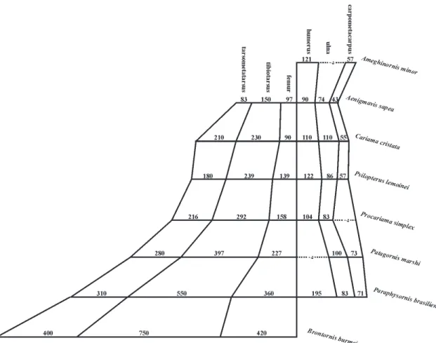
Shapeandproportionsoftheskeletonandtheirimplicationson habits – The Phorusrhacidae birds were unable to fly. This conclusion is easily arrived at by examining the proportional size of the wings (Fig. 3) and the body mass, compared with those of present-day forms.
The reduction in wing-size is more pronounced in the larger-sized species. In the diagram depicted in Fig. 3, it can be seen that Paraphysornis, when compared with certain smaller-sized forms, presents a greater reduction of the ulna (Table 1), this reduction appear-ing to be the main indicator of the loss of the ability to fly. Another example is the comparison of Psilopterus (the smallest Phorusrhacidae) with Cariamacristata, wherein Psilopterus possessed a much larger body-mass, besides an outstanding reduction of the ulna amongst the other wing bones. Moreover, a comparison of the hind-limb bones shows that the shortening of the fe-mur and the relative lengthening of the tarsometatar-sus of Cariama in relation to Psilopterus, associated to a smaller bulk appears to be a greater adaptation to run-ning in Cariama. Thus Psilopterus, contrary to the con-clusions of Tonni & Tambussi (1988), was unable to fly and was slower than Cariamacristata at running, the latter, on the other hand, achieving long gliding flights of up to some hundreds of meters.
The aptitude for speed in birds when running is proportional to the length of the tarsometatarsus in relation to the tibiotarsus. Thus, in the subfamily Brontornithinae, the largest and heaviest of the Phorusrhacidae, the length of the tarsometatarsus is between 50 to 60% of that of the tibiotarsus, indicat-ing they were slow movindicat-ing, walkindicat-ing birds, while in Patagornis this proportion is near to 70%, implying much greater agility and speed. Based on this fact, one can deduce that the representatives of the subfamily Brontornithinae must have had necrophagous habits, eating mainly dead or dying animals. This idea can be supported by the information that, in the Tremembé Formation (Taubaté Basin), a lacustrine formation, where the excellent specimen of Paraphysornisbrasiliensis came from, periods of drought, with high fish motality, occurred. In the same place, the occurrence of a vul-ture (Vulturidae) fossil (Alvarenga, 1985b), was also recorded. It is possible that this Phorusrhacidae was hereabouts looking for fishes and other dead animals when, maybe, it sank into the marshy land and suc-cumbed amongst a plentiful supply of fishes and other dead animals.
Another aspect of the skeleton of the Phorusrhacidae is the expressive narrowing of the pelvis, upper maxilla and thorax. From these charac-teristics we can deduce that these birds often hunted in regions with high vegetation, permitting their greater agility between verticle obstacles. A very narrow up-per maxilla would furthermore facilitate the appre-hension of small animals hidden amongst trunks or stones.
Another inference derived from the skeleton is from the lacrimal bone (= prefrontal), this forming a large caudal expansion (the supra-orbital process), simi-lar to that which nowadays occurs in hawks (Falconiformes), which certainly affords protection for the eyes against sunrays, thus favoring keeness of sight. From this one supposes that these birds inhabited more or less open sunlit regions and not shaded forests, and certainly hunted by sight.
TABLE 1. Measurements (total lenght) of the scapular girdle and the forelimbs of some Phorusrhacidae (mm). Numbers in brackets are estimates on incomplete bones.
Paraphysornis Titanis Patagornis Psilopterus Psilopterus Procariama Mesembriornis brasiliensis walleri Marshi Bachmanni lemoinei simplex milneedwardsis
Specimen DGM- UF- BMNH- PUM- PUM- AMNH- BMNH- FM- (measurements from
1418 30003 A524 15904 15402 9157 A559 P14525 Kraglievich, 1946)
Scapula 215.0 – – – 88.0 95.5 – 87.0 –
Coracoid 245.0 – 150.0 65.5 ( 74.0) 78.5 – 65.0 –
Humerus 195.0 – – – 103.0 – 122.5 104.0 180 a 200
Ulna 83.0 – 110.0 – 79.5 – 86.2 83.0 100 a 114
Radius – – – – 74.0 – 80.0 – 89 a 109
Carpometacarpus 71.5 9.7 76.0 – 47.5 – 56.8 – –
CharacterizationofthePhorusrhacidaefamily – Within the order Ralliformes, the family Phorusrhacidae is char-acterized by: (1) a large or even gigantic build; (2) the shape of the body, especially the premaxila, the pelvis and the thorax being laterally flattened, giving the bird a slim appearance when viewed from the front; (3) a bulky premaxilla, especially high, with a pointed, strong apex, in the form of a hook; (4) a jaw with a very solid symphysis; (5) ample and pervious nostrils (there is no septum); (6) a desmognate palate; (7) the presence of well-developed basipterigoid processes; (8) pterigoids with an evident articulation for the basipterigoid pro-cess in the mid-portion; (9) the absence of uncinate processes on the ribs; (10) the pubis atrophied in the cranial half, the same as occurs in hawks (Accipitridae); (11) the reduction of the wings and loss of the ability to fly; (12) a coracoid with the extreme reduction of the procoracoidal and acrocoracoidal processes, pos-sessing an ample scapular facet, in the form of a groove in the apex (Fig. 4B); (13) a humerus with the internal tuberosity bulging proximally, the proximal half of the diaphysis being strongly bent and the processus flexorius distally prominent (Fig. 5); (14) a tarsometa-tarsus with a triangular-shaped hypotarsometa-tarsus when viewed from below and without tendon grooves; (15) a strongly curved ungual phalanx.
The Cariamidae family, with two extant genera in South America (Cariama and Chunga), and two other very close extinct families that lived in the North American (Bathornithidae) and European (Idiornithidae) MiddleTertiary, are those that present a greater phylogenetic proximity with the Phorusrhacidae (Andrews, 1896; Brodkorb, 1967; Cracraft, 1968 and 1971; Mourer-Chauviré, 1983; Livezey, 1998). They differ, however, through their representatives being of smaller build, the premaxilla not being bulky, the presence of a schizognate palate, the frail mandibular symphysis, the absence of the basipterigoid processes, the relatively wide pelvis, the presence of the uncinate processes on the ribs, the internal tuberosity of the humerus, and a small or non-prominent processusflexorius (Fig. 5).
In the present work, the Ameghinornithinae Mourer-Chauviré, 1981, from Europe, of a size com-parable to the smallest Phorusrhacidae, and known only through the coracoid, humerus and carpometacarpus, are excluded from the family Phorusrhacidae. In spite of the reduction of the acrocoracoidal process (Fig. 4A), the coracoid of Ameghinornis presents an excavated scapular facet, this being egg-shaped and extending towards the procoracoidal process, in a shape very typi-cal of the Idiornithidae, thus reminding one especially
FIGURE 4. The coracoids of some Phorusrhacidae in compari-son with Ameghinornisminor and Cariamacristata. The left coracoid of Paraphysornisbrasiliensis (DGM-1418-R) viewed ventrally (A) and in lateral view (B); the right coracoid of Patagornis marshi (BMNH-A-524) in ventral view (C) and in lateral view (D); the left coracoid in ventral view of Psilopteruslemoinei (E) (PUM-15402, re-drawn from Sinclair & Farr, 1932), and P. bachmanni (F) (PUM-15904, also redrawn from Sinclair & Farr, 1932); the omal extremity of the right coracoid of Mesembriornismilneedwardsi, fused with a part of the clavicle, viewed dorsally (G) (redrawn from Rovereto, 1914); a reproduction of the fused clavicle and coracoid in Mesembriornis milneedwardsi (H) (redrawn from Kraglievich, 1932); the left cora-coid, viewed dorsally, of Ameghinornis minor (cast, MGHN-PQ1200) (I) and Cariamacristata (MHNT-1136) (J); sc = scapularcotyle.
of Idiornis minor or I. itardiensis (cf. Mourer-Chauviré, 1983: pls. 3 and 4). The humerus of Ameghinornis is simi-lar to that of the Phorusrhacidae as to the prominent ventral tubercle (but of a shape distinct from that of the Phorusrhacidae), and differing in other characteris-tics such as the distal part of the deltoid crest being much widened and outstanding and the processusflexorius not being prominent distally (Fig. 5D). The carpometacarpus assigned to Ameghinornis reminds one more of that of the Idiornithidae and Cariamidae than that of the Phorusrhacidae, which in turn present a very variable morphology (Fig. 6). Maybe its shape could be interpreted as being primitive for it further resembles that of the Opisthocomidae and Bathornithidae (Olson, 1985:144). The known bones of Ameghinornis compared with Psilopterus (the smallest Phorusrhacidae) and Cariama, are approximately of the same length. The ulna and radius themselves, much more reduced in Psilopterus than in Cariama, appear to be one more characteristic element of the Phorusrhacidae family, by the greater reduction than that of the other wing bones. Unfortu-nately, the radius and ulna of Ameghinornis are unknown for comparison.
Aenigmavissapea Peters, 1987, another European form described as “Phorusrhacidae” subfamily incertae sedis, and of a much smaller build, is also excluded from the Phorusrhacidae family. Amongst the few osteo-logical characteristics shown by Aenigmavis, the hypotarsus formed by two long and parallel crests dif-fers substancially from that of the Phorusrhacidae. The proportions of the Aenigmavis skeleton are different from those observed in the Phorusrhacidae (Fig. 3): the femur is much longer than the tarsometatarsus, which is only observed in the Phorusrhacidae of the subfamily Brontornithinae (certainly the most derived
phorusrhacids). Such a proportion is also observed as common in birds with arboreal habits (e.g. Columbiformes, Psittaciformes, Coraciiformes), as well as in the Galliformes of the Cracidae family, with ar-boreal habits, such as Pipile and Penelope, thus excluding running habits for Aenigmavis, which must, therefore, remain in insertaesedis. The genera Cunampaia Rusconi, 1946; Smilornis Ameghino, 1891; Pseudolarus Ameghino, 1891; Lophiornis Ameghino, 1891; and Riacama Ameghino, 1899, are also excluded, based on there be-ing few bone segments of very debatable diagnostic value.
Some fossils, still undescribed, coming from the North American Eocene (the Green River Formation), possibly belonging to the Phorusrhacidae (P. Houde, pers. inf.) are also exluded from this family. The exam-ined fossils only consist of wing bones and part of the scapular girdle, of a relatively small-built bird, wherein only the humerus shows some characteristics of the Phorusrhacidae, such as an extremely accentuated bend of the diaphysis, an extremely protruding ventral tu-bercle, besides a distally prominent processusflexorius. In this humerus, the ventral tubercle is much more developed than in any of the known Phorusrhacidae and is projected more in the anconal than proximal direction. The examined carpometacarpus presents some characteristics common to the Psilopterinae, Cariamidae, Bathornithidae and Opisthocomidae, or, in other words, presents several plesiomorph charac-teristics, which are of no assistance in an attempt at classification. The observed similarities appear to be more an adaptive convergence due to the loss of the ability to fly, it being extremely premature to classify such birds amongst the Phorusrhacidae.
The Phorusrhacidae thus consists of five sub-families: the Brontornithinae (Moreno & Mercerat, 1891), Phorusrhacinae (Ameghino, 1889), Patagornithinae (Mercerat, 1897), Psilopterinae (Dolgopol de Saez, 1927) and Mesembriornithinae (Kraglievich, 1932). All the known forms are from South America, except for Titanis walleri, a Phorusrhacinae from North America, which testifies to these birds having emigrated to North America af-ter the elevation of the isthmus of Panama from the Pliocene on.
SYSTEMATICS
The classification herein presented is especially based on the morphology and proportions of the mandibular symphysis and of the tarsometatarsus, for
two main reasons: first because one is dealing with re-sistant bony segments and most often conserved in almost all the species of this family, and also due to the fact that such structures should reflect specializa-tion in food habits as well as differences in the strength and ability of the diverse species.
Order Ralliformes Reichenbach, 1852 Suborder Cariamae Fürbringer, 1888 Family Phorusrhacidae Ameghino, 1889
Phororhacosidae Ameghino, 1889; 1891. Phororhacidae Lydekker, 1893; Ameghino, 1895. Phorusrhacidae Brodkorb, 1963,1967;
Mourer-Chauviré, 1981.
Brontornithidae Moreno & Mercerat, 1891. Devincenziidae Kraglievich, 1932.
Stereornithidae Moreno & Mercerat, 1891. Darwinornithidae Moreno & Mercerat, 1891. Patagornithidae Mercerat, 1897.
Mesembriorniidae Kraglievich, 1932.
Remarks – Ameghino (1887) described Phorusrhacos long-issimus based on an incomplete mandible, afterwards (1889) emending the original generic term to Phororhacos, which was widely accepted (Chiappe & Soria, 1990), proposing for this the criation of the fam-ily Phororhacosidae. As Brodkorb (1963) warned, the original spelling (Phorusrhacos) has a guaranteed prior-ity. Moreover, Brodkorb emended the name of the family, already previously altered by Lydekker (1893), to the Phorusrhacidae, to which almost all the authors since 1963 have acted in accordance. The terms Phorusrhacos and Phorusrhacidae were definitely made official by the International Commission on cal Nomenclature, Opinion 1687: Bulletin of Zoologi-cal Nomenclature, 49(2), June, 1992.
Subfamily Brontornithinae Moreno & Mercerat, 1891
Brontornithidae Moreno & Mercerat, 1891.
Brontornithinae; Brodkorb, 1967; Mourer-Chauviré, 1981.
DiagnosisRevised – Phorusrhacidae of a gigantic build, reaching over two meters high, heavy and robust. The mandibular symphysis is proportionally shorter, wider and higher than in the other Phorusrhacidae (Fig. 7). The tarsometatarsus is proportionally short, widened and
flattened dorso-plantarwise (Fig. 2 and Fig. 8), its length reaching only 50% to 60% of that of the tibiotarsus (Fig. 3). In Brontornis, the dorsomedial extremity of the articular surface of the mesotrochlea of the tarsometa-tarsus is proximally projected, when viewed in profile (Fig. 8C), whilst in Paraphysornis this portion is more medially expanded. Even though the trochlae of Physornis are as yet unknown, one can assume that the dorsomedial portion of the mesotrochlea in the Brontornithinae may be always expanded and could constitute one more char-acteristic of the subfamily.
Included Genera – Brontornis Moreno & Mercerat, 1891, Physornis Ameghino, 1895, and Paraphysornis Alvarenga, 1993.
Remarks – Case etal. (1987) described the fragment of a beak coming from the La Meseta Formation (Upper Eocene ?) from Seymour Island (Antarctic) as a pos-sible portion of the premaxilar region of a Phorusrhacidae. Just by examining the published illus-trations, the appearance of the pieces surmises the man-dibular symphysis of a Brontornithinae, close to Brontornis. More material and information are required in order to arrive at a precise definition of this pos-sible, as yet unknown, Brontornithinae.
Genus Brontornis Moreno & Mercerat, 1891
Brontornis Moreno & Mercerat, 1891:20,37; Brodkorb, 1967.
Rostrornis Moreno & Mercerat, 1891:20,40; Brodkorb, 1967 (syn. of Brontornis).
Type Species – Brontornisburmeisteri Moreno & Mercerat, 1891.
Included Species – Only the type species.
Distribution – Lower to Mid-Miocene in Argentina.
Diagnosis Revised – Certainly the biggest of the Phorusrhacidae, it is the largest bird known from the Americas and one of the largest that has ever existed. The mandibule possesses a proportionally shorter, wider and higher symphysis than Physornis and Paraphysornis (e.g. Alvarenga, 1993: Fig. 1) The internal condyle of the tibiotarsus is medially diverted. The cotyls of the tarsometatarsus are rounded off and the
hypotarsus forming a prominent medial crest, having a slightly pronounced lateral edge (e.g. Alvarenga, 1993: Fig. 2). As regards the remaining Brontornithinae, the hypotarsus of Brontornis is placed on a more distal level in relation to the articular cotyles (Fig. 9).
Remarks – Liornis Ameghino,1895, with the species L. floweri Ameghino, 1895, and L. minor Dolgopol de Saez, 1927, are evident synonyms of Phorusrhacos Ameghino, 1887; Devincenzia Kraglievich 1932, is surely a robust form of Phorusrhacinae. These two genera were dealt with as synonyms of Brontornis by Brodkorb (1967).
Brontornisburmeisteri Moreno & Mercerat, 1891
Brontornis burmeisteri Moreno & Mercerat, 1891:37; Brodkorb, 1967.
Rostrornisfloweri Moreno & Mercerat, 1891:40; Brodkorb 1967 (syn. of B. burmeisteri).
Brontornis platyonyx Ameghino, 1895; Brodkorb 1967
(syn. of B. burmeisteri).
Lectotypes – The left femur, tibiotarsus, fibula and tar-sometatarsus (MLP-88-91), certainly belonging to the same individual, designated by Brodkorb (1967).
Hypodigm – lectotypes; portion of the mandible includ-ing the symphysis and part of the right branch (MHNP-1902-6, Fig. 7); two large fragments of the
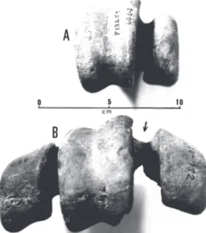
FIGURE 8. Brontornisburmeisteri (FM-P13259), left complete tar-sometatarsus (l) and distal end of the right one (r), in dorsal view (A), plantar view (B), and lateral view (C). This specimen, around 25% smaller than the lectotype (MLP-91), represents an important variation in size in these birds. The arrow in “C” indicates the dorsoproximal spreading of the edge of the articular surface of the middle trochlea, a characteristic of the Brontornithinae.
mandibular symphysis (MLP-94-95), mistakenly attrib-uted to the premaxillas by Moreno & Mercerat (1891); quadrate, several complete and incomplete thoracic and caudal vertebras, phalanges and fragments of the hind-limbs (MLP-92-93 and 96-117, Fig. 10B); the complete left and the distal end of the right tarsometatarsus (FM-P13259, Fig. 10A); a distal extremity of the right tarsometatarsus (BMNH-A578); distal extremity of the left tarsometatarsus (BMNH-A580); 10 podal and un-gual phalanges, the majority belonging to the left foot, apparently from the same specimen (BMNH-A549, Fig. 11); distal end of the left femur (FM-P15309).
Horizon and Locality – Lower and Middle Miocene
(Santacrucian) of Argentina, Province of Santa Cruz: Lago Argentina, Monte Leon, Monte Observación, Kariaken, La Cueva, Rio Gallegos.
Measurements – Table 2.
Illustrations – Moreno & Mercerat (1891).
Remarks – The trochleae of the right tarsometatarsus of specimen MLP-112 (Fig. 10B), which Moreno & Mercerat (1891) conceived and assigned to Rostrornis
floweri, is a mistaken assemblage, wherein the internal trochlea is, in fact, an external left trochlea. Such an error in assemblage also served Dolgopol de Saez (1927) to characterize the genus Rostrornis. An appre-ciable difference in the size of the tarsometatarsus of specimens FM-P13259 and MLP-91 (lectotype) (Fig. 2C and 2D), shows the second to be around 33% larger than the first, which possibly testifies to a sexual dimorphism, seeing that by the characteristics, both are adults. Two distal fragments of tarsometatarsus from the museum in London (BMNH-A578 and A580) are in accordance with this difference in size (Table 2). Ameghino (1895) described Brontornisplatyonyx, basing it on the much smaller build than B. burmeisteri. His measurements are, however, compatible with the abovementioned variation for the species (Fig. 11).
Genus Physornis Ameghino, 1895
Physornis Ameghino, 1895:576; Brodkorb, 1967.
Aucornis Ameghino, 1898:9; Brodkorb, 1967 (syn. of
Physornis).
Type Species – Physornisfortis Ameghino, 1895.
Included Species – Only the type species.
Distribution – Middle and Upper Oligocene of Argen-tina.
Diagnosis Revised – Of a gigantic size, rivaling with Brontornis. A very short and wide mandibular
symphy-FIGURE 10. Brontornisburmeisteri: distal view of the right tarsometa-tarsus. A - cast of specimen FM-P13259, lacking the external tro-chlea; B - specimen MLP-112. Note the error in assembling (B) the “internal” trochlea (arrow), which, in fact, is the left external tro-chlea. This mistake, which was initially made by Moreno & Mercerat (1891), was also passed on by Dolgopol de Saez (1927), serving as a notable difference for the characterization of the genus Rostrornis.
sis, characteristically with an almost flat ventral sur-face in the mid-portion (Fig. 12 and e.g. Alvarenga, 1993: Fig. 1). The lateral cotyl of the tarsometatarsus is al-most quadrangular, when viewed proximally (Fig. 13A). The lateral edge of the hipotarsus, when viewed from the rear, forms a prominent crest which distiguishes it well from Brontornis and Paraphysornis (Fig. 13 and e.g. Alvarenga, 1993: Fig. 2).
Remarks – The genus Physornis and its type species P. fortis, were described by Ameghino in 1895, based on a fragment of the mandible comprehending part
of the symphysis and the right branch, this descrip-tion, unfortunately, not being accompanied by an il-lustration.
Patterson (1941:52) examined the type of Physornisfortis, nowadays deposited in the museum of London (BMNH-A583), arriving at the conclusion that this is a bony fragment without any morphological characteristic, possibly being the iliac crest of a mam-mal, thus proposing the rejection of the terms, both for the genus as well as the species, as being indeter-minate. We had also the opportunity of examining the aforementioned type material (Fig. 14), arriving at the
TABLE 2. Measurements of Brontornisburmeisteri (cm). Numbers in brackets are estimates on incomplete bones.
Mandible MLP-94 MHNP-1902-6
Length of the symphysis on the dorsal surface (14.4) (14.7)
Maximum width at the base of the symphysis ( 9.4) 10.1
Height at the base of the symphysis ( 7.5) 8.4
Femur MLP-88 FM-P15309
Total length (42.0) –
Width in the middle of the diaphysis 7.5 –
Dorsoventral diameter in the middle of the diaphysis 5.8 –
Maximum distal width (15.5) (15.5)
Dorsoventral diameter of the internal condyle – 11.0
Dorsoventral diameter of the lateral condyle – 11.7
Tibiotarsus MLP-89
Total length 75.0
Width in the middle of the diaphysis 6.3
Tarsometatarsus MLP-91 FM-P13259 BMNH-A578 BMNH-A580
Total length 40.0 30.2 – –
Maximum proximal width 13.2 11.5 – –
Width in the middle of the diaphysis 7.4 5.8 – –
Width at the distal foramen level 9.7 8.0 9.9 8.1
Width of the trochlea at the distal end:
internal – 2,5 – –
middle 5.5 4.4 5.5 4.8
lateral 3.7 3.3 3.7 –
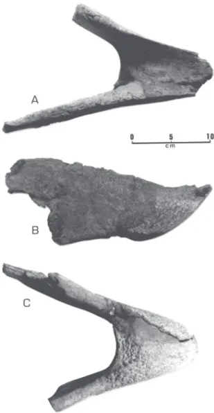
FIGURE 12. Physornisfortis; symphysis with a part of the left man-dibular branch (FM-P13340) in views: A - ventral, B - left lateral, C - dorsal, D - mandibular symphysis (FM-P13619) in dorsal view.
conclusion that this is undoubtedly a fragment of the symphysis and part of the right branch of a large Phorusrhacidae. The experience acquired in the resto-ration of the mandible of Paraphysornis (Alvarenga, 1982), contributed to us for the immediate recogni-tion of the texture of a real mandibular symphysis of a Phorusrhacidae. Further, it is evident that Ameghino had prior experience, having examined and described several other mandibular symphysis of Phorusrhacidae, such as, Phorusrhacos longissimus Ameghino, 1887;
Phororhacos sehuensis Ameghino, 1891; Phororhacos
platygnathus Ameghino, 1891; Tolmodus inflatus
Ameghino, 1891, and others, this showing his intimacy with this anatomical portion, which, apparantly, is of-ten conserved in the Phorusrhacidae fossils.
Although it is an almost shapeless fragment and, thus, very inadequate for typifying a species, the type demonstrates, besides the size and the geographical and stratigraphical source, also the flat portion of the ventral surface of the mandibular symphysis, which is characteristic of this genus (Figs. 12A and 14).
Physornisfortis Ameghino, 1895
Physornisfortis Ameghino, 1895:576; Brodkorb, 1967. Aucorniseuryrhyncus Ameghino, 1898:9; Brodkorb, 1967
(syn. of Physornisfortis).
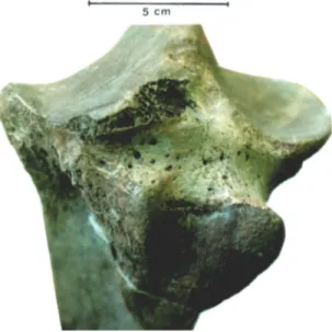
Type – A fragment around 137 cm in length, constitut-ing the ventral portion of the mandibular symphysis and the adjacent part of the right mandibular branch (BMNH-A583: Fig. 14).
Hypodigm – Type; a mandibular symphysis, including a part of the left branch (Figs. 12A-C), associated to the caudal portion of the left quadratojugal (Fig. 15A), and to the atlas (Fig. 16A), besides fragments of vertebras, phalanges etc. (FM-P13340). A well conserved madibular symphysis (Fig. 12D), associated to the cau-dal extremity of the right quadratojugal (FM-P13619).
FIGURE 14. Fragment of the symphysis and right mandibular branch of Physornisfortis (type: BMNH-A583) ventral (A) and dorsomedial (B) views. Compare with Fig. 12A.
FIGURE 15. Caudal portion of the left quadratojugal, in medial view of: A - Physornisfortis (FM-P13340); B - Paraphysornisbrasiliensis (DGM-1418-R).
A proximal portion of the left tarsometatarsus (MACN-A-52-185, Figs. 2A and 13). A rostral extremity of the mandibular symphysis (MACN-A-52-186). A phalanx 1 of the toe II from the left foot (MACN-A-52-187); these last three bones were asso-ciated and served as type for Ancornis euryrhyncus Ameghino, 1898. A phalanx 1 of the toe IV from the left foot (MACN-A-52-188). In the Museum of Amherst College, Massachussetts, U.S.A., there is also a right femur, incomplete on the proximal extremity, without number (?), refered to and figured by Loomis (1914:226).
HorizonandLocality – Middle to Upper Oligocene of Argentina (Deseadan). Province of Santa Cruz, Ar-gentina: Puerto Deseado, Punta Nova, La Flexa, in the levels of occurrence of Pyrotherium. For a better indi-cation of the geological age, see Mac Fadden (1985) and Marshall etal. (1986).
Measurements – Table 3.
Illustrations – Loomis, 1914: Fig. 149.
Remarks – The size of the mandibular symphysis (type: BMNH-A583), originally described by Ameghino (1895), is in perfect agreement with the two measured specimens in the Field Museum of Chicago (Table 3). A flat (not convex) region in the mid-ventral portion of the symphysis is noted in all the specimens.
The branches of the mandible of Physornis, in comparison with those of Phorusrhacos, are really more further apart from the median line, as in the original
description by Ameghino (op. cit.), which implies a skull with a wider base. Ameghino perfectly described the only large built Phorusrhacidae of the stratigraphic layers characterized by the presence of Pyrotherium (Deseadan). Thus, there is no reason for the rejection of the genus Physornis and the species P. fortis, as being indeterminate, as suggested by Patterson (1941).
Although the complete shape of the tarsometa-tarsus of Physornis is unknown, the proximal portion of this is shown to be quite flattened in the dorsoven-tral extent. Associating this to the short and wide man-dibular symphysis, besides the quite large build, it fits in perfectly amongst the Brontornithinae.
Aucornissolidus Ameghino, 1898, considered by Brodkorb (1967) as synonymous of Physornisfortis, was created only based on a proximal extremity of a pha-lanx (MACN-A-52-110), of a much smaller-sized bird than Physor nis fortis, possibly synonymous of Andrewsornisabbotti Patterson, 1941, which should be left as a speciesinquirenda, seeing that one is dealing with an absolutely insufficient segment to diagnose a spe-cies.
The two symphysis examined in the Field Mu-seum of Chicago, showed an interesting difference on the dorsal surface, which, almost flat in FM-P13340, forms a longitudinal channel in FM-P13340 (Figs. 12C and 12D). These differences were interpreted as being individual variation.
Genus Paraphysornis Alvarenga, 1993
Type Species – Physornisbrasiliensis Alvarenga, 1982.
TABLE 3. Measurements of Physornisfortis (cm). Numbers in brackets are estimates on incomplete bones.
Mandible FM-P13340 FM-P13619
Length of the mandible on the dorsal surface (11.0) 11.3
Width of the base of the symphysis 7.0 ( 7.5)
Height at the base of the symphysis ( 5.9) –
Atlas FM-P13340
Condyloid fossa (maximum width) 3.82
Femur*
Smallest transverse diameter of the diaphysis 5.8
Maximum distal width 14.8
Tarsometatarsus MACN-A52-185
Maximum proximal width 10.5
Proximal dorsoventral diameter 6.7
Width of the diaphysis on the level of the fracture (Fig. 13) 5.4
Falanx 1, digit IV, left foot MACN-A52-188
Length of the axis 6.3
Maximum proximal width 4.5
Maximum distal width (3.2)
Included Species – Only the type species.
Distribution – Upper Oligocene or the Lower Miocene of Southeast Brazil.
Diagnosis – Maybe the smallest of the Brontornithinae, the size being comparable to that of a smaller-sized Brontornisburmeisteri (Figs. 1B and 2B). The mandibu-lar symphysis is longer and narrower than in the re-maining Brontornithinae (e.g. Alvarenga, 1993: Fig. 1), being, however, proportionally wider and shorter than in the Phorusrhacinae. The cotyles of the tarsometa-tarsus are slightly quadrangular, especially the inner one (e.g. Alvarenga, 1993: Fig. 2).The lateral edge of the hypotarsus expands until very near to the lateral part of the lateral cotyle, not forming the lateral crest char-acteristic of Physornis.
Paraphysornisbrasiliensis (Alvarenga, 1982)
Physornisbrasiliensis Alvarenga, 1982.
Holotype – The almost complete skeleton of one speci-men (Fig. 17), most of the upper maxilla, braincase, pelvis and sternum being missing (DGM-1418-R).
Hypodigm – Only the type material.
Horizon and Locality – The Tremembé Formation,
Taubaté Basin, State of São Paulo, Brazil. Age near to the Upper Deseadean, certainly the Upper Oligocene or the Lower Miocene (Soria & Alvarenga, 1989, and Alvarenga, 1990).
Measurements – Tables 1 and 4; Alvarenga (1982).
Illustrations – Alvarenga (1982, 1993).
Remarks – A comparison as to the size of the
Brontornithinae, becomes difficult due to the lack of a larger number of individuals and better knowledge of species size variations. The dimension of the tar-sometatarsus of Paraphysornis (the only known speci-men) is equivalent to that of a small Brontornis (FM-P13259), even though a little less bulky (Figs. 2B and 2C). On the other hand, a comparison of the basal portion of the quadratojugal (Fig. 15), of the atlas (Fig. 16), of the third cervical vertebra and of the proxi-mal portion of the tarsometatarsus of Physornis and Paraphysornis, shows a slight superiority in the size of these in Physornis. However, other specimens, as they
come to light, may alter this difference, for the time being established.
Subfamily Phorusrhacinae Ameghino, 1889
Phororhacosidae Ameghino, 1889. Phororhacidae Ameghino, 1895. Phororhacinae Kraglievich, 1932. Phorusrhacinae Brodkorb, 1963; 1967.
DiagnosisRevised – Phorusrhacidae, of a gigantic build, having, however, a more slender and lighter constitu-tion, certainly being more nimble, agile and faster (Fig. 1C), than the Brontornithinae. The mandibular symphysis, relatively longer, narrower and not so high (Figs. 18 and 19), is more than twice as long as the width of the base. The tarsometatarsus (Figs. 2E and 2F), relatively long and slender, is always longer than 60% of the length of the tibiotarsus (Figs. 20 and 21).
Included Genera – Phorushacos Ameghino, 1887, Devincenzia Kraglievich, 1932 and Titanis Brodkorb, 1963.
Genus Phorusrhacos Ameghino, 1887
Phorusrhacos Ameghino, 1887:24; Bordkorb, 1967. Phororhacos Ameghino, 1889:659.
Stereornis Moreno & Mercerat, 1891:21, 45; Brodkorb, 1967 (syn. of Phorusrhacos).
Darwinornis Moreno & Mercerat, 1891:26, 60; Brodkorb, 1967 (syn. of Phorusrhacos).
Owenornis Moreno & Mercerat, 1891:25, 64; Brodkorb, 1967 (syn. of Phorusrhacos).
Mesembriornis Moreno, 1889:29; Brodkrob, 1967 (syn. of Phorusrhacos).
Titanornis Mercerat, 1893:5; Brodkorb, 1967 (syn. of Phorusrhacos).
Callornis Ameghino, 1895:574; Brodkorb, 1967 (syn. of Phorusrhacos).
Eucallornis Ameghino, 1901:78; Brodkorb, 1967 (syn. of Phorusrhacos).
Liornis Ameghino, 1895:570; syn. nov.
Type Species – Phorusrhacoslongissimus Ameghino, 1887.
Included Species – Only the type species.
Distribution – Lower and Middle Miocene of
Argen-tina.
Diagnosis – Of a very big build, being, however, smaller than Devincenzia. The length of the tarsometatarsus is about 70% of that of the tibiotarsus (Figs. 20 and 21),
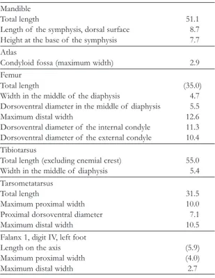
TABLE 4. Measurements of Paraphysornisbrasiliensis (cm), speci-men DGM-1418-R. Numbers in brackets are estimates based on incomplete bones.
Mandible
Total length 51.1
Length of the symphysis, dorsal surface 8.7 Height at the base of the symphysis 7.7 Atlas
Condyloid fossa (maximum width) 2.9
Femur
Total length (35.0)
Width in the middle of the diaphysis 4.7 Dorsoventral diameter in the middle of diaphysis 5.5
Maximum distal width 12.6
Dorsoventral diameter of the internal condyle 11.3 Dorsoventral diameter of the external condyle 10.4 Tibiotarsus
Total length (excluding cnemial crest) 55.0
Width in the middle of diaphysis 5.4
Tarsometatarsus
Total length 31.5
Maximum proximal width 10.0
Proximal dorsoventral diameter 7.1
Maximum distal width 10.5
Falanx 1, digit IV, left foot
Length on the axis (5.9)
Maximum proximal width (4.0)
Maximum distal width 2.7
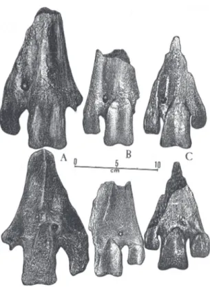
FIGURE 19. Incomplete mandible of Phorusrhacoslongissimus (BMNH-A530) in left lateral view.
FIGURE 20. Phorusrhacoslongissimus (AMNH-9146). Left tarsometa-tarsus from Cañon de Las Vacas, Province of Santa Cruz, Argen-tina. Dorsal view. Maximum length 362 mm, calculated at 370 mm if it were complete.
the distal portion of the mid-trochlea not being later-ally expanded as is the case of Titanis (Fig. 22).
Remarks – Within the subfamily, Phorusrhacos is that which possesses the largest amount of fossils, these coming from the Santacruzian sediments in Argentina. To elaborate a more detailed anatomical diagnosis which would separate Phorusrhacos from both Devincenzia and Titanis becomes, however, difficult through the deficiency in material, seeing that the latter two genera are very badly represented. It is worthwhile pointing out that Devincenzia, with a size which appears to ex-ceed that of Phorusrhacos, comes from more recent lev-els of Argentina and Uruguay, whereas Titanis, with the same size, is only known from a still more recent terrain of North America.
Phorusrhacoslongissimus Ameghino, 1887
Phorusrhacoslongissimus Ameghino, 1887:24; Brodkorb, 1967.
Phororhacoslongissimus Ameghino, 1889:659.
Stereornis rollieri Moreno & Mercerat, 1891:21, 45; Brodkorb, 1967 (syn. of P. longissimus).
Stereornis gaundryi Moreno & Mercerat, 1891:21,47; Brodkorb, 1967 (syn. of P. longissimus).
Mesembriornisstuderi Moreno & Mercerat, 1891:21, 48; Brodkorb 1967 (syn. of P. longissimus).
Mesembriornisquatrefragesi Moreno & Mercerat, 1891:22, 50; Brodkorb, 1967 (syn. of P. longissimus). Darwinorniscopei Moreno & Mercerat, 1891:24, 60;
Brodkorb, 1967 (syn. of P. longissimus). Darwinorniszittelli Moreno & Mercerat, 1891:25, 63; Brodkorb, 1967 (syn. of P. longissimus).
Darwinornissocialis Moreno & Mercerat, 1891:25, 63; Brodkorb, 1967 (syn. of P. longissimus).
Owenornis affinis Moreno & Mercerat, 1891:25, 64; Brodkorb, 1967 (syn. of P. longissimus).
FIGURE 22. Right tarsometatarsus (distal end) of the Phorusrhacinae: A - Devincenzia pozzi (type: MACN-6554); B - Phorusrhacoslongissimus (MLP-132) and C - Titaniswalleri (type: UF-4108). Dorsal (above) and plantar (below) views. Note in Titanis a widening of the transverse diameter on the distal end of the mid-trochlea.
Owenornis lydekkeri Moreno & Mercerat, 1891:25, 64; Brodkorb, 1967 (syn. of P. longissimus).
Phororhacos sehuensis Ameghino, 1891:258; Brodkorb, 1967 (syn. of P. longissimus).
Phororhacosplatygnathus Ameghino, 1891:452; Brodkorb, 1967 (syn. of P. longissimus).
Titanornis mirabilis Mercerat, 1893:5; Brodkorb, 1967 (syn. of P. longissimus).
Callornisgiganteus Ameghino, 1895:78; Brodkorb, 1967 (syn. of P. longissimus).
Eucallornisgiganteus Ameghino, 1901:78; Brodkorb, 1967 (sin. of P. longissimus).
Liornisfloweri Ameghino, 1895; syn. n. Liornisminor Dolgopol de Saez, 1927; syn. n.
Type – A mandibular symphysis, including part of the right mandibular branch, but lacking the rostral extrem-ity, MLP-118 (originally described as the mandible of a toothless mammal).
Hypodigm – Type; several segments of skeletons
which served as the type for the various synonyms of the species (MLP-119 to 139, 171 to 182). The species is known from segments of almost all the skeleton, there lacking, however, a better represen-tation of the skull. Ameghino (1895) tells of the observation and measurements of the skull of a specimen in nature, in fragments and encrusted in crumbling rock. The mandible, the rostral extrem-ity of the upper maxilla and a fragment apparently from the caudal portion of the supra-orbital pro-cess (BMNH-A529, Figs. 18A, 23B, 23C, 24A and 24C), coming from this specimen, were collected. Several other segments of the mandible represent the species (Figs. 18 and 19). For P. longissimus there is still lacking an appropriate number of associated bones for a whole reconstruction of the bird. The specimen AMNH-9146 (Fig. 20) represents an al-most complete left tarsometatarsus, and the AMNH-9497 (Fig. 21) is represented by some asso-ciated bones of a leg.
Horizon and Locality – Lower and Middle Miocene
(Santacrucian) of Argentina; Santa Cruz Formation, the Province of Santa Cruz: La Cueva, Tagua Quemada, Monte Observación, Rio Shehuen.
Measurements – Table 5; Moreno & Mercerat, 1891; Ameghino, 1895.
Illustrations – Moreno & Mercerat, 1891 and Ameghino, 1895.
Genus Devincenzia Kraglievich, 1932
Devincenzia Kraglievich, 1932:323, 338. Onactornis Cabrera, 1939:15; syn. n.
FIGURE 23. A - Restoration of the skull of Devincenziapozzi, modi-fied by Patterson & Kraglievich (1960), based on specimen MLP-37-11-7-8 (type of Onactornispozzi Cabrera, 1939). B and C - Phorusrhacoslongissimus, drawing by Ameghino (1895), based on a “complete skull, but in such a bad state of conservation that it is almost reduced to dust” (BMNH-A529); C - dorsal view of the mandible. The skulls of both Phorusrhacinae are of approximately the same size, and differences in height can, at least partially, be attributed to deformation in fossilization and defects in the reas-sembling of the skull of Devincenziapozzi.
Type Species – Devincenziagallinali Kraglievich, 1932, jun-ior synonym of Devincenziapozzi (Kraglievich, 1931).
Included Species – Only the type species.
Distribution – Upper Miocene to the Lower Pliocene
of Argentina and Uruguay.
Diagnosis – They are the biggest of the known
Phorusrhacinae, with a height comparable to that of the largest Brontornithinae, being, however, lighter. The skull is about 65 cm long, equivalent to that of Phorusrhacos, this being possibly flattened dorso-ven-trally (Fig. 32A). The tarsometatarsus is substantially sturdier than that of Phorusrhacos (Figs. 2F and 22A).
Remarks – The general appearance of the tarsometa-tarsus of Devincenzia (MNHN-189, Fig. 2F), appears to indicate an intermediate position between the Phorusrhacinae and Brontornithinae, as observed by Kraglievich (1932) himself. However, the appearance of the bony cortex makes it clear that one is dealing with a not yet completely adult individual. As happens with several present-day Ralliformes, especially the Rallidae and Cariamidae, besides other groups of birds,
it is normal that the proximal metaphysary portion of the tarsometatarsus reaches, in youngsters, a larger width than that in the adults, associated to a shorter length of the bone. Consequently, one supposes that the tarsometatarsus of Devincenzia, in the adult phase, might be a little longer and with the proximal region slightly narrower than appears in the refered specimen. The origin of this tarsometatarsus (MNHN-189), type of Devincenziagallinali Kraglievich, 1932, and, conse-quently, its geological age, are unknown. From the ap-pearance of the bone (and certainly the remains of the encrusted substract), Kraglievich supposed it to have originated from the Arroyo Roman, Rio Negro, Uruguay, and points out the difference of color, espe-cially when compared with the bones coming from Patagonia, without, however, comparing it with speci-mens from the Huayquerian of north Argentina, stud-ied years before by he himself (1931).
Devincenziapozzi (Kraglievich, 1931). comb. n.
Phororhacospozzi Kraglievich, 1931:306.
Phororhacoslongissimusmendocinus Kragkievich, 1931:314; syn. n.
TABLE 5. Measurements of Phorusrhacoslongissimus (cm). Numbers in brackets are estimates based on incomplete bones.
MLP- BMNH- BMNH-
BMNH-118 A529 A530 A684
Skull*
Total length – (65) – –
Width at base – (30) – –
Height of maxilla – (25) – –
Mandible
Total length – 55 – –
Length of symphysis (16) 17.9 – –
Height- base of the symphysis 5.3 6.2 5.7 5.4
Width- base of symphysis 7.0 6.7 6.6 6.0
BMNH- BMNH- BMNH- MLP- MLP- AMNH-
AMNH-A581 A531 A545 131 76V.1011 9497 9146
Femur
Total length – – – – – – (31.0 )
Width middle of diaphysis – – – – – – 3.62
Maximum distal width – – – – – – 9.2
Tibiotarsus
Total length – – – – – – (50.0)
Smallest width of diaphysis – – – – – – 3.9
Maximum distal width – – – – – – 6.2
Tarsometatarsus
Total length – – – 38.5 (40.0) 37.0 –
Proximal width – – – 8.0 ( 8.4) – –
Width- middle of diaphysis – – – 3.7 3.7 3.6 –
Width at distal foramen level 6.0 7.1 6.5 6.4 6.5 6.3 –
Width of the middle trochea 2.9 – 3.5 3.3 3.6 3.4 –
Devincenziagallinali Kraglievich, 1932:323, 338; syn. n. Onactornisdepressus Cabrera, 1939:15; Brodkorb, 1967
(syn. of O. pozzi).
Onactornispozzi; Brodkorb,1967:165; syn. n. Onactornismendocinus; Brodkorb, 1967:166; syn. n.
Type – Distal portion of the right tarsometatarsus (Fig. 22A), associated to the ungual phalanx of digit II (MACN-6554 and 6681).
Hypodigm – Type; a skull much deformed by dorsoven-tral flattening, most of the upper maxilla being incom-plete (Fig. 23A), together with the proximal phalanges of digits II and III (MLP-37-III-7-8), type of Onactornis depressus Cabrera, 1939; right tarsometatarsus, without the inner trochlea and a large part of the hypotarsus (MNHN-M-189, Fig. 2F), type of Devincenziagallinali (Kraglievich, 1932); fragment of the madibular sym-physis (MACN-6933), described by Kraglievich (1931) as Phororhacos ? aff. platygnathus; proximal portion of a right femur, with the head missing (MACN-6930); dis-tal portion of the left tibiotarsus (MACN-13243).
Horizon andLocality – Upper Miocene to the Lower
Pliocene of Argentina, Provinces of Buenos Aires and Entre Rios (Huayquerian and “Mesopotamian”) and Uruguay (Arroyo Roman ?).
Measurements – Table 6; Kraglievich, 1931; Patterson & Kraglievich, 1960.
Ilustrations – Kraglievich (1931 and 1932) and Cabrera (1939).
Remarks – Tambussi etal. (1999) described an almost com-plete right tibiotarsus (MNHN-M-1563) about 720 mm long, coming from the Raigon Formation (Pliocene) of south Uruguay. In this specimen the cnemial crests are extremely large and cranially projected, in a similar way to what occurrs in Titaniswalleri (UF-7333), and different from what occurs with Brontornithinae. This tibiotarsus should be assigned to the genus Devincenzia and very prob-ably to D. pozzi by the characters of a Phorusrhacinae, the size, age and geographic distribution. Unfortunatelly there is no sufficient material for a definite comparison. Another question would be a better definition of the difference in age between the sediments of the Raigon Formation of Uruguay with the Huayquerian and “Mesopotamian” of Argentina, as well as a new study on the possibility that the tarsometatarsus type of D. gallinali (Kraglievich, 1932), may have also been col-lected in the Raigon Formation and not in the Arroyo Román as supposed by Kraglievich (1932).
Genus Titanis Brodkorb, 1963
Type Species – Titaniswalleri Brodkorb, 1963.
Included Species – Only the type species.
Distribution – Upper Pliocene to Lower Pleistocene of Florida and Texas (U.S.A.).
Diagnosis – Size similar to that of Phorusrhacos, with a less sturdy tarsometatarsus than that of Devincenzia. The mid-trochlea, in the more distal portion, is spread out
TABLE 6. Measuments of Devincenziapozzi and Titaniswalleri (cm). Numbers in brackets are estimates on incomplete bones.
Devincenziapozzi Titaniswalleri
MACN-65541 MHNM-1892 MLP-37.III.7.83 UF-4108 UF-4109
Skull
Total length – – (65.0) – –
Width at base – – (33.0) – –
Height of maxilla – – (17.0) – –
Tarsometatarsus
Total length – (40.0) – – –
Proximal width – (11.0) – – –
Width at the distal foramen level 8.5 8.6 – 6.0 –
Width of the middle trochea 4.8 4.3 – 3.6 –
Falanx 1, digit III
Length (ventral surface) – – (12.5) – 10.4
Proximal height – – 5.0 – 5.8
Proximal width – – 5.1 – 5.4