691 691 691 691 691 Mem Inst Oswaldo Cruz, Rio de Janeiro, Vol. 99(7): 691-696, N ovem ber 2004
Studies of M embrane Fluidity and H eart Contractile Force in
Trypanosoma cruzi
Infected M ice
Julio E Enders+, A Ruth Fernández+ +, H éctor W Rivarola, Patricia A Paglini, José A Palma
Cátedra de Física Biomédica, Facultad de Ciencias Médicas, Universidad Nacional de Córdoba, Santa Rosa 1085, 5000 Córdoba, Argentina
In Chagas disease serious cardiac dysfunction can appear. We specifically studied the cardiac function by evaluating: ventricle contractile force and norepinephrine response, affinity and density of β-adrenergic receptors, dynamic properties of myocardial membranes, and electrocardiography. Albino swiss mice (n = 250) were infected with 55 trypomastigotes, Tulahuen strain and studied at 35, 75, and 180 days post-infection, that correspond to the acute, indeterminate, and chronic phase respectively.
Cardiac β-adrenergic receptors’ affinity, myocardial contractility, and norepinephrine response progressively decreased from the acute to the chronic phase of the disease (p < 0.01). The density (expressed as fmol/mg.prot) of the receptors was similar to non-infected mice (71.96 ± 0.36) in both the acute (78.24 ± 1.67) and indeterminate phases (77.28 ± 0.91), but lower in the chronic disease (53.32 ± 0.71). Electrocardiographic abnormalities began in the acute phase and were found in 65% of the infected-mice during the indeterminate and chronic phases. Membrane contents of triglycerides, cholesterol, and anisotropy were similar in all groups. A quadratic correlation between the affinity to β-adrenergic receptors and cardiac contractile force was obtained. In conclusion the changes in cardiac β-adrenergic receptors suggests a correlation between the modified β-adrenergic receptors affinity and the cardiac contractile force.
Key words:Chagas disease - cardiomyopathy - contractile function - cardiac β-receptors
Chagas disease is caused by the parasite Trypano-soma cruzi and during the course of the disease charac-teristically develops cardiomyopathy and digestive com-plications (Tanowitz et al. 1992, Andrade 1999).
The mechanisms proposed to explain the cardiac dam-age are related to the persistence of T. cruzi at specific sites of the infected host and to the immune response induced by the infection. A complete understanding of the pathogenesis of the disease, however, needs to be elucidated (Brener & Gazzinelly 1988, Petri & Eissen 1989, Salomone et al. 2001).
Antibodies against different parasite components and other cardiac structures have been identified (Ferrari et al. 1995) and specific antibodies anti-cardiac β-adrenergic and muscarinic receptors (Borda & Sterin Borda 1996) seem to play an important role in the pathogenesis of the typi-cal Chagas cardiopathy disorders: brady-arrhythmias and tachy-arrythmias (Elizari 1999). Several evidences have shown that the neurohumoral catecholamine system plays a crucial role in cardiac diseases (Brodde 1996). Changes in β-adrenergic receptors such as down regulation, un-coupling from G-proteins and internalization or degrada-tion of the receptors may contribute to the abnormalities of the contractile function (Chakraborti et al. 2000).
Financial support: Universidad National de Córdoba
+Corresponding author. Fax: +54-351-4332019. E-mail:
++Senior investigator from Consejo Nacional de Investigaciones
Científicas y Técnicas de la República Argentina Received 8 March 2004
Accepted 14 September 2004
Additionally, the chronic activation of the sympathetic nervous system leads to a decrease of the function of adrenergic receptors in patients with chronic heart dis-eases. The number of β1-receptors is lower in dilated car-diopathies of any origin, and this fact is directly related to the severity of the disease (Brodde 1991, 1996, Bristow 1993, Harding et al. 1994).
Chagas cardiopathy has been described as a myo-neurocardiopathy with inflammatory involvement, micro-circulation alterations (Madoery & Madoery 1992) asso-ciated to modifications in catecholamine response and myocardial contractility (Paglini-Oliva et al. 1987, Enders et al. 1995, Peres Leiros et al. 1997, Sterin Borda et al. 1999). We have analyzed the dynamic properties of the myocardial membrane, cardiac electric function as well as density, and affinity of cardiac β-adrenergic receptors in TC-infected mice, and correlated them with the cardiac contractile force in different stages of experimental Chagas disease.
MATERIALS AND METHODS
Infection - We inoculated 250 mice intraperitoneally (Swiss albino males, weigh 30 ± 1 g each) with heparinized blood from infected mice with Tulahuen strains of T. cruzi. The number of parasites per milliliter of blood was counted in a Neubauer haemocytometer, so that each mouse was inoculated with blood containing 55 trypomastigotes. Tail-vein blood samples were collected from each infected mouse once a week from day 7 post-infection (PI) to fol-low the parasitemia level by using a Neubauer haemo-cytometer.
692 692 692 692
692 Chagas Cardiomyopathy • Julio E Enders et al.
indeterminate, and chronic stages of the experimental in-fection, respectively. A group of non-infected mice were used as control. The investigation was performed accord-ing to the Guides for the Care and Use of Laboratory Ani-mals published by the US National Institute of Health (NIH 1985).
Histopathological studies - The animals were killed by ether anesthesia and the hearts dissected. The organ was fixed in buffered (pH 7.0) 10% formalin and embed-ded in paraffin. The tissue was cross sliced from the apex to the auricles. The slices (5 µm thick) were stained with the haematoxylin-eosin technique. A total of 50 slices from each group were analyzed. At least areas from each slice were examined with a × 40 objective.
Membrane extraction - The extraction of membranes was performed from the right ventricle of the mice at 35, 75, and 180 days PI. A pool of two ventricles was homog-enized in 10 volumes of ice cold homogenization buffer (250 mM Sucrose, 1 mM MgCl2 , and 20 mM TRIS-HCL, pH 7.4). Homogenates were centrifuged at 2000 x g for 10 min. Pellets were homogenized again and centrifuged at 40,000 x g for 30 min and then centrifuged two more times with KCl 0.6 M in homogenization buffer only. The final pellet was suspended in incubation buffer (mM composi-tion: 125 MgCl2; 1.5 EDTA; 75 TRIS-HCL; pH 7.65) in a volume of 1 ml/g of wet tissue.
Determination of lipid composition - To determine the triglycerides and cholesterol we used enzymatic meth-ods (Abell et al. 1952, Mcgowan et al. 1983) with reagents from Weiner Lab. The quantification of triglycerides con-tained in the suspension samples of sarcoplasmic mem-branes was read in a spectrophotometer (Metrolab 1600 UV-Vis) at 505 nm and the concentration was obtained using a 2.26 mmol/l glycerol witness, equivalent to a 2 g/l triolein. To determine the triglycerides we used the following reagents: “buffer” Goods 50 mmol/l, chlorophe-nol 2 mmol/l, lipase lipoprotein ≥ 800 U/L, glycerol kinase ≥ 500 U/L, oxidase glycerol phosphate ≥ 1500 U/L, peroxi-dase ≥ 900 U/L and ATP 2 mmol/l, in a pH of 7.5.
The quantification of cholesterol contained in the sus-pension of sarcoplasmic membranes was read in a spec-trophotometer (Metrolab 1600 UV-Vis) at 505 nm and the concentration was obtained using a 2 g/l cholesterol wit-ness. To determine cholesterol we used a reagent formed with 4-AF 1.25 mmol/l, phenol 2.75 mmol/l, lipase ≥ 6000 U/L, cholesterol oxidase ≥ 60 U/L and peroxidase ≥ 400 U/L, in a pH 7.4 solution.
Fluorescence anisotropy measurement - Diphenyl hexatriene (DPH) fluorescence emission anisotropy (1,6-diphenyl 1,3,5-hexatriene) in the cardiac membrane sus-pension was determined using a SLM 4800 C spectrofluo-rometer controlled by a T format microprocessor. The rDPH parameter showed the degree of impediment to the rota-tion of DPH molecules inserted in the membranes, provid-ing a notion of the relative fluidity of these membranes. The term “membrane fluidity” was used to refer to the structural and dynamic properties that determined the order and relative movements of the lipids in the mem-brane. To determine the fluorescence, the suspension of cardiac membranes was diluted in 1000 µl of incubation “buffer” (pH: 7.65), a 10 µl DPH solution was added
(dis-solved in chloroform) at the rate of 1% of the final volume of the incubation solution, and after being shaken, was left to settle for an hour at 37ºC. The sample was excited at 360 nm and the emission was measured at 420 nm. In all determinations, a 10 x 10 x 45 mm quartz cuvette was used. The cuvette was kept at a constant temperature. The DPH “rDPH” anisotropy was calculated by the following equa-tion: r = [lVV – (lHV ÷ lHH) lVH] ÷ [lVV + 2 (lHV ÷ lHH) lVH]. The letter “l” represents the intensity of fluorescence. The first and second sub indexes indicate the position (V: vertical, H: horizontal) of the excitation and emission po-larizer, respectively.
β-adrenergic receptors - 3H/Dihydroalprenolol (3H/ DHA, specific activity 3.515×10¹⁵ Bq/mol from NEN, US) was used as radioligand in β-adrenergic receptors bind-ing assays. The experiments were carried out in triplicate with 100 µl of membrane suspension (480 mg protein) and
3H/DHA (2.4-11.5 nM) and incubated at 37ºC for 10 min in
a final volume of 1 ml. The incubation process was con-cluded by adding 1 ml of cold incubation buffer to each tube and rapidly filtering the contents under reduced pres-sure through Whatman GF/B filters. The filters were dried and transferred to vials to count radioactivity in Aquasol Universal LSC cocktail-NEN.
Specific binding was defined as the difference in ra-dioactivity bound in the absence or presence of propanolol 1 µM. Dissociation constant (Kd) and maximum 3H/DHA binding (Bmax) were determined by a saturation curve and Scatchard analysis using GraFit (Erithacus Software, Staines, UK).
Adrenoceptor functional studies - Non-infected and infected animals were sacrificed at 35, 75, and 180 days PI and right ventricles were dissected and placed in a glass chamber containing KRB – Ringer solution, saturated with 95% O2 and 5% CO2, glucose 11 mM, pH 7.4 and kept at 32ºC. Control values for tension were recorded using a force transducer coupled to an ink writing oscillograph, as previously described (Paglini-Oliva et al. 1987).
Temperature and pH were kept constant throughout the experimental period. One end of the ventricle was hooked to a stimulating electrode and the other to a strain gauge (Statham, Universal Cell, model UC5). It was ap-plied a resting tension of 5 mN (milliNewton).
Prior to the initiation of the experimental period, the tissue was allowed to equilibrate for 60 min, during the last 30 min of this period, the ventricle was stimulated with a 15 V current at a frequency of 30 pulses/minute and duration of 12 msec each. This stimulation pattern was maintained throughout the experimental period.
The effect of norepinephrine was studied by means of cumulative dose response curve; a progressively increas-ing dose of drug was added, producincreas-ing a measurable ef-fect before the addition of the next higher dose, until a maximum response was obtained. L-artenerol bitartrate (norepinephrine, Sigma Chemical Co.), 10–5 M was used.
693 693 693 693 693 Mem Inst Oswaldo Cruz, Rio de Janeiro, Vol. 99(7), N ovem ber 2004
leads D1, D2, D3 and unipolar leads aVR, aVL, aVF). The tracings were recorded at a paper speed of 50 mm/second and a calibration amplitude of 1 mV = 10 mm.
Statistical analyses - We used a linear model of vari-ance analysis to analyze the results. After that, a compari-son of all possible combinations of pairs of means was performed by the REGWQ multiple range test (Ryan-Einot-Gabriel-Welsch). A 0.05 significance level was established for all cases. To determine a possible correlation between the affinity (Kd) and density (Bmax) of cardiac β -recep-tors with the cardiac contractile force, a polynomic re-gression model was proposed, as follows: Y¡ = Co + C1 Xj + C2 X2j + ... + Ck X kj + ε¡. “Y¡” were the observations of the response variable, “Ck” were the model parameters, “Xkj” were the observations of the regressive variables and “εj” were aleatory errors with zero means and “σ2” variance. The regressive variables were “Kd” and “Bmax” and the response variable was the cardiac contractile force. Hypothesis test procedures of this model were per-formed by means of a generalized ANOVA for a simple linear regression.
RESULTS
Parasitemia and histopathological studies - The para-sitemia from infected mice reached a value of 300 ± 50 parasites/µl by day 21st PI. Since day 50 until the end of the experiments no parasites were detected. Histological sections from hearts of T. cruzi infected mice 35 days PI showed mononuclear cell infiltrate and amastigotes nests, 75 days PI (indeterminate stage) hearts presented infil-trates and isolated fibrosis focus. In the chronic phase (180 days PI) fibber deorganization, necrosis, and fibrosis were observed.
Density and affinity of cardiac β-adrenergic recep-tors - Table I shows a progressive decrease in the affinity of cardiac β-adrenergic receptors (Kd) from the acute to the chronic stage of experimental disease when compared to the non-infected group (p < 0.001). Hearts from mice in the acute stage (35 days PI) presented a greater β
-recep-tor affinity than mice in the indeterminate (75 days PI) and chronic (180 days PI) phases (p < 0.01 and p < 0.001, re-spectively).
The density of cardiac β-adrenergic receptors (Bmax) is shown in Table I. Bmax were similar in mice from the acute and indeterminate stages, but higher than in non-infected mice (p < 0.01) and those with chronic disease (p < 0.001).
Measurements of lipid composition and determina-tion as well as fluorescence anisotropy - The triglycer-ides and cholesterol contents obtained from the myocar-dium membrane of the infected groups resulted similar to non-infected ones, as shown in Table II.
The determinations of the emission anisotropies of the DPH fluorescence tested in suspensions of cardiac sarcoplasm membrane (Table II) were also similar in all the experimental groups.
Cardiac contractile force, and norepinephrine re-sponse - The cardiac contractile force measured as metric developed tension (IDT) of right ventricles iso-lated from normal mice (n = 15) was 1.76 ± 0.14 mN. The addition of 10-5 M norepinephrine (NE) induced an in-crease of 2.10 ± 0.25 mN. This value as well as all drug effects was obtained as IDT, between basal IDT and the response reached with the drug used. The IDT value in ventricles from mice with acute Chagas disease (n = 15) was 2.84 ± 0.59 mN and significantly higher than the value measured in the control group (p < 0.01).
The IDT value found during the indeterminate stage (n = 15) was similar to the one observed in myocardium from non-infected mice, the addition of NE provoked atypi-cal responses, in 35% of the ventricles studied, NE had no effect and in 50% and 15% induced a negative and positive inotropic effect, respectively. This IDT increment in the last group was significantly lower than in control tissues (p < 0.01). The basal IDT in ventricles isolated from mice in the chronic stage (n = 15) was lower than the one observed in the control group and the contractility was significantly lower when NE was added (0.84 ± 0.33 mN, p < 0.01).
TABLE I
Quantification of affinity (Kd) and density (Bmax) of cardiac β-adrenergic receptors during the evolution of the disease
Groups n Kd (nM) Test Bmax (fmol/mg.prot) Test
Non-infected 15 3.610 ± 0.05 A 71.965 ± 0.36 A
Acute 15 5.632 ± 0.26 B 78.245 ± 1.67 B
Indeterminate 15 6.859 ± 0.20 C 77.282 ± 0.91 B
Chronic 15 11.208 ± 0.25 D 53.325 ± 0.71 C
Results are expressed as means ± ES; the means that have the same letter are not significantly different; REGWQ: multiple range tests
TABLE II
Triglyceride, cholesterol, and anisotropy values in animals infected or not with Trypanosoma cruzi
Groups n Triglyiceride (g/l) Cholesterol (mg%) r (anisotropy) Test
Non-infected 25 0.25 ± 0.01 40.46 ± 4.07 0.117 ± 0.008 A
Acute 25 0.26 ± 0.02 45.13 ± 2.42 0.136 ± 0.003 A
Indeterminate 25 0.18 ± 0.03 36.59 ± 3.03 0.123 ± 0.002 A
Chronic 25 0.20 ± 0.03 37.50 ± 4.10 0.126 ± 0.002 A
694 694 694 694
694 Chagas Cardiomyopathy • Julio E Enders et al.
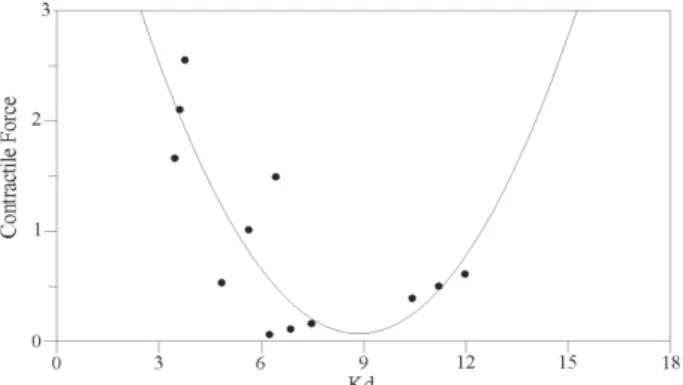
Correlation between affinity and density of β -adren-ergic receptors (Kd and Bmax) and cardiac contractile force - A quadratic order model was obtained when myo-cardial contractile force and affinity were correlated, pro-viding as a result, the coefficients corresponding to the function Y = Co + C1X + C2X2. This fact explained the behavior of the “affinity” (Kd) as a regressive variable, when compared to myocardial contractile force as re-sponse variable. The coefficient values corresponding to the second-degree model were: Co = +5.692 ± 0.712;C1 = –1.269 ± 0.209; C2 = +0.072 ± 0.014 (p < 0.001) (Figure).
A correlation between myocardial contractile force and density of β-adrenergic receptors could not be demon-strated.
Electrocardiography - Table III shows how the elec-trocardiographic findings of the infected mice worsened from the acute to the chronic (180 days PI) phases of the disease.
In our study, we confirmed that T. cruzi infected mice developed cardiac functional disorders such as: decreased contractility and modifications to norephinephrine re-sponse in right ventricles as well as electric conduction disturbances all along the acute, indeterminate and chronic stages of Chagas disease.
Cardiac hyporeactivity to neurotransmitters, a decrease in the contractile force, an increment in electrocardio-graphic changes and modifications in the affinity and density of β-adrenoceptors were typical findings of the acute period of T. cruzi infection (35 days PI). The level of the neurotransmitters become relevant during this acute stage, since it was demonstrated that in the hearts of rats the concentration of norepinephrine is reduced to non-detectable levels. It has been suggested that the deple-tion of this neurotransmitter produced by the destrucdeple-tion of the sympathetic cardiac innervation (Lohse et al. 1996) is the main cause of heart failure in the acute stage of the disease. Our results agree with this mechanism since we observed in the infected mice an increased number of bind-ing sites and a decrease in the affinity values, demon-strating that the mechanism that regulates the affinity and density was maintained, although in different values com-pared to non-infected animals.
During the indeterminate stage (75 days PI) the right ventricle myocardium presented an atypical response to norepinephrine, such as negative inotropic effect or a complete lack of response to the neurotransmitter. Addi-tionally, we were able to verify a decrease of the affinity when comparing the non-infected and acute groups. On the other hand, the number of β-adrenergic receptors was similar in mice in the acute stage, but higher than the number found in the non-infected group. Atrioventricular and intraventricular blocks were detected during this in-determinate stage, demonstrating that this stage of the disease is not silent and that these findings would prob-ably determine the severity of the cardiopathy observed in the chronic stage. The results found on functional ac-tivity of β-adrenergic receptors are not enough to explain the absence of response to norepinephrine or the nega-tive inotropic response described in myocardium of mice in the indeterminate stage. They might probably be ex-plained after studying AMPc or other messengers that are currently under analysis in our laboratory.
The studies performed on myocardium contractility
TABLE III
Results of the electrocardiographic studies of 10 non-infected and 250 Trypanosoma cruzi infected mice
35 days 75 days 180 days
Electrocardiography Non-infected post-infection post-infection post-infection
n = 90 n = 80 n = 80
Mean (S.E.) pulse rate (beats/min) 446 (13.5) 468 (12.1) 575 (4.8) 526 (16.9)
Mean (S.E.) axes (grade) 66 (4.7) 51 (4.7) 44 (2.3) 55 (2.9)
PQ interval (s) 0.02 0.02-0.04 0.03-0.04 0.02-0.05
QRS interval (s) 0.02-0.03 0.02-0.03 0.02-0.04 0.02-0.06
% of mice showing abnormalities 2 12.5 a 66 b 60 b
a: p < 0.01 when compared with non-infected and p < 0.001 when compared with the acute (35 days post-infection) and indeterminate stage (75 days post-infection); b: p < 0.001 when compared with non-infected and acute groups
Correlation between the affinity (Kd) of cardiac β-adrenergic re-ceptors and myocardial contractile force.
DISCUSSION
695 695 695 695 695 Mem Inst Oswaldo Cruz, Rio de Janeiro, Vol. 99(7), N ovem ber 2004
and pharmacological response during the chronic stage of Chagas disease showed a significant decrease in the contractile force, a marked hyporeactivity to norepineph-rine and a significant increase of electrocardiographic abnormalities.
These results are related with findings in patients stud-ied with specific functional tests, which have evidenced during the chronic phase of the disease alterations of the autonomous nervous system secondary to neurolysis (Madoery & Madoery 1992). This process can destroy up to 80% of heart nervous structures (Laucella et al. 1996). Similar results have been described by other authors in infected mice with a non-lethal strain of T. cruzi at nine weeks PI (Sterin Borda et al. 1999). Several non-excluding mechanisms have been proposed to explain the develop-ment of the chronic chagasic cardiomyopathy: the au-tonomous nervous system denervation (Caeiro 1994, Laucella et al. 1996), microvasculature disorders (Madoery & Madoery 1992), and immunological mechanisms (Tarleton 2001).
The microvascular and immunological theories, or the combination of both could explain the results of the marked decrease of affinity and density of the cardiac β -adrener-gic receptors in the chronic stage. Besides, similar modifi-cations have been described in dilated cardiomyopathies due to other etiologies (Bristow 1993), in which the reduc-tion in the number of β-adrenergic receptors has been directly related to the severity of the disease (Brodde 1991, 1996, Harding et al. 1994).
The down regulation system of cardiac β-adrenergic receptors is a common finding of different diseases. The heart failure (Brodde 1996, Chakraborti et al. 2000) can be produced by an increment of catecholamine levels; how-ever, these are not elevated in chronic chagasic patients (Iosa et al. 1989). It has been proposed that in chagasic patients, the down regulation may be induced by the spe-cific circulating antibodies (Sterin Borda et al. 1999).
Having in mind that there is a cause-effect correlation between the function of β-adrenergic receptors and the cardiac contractile force, we suggest a linear model repre-sented by a quadratic correlation between the affinity of β-adrenergic receptors and contractile force. This implies the dependence of the myocardial contractility on the af-finity of the receptors, showing the minimum ability of contractile force after 75 days PI, with slightly recovery in the chronic stage of the disease.
These results may indicate that the changes in the number of β-adrenergic receptor binding sites would gen-erate compensating mechanisms in the affinity of these receptors, with a correlation with the contractile force, that would be effective only until reaching the indetermi-nate stage of the chagasic cardiomyopathy. Notwithstand-ing, all the functional modifications observed in β -adren-ergic receptors are neither followed by alterations in the membrane lipid composition nor in its fluidity, as it has been described in other cardiopathies (Villar et al. 1996).
ACKNOWLEDGEMENTS
To Prof. Dr Gerardo Fidelio for his contribution in the mea-surements of membrane fluidity, and to Drs A Casas and C Tulian for their help in the measurement membrane compo-nents.
REFERENCES
Abell LL, Levy BB, Brodie BB, Kendall FE 1952. A simplified method for the estimation of total cholesterol in serum and demonstration of its specificity. J Biol Chem 195: 357-366. Andrade Z 1999. Inmmunopathology of Chagas’ disease. Mem
Inst Oswaldo Cruz 94: 71-80.
Borda ES, Sterin Borda L 1996. Antiadrenergic and muscarinic receptor antibodies in Chagas’ cardiomyopathy. Int J Cardiol 54: 149-156.
Brener Z, Gazzinelly RT 1988. Immunological control of Try-panosoma cruzi infection and pathogenesis of Chagas’ dis-ease. Int Arch Allergy Imm 114: 103-110.
Bristow MR 1993. Changes in myocardial and vascular recep-tors in heart failure. J Am Coll Cardiol22: 61-71. Brodde OE 1991. β1- and β2- adrenoceptors in the human heart:
properties, function and alterations in chronic heart failure.
Pharmacol Rev 43: 203-242.
Brodde OE 1996. β- adrenergic receptors in failing human myo-cardium. Basic Res Cardiol91: 35-40.
Caeiro T 1994. Alteración del sistema nervioso autónomo. In R Storino, J Milei (eds), Enfermedad de Chagas, Doyma Ar-gentina Press, Buenos Aires, p. 321-329.
Chakraborti S, Chakraborti T, Shaw G 2000. β-adrenergic mecha-nisms in cardiac diseases. A perspective. Cell Signal 12: 499-513.
Elizari MV 1999. La cardiomiopatía chagásica: perspectiva histórica. Medicina (Bs As) 59: 25-40.
Enders JE, Paglini P, Fernández AR, Marco F, Palma JA 1995. Cardiac β receptors in experimental Chagas’ disease. Rev Inst Med Trop São Paulo 37: 59-62.
Ferrari I, Levin M, Wallukat G, Elies R, Lebesgue D, Chiale P, Elizari M, Rosenbaum M, Hoebeke J 1995. Molecular mim-icry between the inmonodominant ribosomal protein; of
Trypanosoma cruzi and a functional epitope on the human β1- adrenergic receptor. Exp Med 182: 59-65.
Harding SE, Brown LA, Wynne DG, Davies CH, Poole-Wilson PA 1994. Mechanisms of β-adrenoceptor desensitization in the failing human heart. Cardiovasc Res 28: 1451-1460. Iosa D, De Quatro V, DePink Lee D, Elkayan V, Palmero H 1989. Plasma norepinephrine in Chagas’ cardioneuromy-opathy: a marker of progressive dysautonomia. Am Heart J 117: 882-887.
Laucella SA, Rottemberg ME, De Titto EH 1996. Papel de las citoquinas en la resistencia y patologia durante la infección con Trypanosoma cruzi. Rev Arg de Microbiol 28: 99-109. Lohse MJ, Engelhart S, Danner S, Bohm M 1996. Mechanisms of β-adrenergic receptor desenzitation: from molecular bi-ology to heart failure. Basic Res Cardiol 91: 29-34. Madoery RJ, Madoery C 1992. Periodo intermedio de la
enfermedad de Chagas. In RJ Madoery, C Madoery, R Ca-mera (eds), Actualización de la Enfermedad de Chagas, Doyma Press, Buenos Aires, p. 51-56.
Mcgowan MW, Artiss JD, Strndbergh DR, Bernie Z 1983. A peroxidase-coupled method for the colorimetric determina-tion of serum triglycerides. Clin Chem 29: 538-542. NIH 1985. Guide for the Care and Use of Laboratory Animals.
Office of Science and Heath report, DRR/NIH. DHEW Publication (NIH) 85-23.
Paglini-Oliva P, Fernández AR, Lacuara JL 1987. Pharmaco-logical and contractile response of myocardium of chagasic albino swiss mice. Acta Physiol Pharmacol et Therapeutica Latinoam 35: 395-401.
696 696 696 696
696 Chagas Cardiomyopathy • Julio E Enders et al.
Paglini-Oliva P, Palma JA, Gonzalez Cremer M, Segura EL, Lacuara JL 1985. Acute Chagas disease in the mouse. Con-tractile response of the isolated myocardium. Trop Med Hyg 34: 1149-1152.
Peres Leiros C, Sterin-Borda L, Borda ES, Goin JC, Hosey M 1997. Desensitization and sequestration of human M2 mAChRs by autoantibodies from patients with Chagas’ disease. J Bio Chem 272: 12989-12993.
Petry K, Eisen H 1989. Chagas’ disease: a model for the study of autoimmune disease. Parasitol Today 5: 11.
Salomone O, Caeiro T, Madoery R, Amuchástegui M, Omelinauk M, Juri D, Kaski J 2001. High plasma immunoreactive endothelin levels in patients with Chagas’ cardiomyopathy.
Am J Cardiol 87: 1217-1220.
Sterin Borda L, Gorelik G, Postan M, Gonzalez Cappa S, Borda E 1999. Alterations in cardiac beta-adrenergic receptors in chagasic mice and their association circulating beta-adrenoceptor-related autoantibodies. Cardiovasc Res 41: 116-125.
Tanowitz HB, Kirchhoff LV, Simon D, Morris SA, Weiss LM 1992. Chagas disease. Clin Microbiol Rev 5: 400-419. Tarleton RL 2001. Parasite persistence in the etiology of Chagas’
disease. Int J Parasitol 31: 550-554.