Expression and production of cardiac angiogenic mediators depend on
the
Trypanosoma cruzi
-genetic population in experimental C57BL/6
mice infection
☆
Deena Shrestha
b, Bijay Bajracharya
b, Guilherme Paula-Costa
b, Beatriz C Salles
b, Ana Luísa J Leite
b,
Ana Paula J Menezes
b, Débora MS Souza
b, Laser AM Oliveira
a,b, André Talvani
a,b,c,d,⁎
aDepartamento de Ciências Biológicas, Brazil
bPrograma de Pós-Graduação em Ciências Biológicas/NUPEB, Brazil cPrograma de Pós-Graduação em Saúde e Nutrição, Brazil
dPrograma de Pós-Graduação em Biomas Tropicais, Universidade Federal de Ouro Preto, Ouro Preto, MG, Brazil
a b s t r a c t
a r t i c l e
i n f o
Article history:
Received 1 September 2016 Revised 5 December 2016 Accepted 6 December 2016 Available online 9 December 2016
Mammalian cardiac cells are important targets to the protozoanTrypanosoma cruzi. The inflammatory reaction in the host aims at eliminating this parasite, can lead to cell destruction,fibrosis and hypoxia. Local hypoxia is well-defined stimulus to the production of angiogenesis mediators. Assuming that different geneticT. cruzi popula-tions induce distinct inflammation and disease patterns, the current study aims to investigate whether the pro-duction of inflammatory and angiogenic mediators is a parasite strain-dependent condition. C57BL/6 mice were infected with the Y and Colombian strains ofT. cruziand euthanized at the 12th and 32nd days, respectively. The blood and heart tissue were processed in immune assays and/or qPCR (TNF, IL-17, IL-10, CCL2, CCL3, CCL5, CCR2, CCR5 and angiogenic factors VEGF, Ang-1, Ang-2) and in histological assays. TheT. cruziincreased the infl amma-tory and angiogenic mediators in the infected mice when they were compared to non-infected animals. However, the Colombian strain has led to higher (i) leukocyte infiltration, (ii) cardiac TNF and CCL5 production/expression, (iii) cardiac tissue parasitism, and to higher (iv) ratio between heart/body weights. On the other hand, the Co-lombian strain has caused lower production and expression VEGF, Ang-1 and Ang-2, when it was compared to the Y strain of the parasite. The present study highlights that theT. cruzi-genetic population defines the pattern of angiogenic/inflammatory mediators in the heart tissue, and that it may contribute to the magnitude of the car-diac pathogenesis. Besides, such assumption opens windows to the understanding of the angiogenic mediator's role in association with the experimentalT. cruziinfection.
© 2016 Elsevier Inc. All rights reserved.
Keywords:
Inflammation
Trypanosoma cruzi
Angiogenic factors, chemokines VEGF
1. Introduction
Inflammation is a multifactorial process involving cellular activation and migration, inflammatory mediators' production, vascular responses and local and/or systemic reactions. Angiogenesis emerges as an event cross-linked to inflammation and it aims at keeping homeostasis and repairing the tissue through the formation of new capillary networks from a preexisting vasculature. These networks are tightly regulated by the production and release of angiogenic factors (Medzhitov, 2008;
Folkman, 2006).
The protozoanTrypanosoma cruzitriggers acute and chronic infl am-matory processes in smooth, skeletal and cardiac muscles and promotes a progressive damage that causes localfibrosis prevalence and function-ality deficit (Lannes-Vieira et al., 2009; Talvani and Teixeira, 2011;
Penitente et al., 2015). This parasite has a well-adapted mechanism
that favors its survival in mammalian host cells and drives an infl amma-tory response in the host according to theT. cruzihigh genetic variabil-ity, which is evidenced through six distinct Discrete Typing Units—DTU's expressed asT. cruziI (TcI) toT. cruziVI (TcVI). Each of these units present a biological feature based on their geographic distri-bution, and eco-epidemiological and clinical associations (Zingales et
al., 2012). The inflammatory response aims at eliminating the parasites
in the acute phase; whereas, in the chronic phase, such response is rath-er kept undrath-er control in ordrath-er to keep the parasites and the immune re-sponse under control as a way to prevent excessive tissue damage
(Nagajyothi et al., 2012; Cardoso Reis-Cunha and Bartholomeu, 2015).
When these protective strategies fail, the pathophysiological ☆ Angiogenic mediators inT. cruziinfection
⁎ Corresponding author at: Universidade Federal de Ouro Preto, Laboratório de Imunobiologia da Inflamação, DECBI/NUPEB, Campus Morro do Cruzeiro, 35400-000 Ouro Preto, MG, Brazil.
E-mail address:talvani@nupeb.ufop.br(A. Talvani).
http://dx.doi.org/10.1016/j.mvr.2016.12.002
0026-2862/© 2016 Elsevier Inc. All rights reserved.
Contents lists available atScienceDirect
Microvascular Research
manifestations and the disease emerge in the host, in part, in a close-de-pendence of the parasite DTUs.
It is likely that the immune response and theT. cruzimolecules trig-ger the angiogenic factors since they are required to tissue repair
(Ferreira et al., 2005, Guedes-da-Silva et al., 2015). In turn, infl
amma-tion may lead to condiamma-tions favorable for angiogenesis, partly due to the hypoxia that emerges afterfibrosis generation and, partly, to the in-flammatory mediators that are further amplified by pro-angiogenic fac-tors released around the inflammatory site (Yamakawa et al., 2003, Van
Nieuwenhoven and Turner, 2013). Besides the conventional roles
played by cytokines and chemokines in the activation and recruitment of leukocytes to the inflammatory foci, some other studies have shown that these soluble mediators can directly or indirectly increase the vascular growth by stimulating the production of the vascular endo-thelial growth factor (VEGF) (Barcelos et al., 2009; Pickens et al., 2010;
Maloney and Gao, 2015). The VEGF is an essential factor responsible
for angiogenesis after its binding in the VEGF receptor, which is found in many cells. It leads to the proliferation, migration and survival of new endothelial cells (Leung et al., 1989; Bao et al., 2009; Zhang et al.,
2012; Wang et al., 2013). Furthermore, the angiopoietins can inhibit
the tumor necrosis factor (TNF)-stimulated leukocyte transmigration and act as an important angiogenesis and inflammation regulator
(Gamble et al., 2000; Seok et al., 2013).
Since distinct geneticT. cruzipopulations may cause different in-flammation patterns in experimental models, the aim of the present study is to show, for thefirst time, that the Y (DTU II) and Colombian (DTU I) strains ofT. cruziperform different interactions with angiogenic and inflammatory mediators in the plasma and cardiac tissue of infected C57BL/6 mice.
2. Materials and methods
2.1. Ethics statement
All procedures in the current study meet the guidelines issued by the Brazilian College of Animal Experimentation (COBEA); the research was previously approved by the Ethics Committee on Animal Research of UFOP (Protocol number 043/2010).
2.2. Experimental animals, parasites and infection
Ten week-old male C57BL/6 mice were bred and housed at the Ani-mal Sciences Center at Universidade Federal de Ouro Preto—UFOP, Bra-zil. The mice (n = 10) were infected with 100 blood trypomastigote forms ofT. cruziusing the following strains: (i) Y and (ii) Colombian. The Y strain ofT. cruzi, classified as DTU II, is characterized by a rapid parasite multiplication with very high peaks between 9 and 10 days of infection, presenting high virulence and mortality between 10 and 14 days of infection. The Colombian strain, classified as DTU I, presents a very slow parasitic multiplication, reaching very high parasitemic peaks between 20 and 30 days of infection, low virulence with no mor-tality up to 50 days (Andrade and Magalhães, 1996; Zingales et al., 2012;
Zingales et al., 2009). Parasitemia was daily determined according to the
method described byBrener (1962). The mice were euthanized after the parasitemia peak, at the 12th and 32nd days of infection in the Y and Colombian strains, respectively. Blood was collected for immunoas-say; half heart (i) wasfixed in 10% formalin for histological analysis, the other half (ii) was used in molecular and immunoassay parameter assessments.
2.3. Heart mass measurement
The heart from each animal was carefully excised after blood collec-tion. The vessels and heart chambers were washed in phosphate buffer solution. The wet organ was weighed and the relative heart weight was calculated using the heart weight in milligram/body weight in gram
(mg/g). This mg/g value was used to determine the cardiac mass mea-surement at the time of the euthanasia. In addition, both ventricles were split in two fragments: the upper part was used in the homoge-nate (immune and molecular assays); and the lower (apex) one, in the histopathological analysis.
2.4. Immunoassay
The circulating levels of TNF, VEGF, IL-10, IL-17, the macrophage in-flammatory protein alpha (MIP-alpha/CCL3) regulated upon activation, normal T cell expressed and secreted normal T cells (CCL5/RANTES), and the monocyte chemoattractant proteins (CCL2/MCP-1) were de-tected in plasma. Blood from the orbital venous sinus (0.5 mL) was col-lected during euthanasia and centrifuged (1500 g for 15 min at 4 °C). The plasma was stored at−80 °C. In parallel, a 10 mg cardiac tissue fragment collected from each animal was homogenized in cold radioimmunoprecipitation assay (RIPA) buffer using a protease inhibi-tor; the supernatant was stored at−80 °C. Next, these samples were used to measure TNF, CCL2, CCL3, CCL5, IL-10, IL-17 and the angiogenic factor VEGF (Peprotech, Ribeirão Preto, Brazil), according to the proto-col recommended by the manufacturer. The samples were simulta-neously measured in duplicates.
2.5. Morphometric and histopathological analysis
Cardiac tissue fragments werefixed in 10% buffered-formalin solu-tion; then, dehydrated, cleared and embedded in paraffin in order to an-alyze and quantify the inflammatory infiltration and the amastigote nests. Blocks were cut in 4 mm-thick sections and stained in hematoxyllin and eosin (HE). Twentyfields from each HE stained sec-tion were randomly chosen at 40 × magnification, thus totaling 74,931μm2
—the equivalent area of 50fields of the analyzed myocardi-um. Images were obtained in a Leica DM 5000 B micro chamber (Leica Application Suite, UK, version 2.4.0 R1) and processed in the Leica Quinn (V3) image analyzer software. The inflammatory process was assessed through the number of cellular nuclei found in the infected heart tissue and compared to the background of the cardiac cellular nu-clei found in the non-infected mice. Amastigote nests were quantified in the Image J 1.45s software, at the National Institute of Health, USA
(www.imagej.nih.gov/ij). The area occupied by parasites was assumed
Table 1
Primer sequences according to the GenBank database.
Sequences (forward and reverse)
VEGF 5′AAAAACGAAAGCGCAAGAAA 3′
5′TTTCTCCGCTCTGAACAAGG 3′
ANG-1 5′GGGGGAGGTTGGACAGTAA 3′
5′CATCAGCTCAATCCTCAGC 3′ ANG-2 5′GATCTTCCTCCAGCCCCTAC 3′
5′TTTGTGCTGCTGTCTGGTTC 3′
TNF 5′TGAGTGACCAAGGGACAGAACC 3′
5′AGCCAGGAGGGAGAACAG 3′ IL-10 5′ACTACCAAAGCCACAAGG 3′
5′AAGAGCAGGCAGCATAG 3′
CCL2 5′AACTGCATCTGGCTGAGC 3′
5′CAGCACCAGCCAACTCTC 3′ CCL5 5′ACCCTCTATCCTAGCTCATC 3′
5′CGTGTTTGTCACTCGAAG 3′ CCR2 5′CCTGTCCACTAATGCGTTTC 3′
5′GCAAAGCCAGACCACAATG 3′ CCR5 5′CCCTGTCATCTATGCCTTTG 3′ 5′GCTTGCACGATCAGGATTG 3′ β-actin 5′CCACTTTCCTGTCTTACCCAA 3′
5′AATTAACCACCCACGGTGTT 3′
Fig. 2.Concentrations of inflammatory mediators in the plasma. CCL2/MCP-1 (A), CCL5/RANTES (B) and CCL3/MIP-1 alpha (C) TNF (D), IL-17 (E) and IL-10 (F) were measured through immunoassay (ELISA) in the plasma of C57BL/6 mice infected with the Colombian and Y strains ofT. cruzi. Data are shown as the mean of 10 animals ± SEM and different letters denote difference (pb0.05).
to be the same area previously used to quantify the inflammatory process.
2.6. Expression of the inflammatory and angiogenic mediators
We quantified the expression of inflammatory and angiogenic genes. Thus, we extracted the total RNA from the cardiac tissue of the animals with theSV Total RNA Isolation System Kit(Promega, USA) fol-lowing the manufacturer's protocol. Subsequently, we made the com-plementary DNA (cDNA) using the high-capacity cdna reverse transcription kit(Applied Biosystems, USA). Then, for the angiogenic (VEGF, Ang-1, Ang-2) and inflammatory (CCL2, CCL5, CCR2, CCR5, TNF and IL-10) genes, a standard curve from serial dilutions of a known con-centration of purified DNA was achieved. This quantified DNA consisted of the target PCR product prepared by conventional PCR from cDNA pos-itive for the corresponding target mRNA. Threefold measurement for each standard dilution point over the whole standard curve range was produced to generate a reliable standard curve. Then, real-time PCR quantitative mRNA analyses were performed using an ABI Prism 7000 SDS unit (Applied Biosystems) through the Platinum® SYBR® Green qPCR SuperMix UDG with ROX reagent (Invitrogen) for quantification of amplicons. The standard PCR conditions were as follows: 50 °C (2 min), 95 °C (10 min); 40 cycles of 94 °C (30 s), 58 °C (30 s), and 72 °C (1 min); followed by the standard denaturation curve. The se-quences of the primers were designed using the Primer Express soft-ware (Applied Biosystems) assuming the nucleotide sequences available in the GenBank database (Table 1). In each reaction the Plati-num® SYBR® Green qPCR SuperMix UDG with ROX reagent (Invitrogen), 1μg/μL of each specific primer, and cDNA diluted 20 times were used. In this study, all data were normalized to beta-actin mRNA. Relative increase in CK and CKR were plotted in comparison to the non-infected control group using 2−Δ/ΔCT method.
2.7. Statistical analysis
Data are expressed as the mean ± standard error of the mean (SEM) and were analyzed using theKolmogorov-Smirnovnormality test and One-Way analysis of variance. All analyses were performed using PRISM 5.01 software (GraphPad, San Diego, CA, USA) and the level of significance was accepted atpb0.05.
3. Results
3.1. Parasitemia curve and the relative heart weight
The parasitemia curves are represented inFig. 1A and show the bio-logical feature of the Y and Colombian strains ofT. cruziused in the pres-ent study. The pre-patpres-ent period ranged from 6 days, in the Y strain, to 10 days, in the Colombian strain ofT. cruzi. Throughout the current study, both strains have led to high parasite load in the infected C57BL/6 mice. However, in the end of the acute phase, only mice infect-ed with the Colombian strain ofT. cruzihave presented increased heart/ body weight ratios. The animals infected with the Y strain kept heart/ body weight ratio similar to that of the non-infected animals (Fig. 2B).
3.2. The production and expression of plasma and heart inflammatory, reg-ulatory and angiogenic mediators
The plasma cytokines (IL-10, IL-17 and TNF) and chemokines (CCL2, CCL3, CCL5) production was quantified in order to be associated with the inflammatory pattern of eachT. cruzistrain, since theT. cruzi infec-tion develops systemic inflammatory mediators and since these soluble factors play a potential role in the release of angiogenic factors. All fected animals have presented plasma elevation in the production of in-flammatory/regulatory cytokines and chemokines when they were compared to the uninfected mice (Fig. 2C). There was inverse relation
between CCL2 and CCL5 production; the Y and Colombian strains have induced more CCL2 (Fig. 2A) and CCL5 (Fig. 2B) in infected animals, re-spectively. Interestingly, besides the pattern observed to the chemokines, no differences were observed between both parasite strains in TNF (Fig. 2D), IL-17 (Fig. 2E) and IL-10 (Fig. 2F) production after the day of their respective parasitemia peaks.
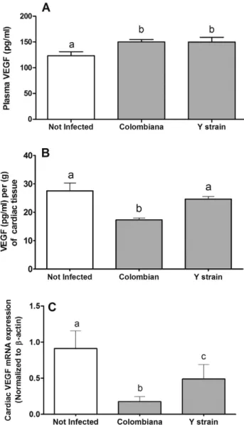
In parallel, the vascular endothelial growth factor (VEGF), the main representative among the angiogenic mediators, increased in the plas-ma when it was associated with bothT. cruzistrains (Fig. 3A). However, the DTU I, Colombian strain, was able to inhibit the VEGF production in the heart tissue, which was measured in the tissue homogenate (Fig. 3B).
By following the plasma cytokines and chemokines production, the cardiac expression of the CCL2 (Fig. 4A), CCL5 (Fig. 4B), TNF (Fig. 4C), IL-10 (Fig. 4D), and the CCL2 and CCL5 receptors, respectively, CCR2
(Fig. 4E) and CCR5 (Fig. 4F) also increased in the presence ofT. cruzi.
However, the Colombian strain was able to increase by 200 and by 6 times the expression of CCL5 (Fig. 4B) and TNF (Fig. 4C), respectively, in the heart tissue of infected mice when it was compared to the Y strain.
In parallel, the VEGF expression in cardiac tissue was also measured through qPCR (Fig. 3C). The Colombian strain was, once more, capable of inhibiting this angiogenic factor more intensely at molecular level when it was compared to the Y strain of the parasite. Moreover, the ex-pression of angiopoetin-1 (Ang-1) (Fig. 5A) and angiopoetin-2 (Ang-2)
(Fig. 5B), which are other angiogenic mediators, just decreased in
ani-mals infected with the Colombian strain ofT. cruzi.
3.3. Inflammatory infiltration and parasitism in cardiac tissue
Although all animals have received the same load of parasites during the infection, each DTU has promoted specificities in the inflamed cardi-ac tissue. With regard to the present study, the Colombian strain was as-sociated with higher leukocytes influx, followed by the Y strain (Fig.
6A).Fig. 6(left side) shows a representative photomicrography of the
non-infected cardiac tissue and the presence of amastigote nests associ-ated with theT. cruzi(Colombian strain). These images are reinforced by the quantification of the amastigote nest area (Fig. 6B). Tissue parasites associated with the Y strain were not detected in the current study.
4. Discussion
There is no way to escape from the close relation between the genet-ic variability of theT. cruzi, currently classified as DTU's, and the gener-ation of the pathology and of cardiac diseases in humans and in experimental animals (Zingales et al., 2012, 2009). However, the dis-tinct genetic variability between mammalians is also another highlight that must be considered in theT. cruzipathogenic puzzle. The para-site/host interaction dictates the immune balance or imbalance and leads to serious disturbances in the affected organs (Penitente et al., 2015; Guedes et al., 2010; Oliveira et al., 2012; Martins et al., 2013;
Bryan et al., 2016; de Oliveira et al., 2016).Some studies have proposed
that a panel of inflammatory mediators is related to the protection against circulating or tissue infectingT. cruzi(Lannes-Vieira et al., 2009; Talvani and Teixeira, 2011; Penitente et al., 2015; Guedes et al.,
2010; Gomes et al., 2003). Soluble mediators such as IFN-gamma,
IL-12, TNF and IL-17 were previously described to activate macrophages and to circulate mononuclear cells in reactive oxygen species—ROS
(Guedes et al., 2010; Gupta et al., 2011; Magalhães et al., 2013; Costa
et al., 2006; Machado et al., 2000) in order to eliminate parasites.
Some other regulatory proteins are also released in order to control the immune response intensity (e.g. IL-10, IL-4, IL-22) and, consequent-ly, to contribute to pathogenic process decay of Chagas disease (Gomes
et al., 2003; Flórez et al., 2011; Poveda et al., 2014) in humans and in
ex-perimental animals infected withT. cruzi(Abrahamsohn, 1998; Hiyama
et al., 2001). Part of this pathogenic process is driven by the chemokines
(e.g. CCL2, CCL3, CCL5 and others) and was previously evidenced in human and experimental T. cruzi infection (Talvani et al., 2000;
Teixeira et al., 2002; Talvani et al., 2004; Nogueira et al., 2012). These
small and soluble molecules are capable of recruiting leukocytes in the bone marrow and of sending them to the blood, and/or of sending them from the blood to the tissue, thus intensifying local inflammation and parasite elimination.
This parasite-dependent inflammatory process is usually persistent and contributes to tissue destruction, to toxic products release and to local tissue hypoxia, thus culminating in a heart repair or remodeling process (Rossi and Carobrez, 1985; Melo et al., 2011). This hypoxic en-vironment is a required stimulus to the release of a new set of angiogen-ic mediators such as VEGF, angiopoietin (Ang)-1 and Ang-2. These mediators are involved in the angiogenesis process and in the modula-tion of inflammatory activities (McCarter et al., 2006; Scholz et al., 2015). The VEGF acts through tyrosine kinase receptors (VEGFRs) expressed in myelomonocytic inflammatory cells, as well as in vascular endothelial cells. The inhibition of the VEGF signaling has been assumed to induce anti-inflammatory properties through the blockage of STAT-3
(Wang et al., 2013; Waldner et al., 2010). The angiopoietins are
oligo-meric-secreted glycoprotein ligands that bind to a Tie family of recep-tors primarily expressed in the vascular endothelium. The expression of the leukocyte adhesion molecules E-selectin, ICAM1, and VCAM1 is usually suppressed through Ang-1; whereas, Ang-2 appears to be a
key regulator in vascular inflammations. The signaling mechanism in these immune regulatory mechanisms remains unclear, although some studies point out PI3K and Akt as required in this pathway (Brindle et al., 2006).
The magnitude of the regulatory, inflammatory or angiogenic medi-ators appears to depend on the DTU's of the protozoan, which demands care at the interpretation of the data on experimental and human infec-tions. Indeed, according to the literature, the presence of the parasite or of its antigen molecules (e.g. mucin-glycosylphosphatidylinositol) in experimental animals is sufficient to trigger inflammatory protein/ lipid mediators (Talvani and Teixeira, 2011; Golgher and Gazzinelli,
2004; Rodrigues et al., 2012). However, it is now suggested that the
magnitude depends on the parasite strain.
The Colombian strain ofT. cruziis an inducer of high murine TNF levels and, consequently, it is responsible for releasing high levels of dis-tinct chemokines and of their receptors in the immune and cardiac cells
(Medeiros et al., 2009). The DTU I is an adaptive strain to infected heart
tissue (Oliveira et al., 2012) and it also presents biological features concerning the resistance against available nitro derivative drugs
anti-T. cruzisuch as benznidazole (Romanha et al., 2010; Gruendling et al., 2015). This genetic parasite population led to reduction in the expres-sion of the following angiogenic factors in the present study: VEGF, Ang-1 and Ang-2; however, in the cardiac tissue only, not in the plasma context. On the other hand, the Colombian strain promoted TNF, CCL5 and CCL3 increase in the plasma and/or in the cardiac tissue. The persis-tence of the tissue amastigote forms, the increase of other inflammation mediators (TNF, IL-17, chemokines) and the reduction of Ang-1 and Ang-2 may have reinforced the magnitude of the local inflammatory re-sponse due to the presence of greater leukocyte infiltration.
On the other hand, the Y strain ofT. cruzihas also induced TNF and other inflammatory mediators, but it was less intense than in the Co-lombian strain. This genetic population of the parasite was homoge-neous throughout different organs, including the heart (Oliveira et al., 2012). It is partially resistant against derivative drugs anti-T. cruzi
(Romanha et al., 2010; Gruendling et al., 2015). The Y strain ofT. cruzi
in the current study has induced higher expression of cardiac VEGF, Ang-1 and Ang-2 in comparison to the lower production and/or expres-sion of TNF, CCL5, amastigotes and leukocytes infiltration, which were observed in the Colombian strain. The chemokine receptors were herein higher expressed during the expression of bothT. cruzistrains, as well as
Fig. 6.Cardiac inflammatory cells and amastigote nests. Cellular infiltration in the cardiac tissue of C57BL/6 mice infected with the Colombian and Y strains ofT. cruziwas quantified in (A), and the area of amastigote nests in (B). Data are shown as the mean from 10 animals ± SEM. Different letters denote difference in expression (pb0.05). Illustrations from the uninfected cardiac tissue or of tissue infected byT. cruzi(left) present 40× magnification. Black arrows = amastigote nests.
of other soluble mediators such as IL-17 and CCL2, when they were compared to animals who had no contact with the protozoan.
It is plausible that both polar populations ofT. cruzihave led to in-creased, but distinct, patterns of inflammatory and angiogenic media-tors in the experimental infection model. The persistence of parasites triggering inflammation in the cardiac tissue may cause local oxygen re-duction, hypoxia, and it may trigger favorable conditions to VEGF secre-tion (Liu et al., 1995; Krock et al., 2011). The local inflammatory process and the new tissue repair environment activate the platelet used as the first vascular component to release VEGF after thrombin stimulation
(Möhle et al., 1995). The monocytes expressing the Flt-1 receptor can
be attracted by this VEGF, which, in part, is activated by TNF. In turn, the TNF may induce more VEGF expression through the local cells
(Clauss et al., 2001; Lu et al., 2012). As for the present study, VEGF,
Ang-1 and Ang-2 have presented low expression in the heart tissue of animals infected with the Colombian strain and presented higher amastigote nests. Accordingly, the proinflammatory activity of VEGF seems to have been balanced by the low activity of Ang-1 and Ang-2 during the infection with both genetic populations ofT. cruzi.
5. Conclusion
The present study has demonstrated that DTU I (Y strain) and DTU II (Colombian strain)T. cruziparasites promote the release and the ex-pression of different levels of angiogenic and inflammatory mediators in the acute phase of the experimental infection. In this particular case, the angiogenic mediators may work side by side with chemokines and inflammatory cytokines in order to switch“on/off”the cardiac tis-sue pathogenesis development, depending on the features dictated by each DTU of the parasite.
Funding sources
The present study was granted by Conselho Nacional de Desenvolvimento Científico e Tecnológico/CNPq (#476229/2009-0), Fundação de Amparo à Pesquisa do Estado de Minas Gerais (FAPEMIG APQ-01698-13), International Society for Infectious Diseases (ISID/ EUA—Small Grant/2009) and Universidade Federal de Ouro Preto/ UFOP (PROPP # 67/2016). AT thanks CNPq for the fellowship granted to the research in productivity and for the PhD fellowships of DS and BB (both CNPq/TWAS Program).
Disclosures
Authors declare no conflict of interest regarding the publication.
References
Abrahamsohn, I.A., 1998.Cytokines in innate and acquired immunity toTrypanosoma cruziinfection. Braz. J. Med. Biol. Res. 31, 117–121.
Andrade, S.G., Magalhães, J.B., 1996.Biodemes and zymodemes ofTrypanosoma cruzi
strains: correlations with clinical data and experimental pathology. Rev. Soc. Bras. Med. Trop. 30, 27–35.
Bao, P., Kodra, A., Tomic-Canic, M., Golinko, M.S., Ehrlich, H.P., Brem, H., 2009.The role of vascular endothelial growth factor in wound healing. J. Surg. Res. 153, 347–358.
Barcelos, L.S., Coelho, A.M., Russo, R.C., Guabiraba, R., Souza, A.L., Bruno-Lima Jr., G., Proudfoot, A.E., Andrade, S.P., Teixeira, M.M., 2009.Role of the chemokines CCL3/ MIP-1alpha and CCL5/RANTES in sponge-induced inflammatory angiogenesis in mice. Microvasc. Res. 78, 148–154.
Brener, Z., 1962.Therapeutic activity and criteria on of cure on mice experimentally in-fected withTrypanosoma cruzi. Rev. Inst. Med. Trop. 4, 389–396.
Brindle, N.P., Saharinen, P., Alitalo, K., 2006.Signaling and functions of angiopoietin-1 in vascular protection. Circ. Res. 98, 1014–1023.
Bryan, L.K., Hamer, S.A., Shaw, S., Curtis-Robles, R., Auckland, L.D., Hodo, C.L., Chaffin, K., Rech, R.R., 2016.Chagas disease in a Texan horse with neurologic deficits. Vet. Parasitol. 216, 13–17.
Cardoso Reis-Cunha, J.L., Bartholomeu, D.C., 2015.Evasion of the immune Response by
Trypanosoma cruziduring acute infection. Front. Immunol. 44, 84–90.
Clauss, M., Sunderkötter, C., Sveinbjörnsson, B., Hippenstiel, S., Willuweit, A., 2001.A per-missive role for tumor necrosis factor in vascular endothelial growth factor-induced vascular permeability. Blood 97, 1321–1329.
Costa, V.M., Torres, K.C., Mendonça, R.Z., Gresser, I., Gollob, K.J., Abrahamsohn, I.A., 2006.
Type I IFNs stimulate nitric oxide production and resistance toTrypanosoma cruzi in-fection. J. Immunol. 177, 3193–3200.
de Oliveira, L.F., Romano, M.M., de Carvalho, E.E., Cabeza, J.M., Salgado, H.C., Fazan Júnior, R., Costa, R.S., da Silva, J.S., Higuchi Mde, L., Maciel, B.C., Cunha-Neto, E., Marin-Neto, J.A., Simões, M.V., 2016.Histopathological correlates of global and segmental left ven-tricular systolic dysfunction in experimental chronic Chagas cardiomyopathy. J. Am. Heart Assoc. 5, e002786.
Ferreira, V., Molina, M.C., Schwaeble, W., Lemus, D., Ferreira, A., 2005.DoesTrypanosoma cruzicalreticulin modulate the complement e system and angiogenesis? Trends Parasitol. 21, 169–174.
Flórez, O., Martín, J., González, C.I., 2011.Interleukin 4, interleukin 4 receptor-αand inter-leukin 10 gene polymorphisms in Chagas disease. Parasite Immunol. 33, 506–511.
Folkman, J., 2006.Angiogenesis. Annu. Rev. Med. 57, 1–18.
Gamble, J.R., Drew, J., Trezise, L., Underwood, A., Parsons, M., Kasminkas, L., Rudge, J., Yancopoulos, G., Vadas, M.A., 2000.Angiopoietin-1 is an antipermeability and
anti-in-flammatory agent in vitro and targets cell junctions. Circ. Res. 29, 603–607.
Golgher, D., Gazzinelli, R.T., 2004.Innate and acquired immunity in the pathogenesis of Chagas disease. Autoimmunity 37, 399–409.
Gomes, J.A., Bahia-Oliveira, L.M., Rocha, M.O., Martins-Filho, O.A., Gazzinelli, G., Correa-Oliveira, R., 2003.Evidence that development of severe cardiomyopathy in human Chagas' disease is due to a Th1-specific immune response. Infect. Immun. 71, 1185–1193.
Gruendling, A.P., Massago, M., Teston, A.P., Monteiro, W.M., Kaneshima, E.N., Araújo, S.M., Gomes, M.L., Barbosa, M., Toledo, M.J., 2015.Impact of benznidazole on infection course in mice experimentally infected withTrypanosoma cruziI, II, and IV. Am. J. Trop. Med. Hyg. 92, 1178–1189.
Guedes, P.M., Veloso, V.M., Talvani, A., Diniz, L.F., Caldas, I.S., Do-Valle-Matta, M.A., Santiago-Silva, J., Chiari, E., Galvão, L.M., Silva, J.S., Bahia, M.T., 2010.Increased type 1 chemokine expression in experimental Chagas disease correlates with cardiac pa-thology in beagle dogs. Vet. Immunol. Immunopathol. 138, 106–113.
Guedes-da-Silva, F.H., Shrestha, D., Salles, B.C., Figueiredo, V.P., Lopes, L.R., Dias, L., Barcelos, L.S., Moura, S., Andrade, S.P., Talvani, A., 2015.Trypanosoma cruziantigens induce inflammatory angiogenesis in a mouse subcutaneous sponge model. Microvasc. Res. 97, 130–136.
Gupta, S., Dhiman, M., Wen, J.J., Garg, N.J., 2011.ROS signalling of inflammatory cytokines duringTrypanosoma cruziinfection. Adv. Parasitol. 76, 153–170.
Hiyama, K., Hamano, S., Nakamura, T., Nomoto, K., Tada, I., 2001.IL-4 reduces resistance of mice toTrypanosoma cruziinfection. Parasitol. Res. 87, 269–274.
Krock, B.L., Skuli, N., Simon, M.C., 2011.Hypoxia-induced angiogenesis: good and evil. Genes Cancer 2, 1117–1133.
Lannes-Vieira, J., Silverio, J.C., Pereira, I.R., Vinagre, N.F., Carvalho, C.M., 2009.Chronic
Trypanosoma cruzi-elicited cardiomyopathy: from the discovery to the proposal of ra-tional therapeutic interventions targeting cell adhesion molecules and chemokine receptors—how to make a dream come true. Mem. Inst. Oswaldo Cruz 104, 226–235.
Leung, D.W., Cachianes, G., Kuang, W.J., Goeddel, D.V., Ferrara, N., 1989.Vascular endothe-lial growth factor is a secreted angiogenic mitogen. Science 246, 1306–1309.
Liu, Y., Cox, S.R., Morita, T., Kourembanas, S., 1995.Hypoxia regulates vascular endothelial growth factor gene expression in endothelial cells: identification of a 5′enhancer. Circ. Res. 77, 638–643.
Lu, P., Li, L., Liu, G., Baba, T., Ishida, Y., 2012.Critical role of TNF-α-induced macrophage VEGF and iNOS production in the experimental corneal neo vascularization. Invest. Ophthalmol. Vis. Sci. 53, 3516–3526.
Machado, F.S., Martins, G.A., Aliberti, J.C., Mestriner, F.L., Cunha, F.Q., Silva, J.S., 2000. Trypanosoma cruzi-infected cardiomyocytes produce chemokines and cytokines that trigger potent nitric oxide-dependent trypanocidal activity. Circulation 102, 3003–3008.
Magalhães, L.M., Villani, F.N., Nunes, M.C., Gollob, K.J., Rocha, M.O., Dutra, W.O., 2013.High interleukin 17 expression is correlated with better cardiac function in human Chagas disease. J. Infect. Dis. 07, 661–665.
Maloney, J.P., Gao, L., 2015.Proinflammatory cytokines increase vascular endothelial growth factor expression in alveolar epithelial cells. Mediat. Inflamm. 2015, 1–7.
Martins, R.F., Martinelli, P.M., Guedes, P.M., da Cruz Pádua, B., Dos Santos, F.M., Silva, M.E., Bahia, M.T., Talvani, A., 2013.Protein deficiency alters CX3CL1 and endothelin-1 in experimentalTrypanosoma cruzi-induced cardiomyopathy. Tropical Med. Int. Health 18, 466–476.
McCarter, S.D., Lai, P.F., Suen, R.S., Stewart, D.J., 2006.Regulation of endothelin-1 by angiopoietin-1: implications for inflammation. Exp. Biol. Med. 231, 985–991.
Medeiros, G.A., Silvério, J.C., Marino, A.P., Roffê, E., Vieira, V., Kroll-Palhares, K., Carvalho, C.E., Silva, A.A., Teixeira, M.M., Lannes-Vieira, J., 2009.Treatment of chronically
Trypanosoma cruzi-infected mice with a CCR1/CCR5 antagonist (Met-RANTES) results in amelioration of cardiac tissue damage. Microbes Infect. 11, 264–273.
Medzhitov, R., 2008.Origin and physiological roles of inflammation. Nature 454, 428–435.
Melo, L., Caldas, I.S., Azevedo, M.A., Gonçalves, K.R., da Silva do Nascimento, A.F., Figueiredo, V.P., de Figueiredo Diniz, L., de Lima, W.G., Torres, R.M., Bahia, M.T., Talvani, A., 2011.Low doses of simvastatin therapy ameliorate cardiac inflammatory remodeling inTrypanosoma cruzi-infected dogs. Am. J. Trop. Med. Hyg. 84, 325–331.
Möhle, R., Green, D., Moore, M.A.S., Nachman, R.L., 1995.Constitutive production and thrombin-induced release of vascular endothelial growth factor by human megakar-yocytes and platelets. Proc. Natl. Acad. Sci. 94, 663–668.
Nagajyothi, F., Machado, F.S., Burleigh, B.A., Jelicks, L.A., Scherer, P.E., Mukherjee, S., Lisanti, M.P., Weiss, L.M., Garg, N.J., Tanowitz, H.B., 2012.Mechanisms ofTrypanosoma cruzi
persistence in Chagas disease. Cell. Microbiol. 14, 634–643.
expression and intensity of myocarditis in Chagas cardiomyopathy are controlled by polymorphisms in CXCL9 and CXCL10. PLoS Negl. Trop. Dis. 6, e1867.
Oliveira, L.R.C., Picka, M.C.M., Nicolete, V.C., Calvi, S.A., Marcondes-Machado, J., 2012.
Organ tropism during the acute and chronic phases ofTrypanosoma cruziinfection in BALB/c mice. J. Venom. Anim. Toxins Incl. Trop. Dis. 18, 34–43.
Penitente, A.R., Leite, A.L., Paula Costa, G., Shrestha, D., Horta, A.L., Natali, A.J., Neves, C.A., Talvani, A., 2015.Enalapril in combination with benznidazole reduces cardiac infl am-mation and creatine kinases in mice chronically infected withTrypanosoma cruzi. Am.J.Trop. Med. Hyg. 93, 976–982.
Pickens, S.R., Volin, M.V., Mandelin, A.M., Kolls, J.K., Pope, R.M., Shahrara, S., 2010.IL-17 contributes to angiogenesis in rheumatoid arthritis. J. Immunol. 184, 3233–3241.
Poveda, C., Fresno, M., Gironès, N., Martins-Filho, O.A., Ramírez, J.D., Santi-Rocca, J., Marin-Neto, J.A., Morillo, C.A., Rosas, F., Guhl, F., 2014.Cytokine profiling in Chagas disease: towards understanding the association with infectingTrypanosoma cruzidiscrete typ-ing units (a BENEFIT TRIAL sub-study). PLoS One 2014 (9), 1–8.
Rodrigues, M.M., Oliveira, A.C., Bellio, M., 2012.The immune response toTrypanosoma cruzi: role of Toll-like receptors and perspectives for vaccine development. J. Parasitol. Res. 2012, 1–12.
Romanha, A.J., Castro, S.L., Soeiro, M.N., Lannes-Vieira, J., Ribeiro, I., Talvani, A., Bourdin, B., Blum, B., Olivieri, B., Zani, C., Spadafora, C., Chiari, E., Chatelain, E., Chaves, G., Calzada, J.E., Bustamante, J.M., Freitas-Junior, L.H., Romero, L.I., Bahia, M.T., Lotrowska, M., Soares, M., Andrade, S.G., Armstrong, T., Degrave, W., Andrade, Z.A., 2010.In vitro
andin vivoexperimental models for drug screening and development for Chagas dis-ease. Mem. Inst. Oswaldo Cruz 105, 233–238.
Rossi, M.A., Carobrez, S.G., 1985.ExperimentalTrypanosoma cruzicardiomyopathy in BALB/c mice: histochemical evidence of hypoxic changes in the myocardium. Br. J. Exp. Pathol. 66, 155–160.
Scholz, A., Plate, K.H., Reiss, Y., 2015.Angiopoietin-2: a multifaceted cytokine that func-tions in both angiogenesis and inflammation. Ann. N. Y. Acad. Sci. 1347, 45–51.
Seok, S.H., Heo, J.I., Hwang, J.H., Na, Y.R., Yun, J.H., Lee, E.H., Park, J.W., Cho, C.H., 2013.
Angiopoietin-1 elicits pro-inflammatory responses in monocytes and differentiating macrophages. Mol. Cells 35, 550–556.
Talvani, A., Teixeira, M.M., 2011.Inflammation and Chagas disease: some mechanisms and relevance. Adv. Parasitol. 76, 171–194.
Talvani, A., Ribeiro, C.S., Aliberti, J.C., Michailowsky, V., Santos, P.V., Murta, S.M., Romanha, A.J., Almeida, I.C., Farber, J., Lannes-Vieira, J., Silva, J.S., Gazzinelli, R.T., 2000.Kinetics of cytokine gene expression in experimental chagasic cardiomyopathy: tissue parasit-ism and endogenous IFN-gamma as important determinants of chemokine mRNA ex-pression during infection withTrypanosoma cruzi. Microbes Infect. 2, 851–866.
Talvani, A., Rocha, M.O., Barcelos, L.S., Gomes, Y.M., Ribeiro, A.L., Teixeira, M.M., 2004. El-evated concentrations of CCL2 and tumor necrosis factor-alpha in chagasic cardiomy-opathy. Clin. Infect. Dis. 38, 943–950.
Teixeira, M.M., Gazzinelli, R.T., Silva, J.S., 2002. Chemokines, inflammation and
Trypanosoma cruziinfection. Trends Parasitol. 18, 262–265.
Van Nieuwenhoven, F.A., Turner, N.A., 2013.The role of cardiacfibroblasts in the transi-tion from inflammation tofibrosis following myocardial infarction. Vasc. Pharmacol. 58, 182–188.
Waldner, M.J., Wirtz, S., Jefremow, A., Warntjen, M., Neufert, C., Atreya, R., Becker, C., Weigmann, B., Vieth, M., Rose-John, S., Neurath, M.F., 2010.VEGF receptor signaling links inflammation and tumorigenesis in colitis-associated cancer. J. Exp. Med. 207, 2855–2868.
Wang, J., Ikeda, R., Che, X.F., Ooyama, A., Yamamoto, M., 2013.VEGF expression is aug-mented by hypoxia-induced PGIS in humanfibroblasts. Int. J. Oncol. 43, 746–754.
Yamakawa, M., Liu, L.X., Date, T., Belanger, A.J., Vincent, K.A., Akita, G.Y., 2003. Hypoxia-in-ducible factor-1 mediates activation of cultured vascular endothelial cells by inducing multiple angiogenic factors. Circ. Res. 93, 664–673.
Zhang, L., Wang, J.N., Tang, J.M., Kong, X., Yang, J.Y., 2012.VEGF is essential for the growth and migration of human hepatocellular carcinoma cells. Mol. Biol. 39, 5085–5093.
Zingales, B., Andrade, S.G., Briones, M.R.S., Campbell, D.A., Chiari, E., 2009.A new consen-sus forTrypanosoma cruziintraspecific nomenclature: second revision meeting rec-ommends TcI to TcVI. Mem. Inst. Oswaldo Cruz 104, 1051–1054.