915 915 915 915 915 Mem Inst Oswaldo Cruz, Rio de Janeiro, Vol. 100(8): 915-923, D ecem ber 2005
M orphobiological aspects of
Rhodnius brethesi
M atta, 1919
(H emiptera:Reduviidae) from the Upper and M iddle Negro River,
Amazon region of Brazil. I - Scanning electron microscopy
Jacenir Reis dos Santos-M allet/+, Angela Cristina Verissimo Junqueira* , Carlos José de Carvalho M oreira* , Zelia Andrade, José Rodrigues Coura* ,
Teresa Cristina M onte Gonçalves
Departamento de Entomologia *Departamento de Medicina Tropical, Instituto Oswaldo Cruz-Fiocruz, Av. Brasil 4365, 21040-900 Rio de Janeiro, RJ, Brasil
The occurrence of autochthonous cases of Chagas disease in the Amazon region of Brazil over recent decades has motivated an intensification of studies in this area. Different species of triatomines have been identified, and ten of these have be proven to be carriers of the parasite Trypanosoma cruzi or “cruzi-like” parasites. Studies conducted in the municipalities of Santa Isabel do Rio Negro and Barcelos, located on the Upper and Middle of the Negro River, microregion of Negro River, state of Amazonas have confirmed not only that Rhodnius brethesi is present in the palm tree Leopoldinia piassaba, but also that this insect was recognized by palm fiber collectors. A morphological study of eyes, inter-ocular and inter-ocellar regions, antennae, buccula, labrum, rostrum, stridulatory sulcus and feet, including the apex of the tibia, spongy fossette and ctenidium was conducted by scanning electron microscopy. The buccula and the stridulatory sulcus presented notable differences in specimens of different genera and also of different species. These data make it possible to suggest that the details presented in these structures can be included as diagnostic characteristics to be used in new dichotomous keys, thereby contributing towards studies of taxonomy and systematics and furnishing backing for comparative analysis of specimens collected from different localities.
Key words: Rhodnius brethesi - external morphology - taxonomy - scanning electron microscopy - Amazon region - Brazil
Over recent years, attention has been drawn to Chagas disease infection in the Amazon region of Brazil because of increased numbers of reports of acute cases and the presence of individuals who are serologically positive for this infection (Coura et al. 1995, 1999, 2002a, Fraiha Neto et al. 1995, Valente et al. 1999, Dias et al. 2002). So far, it is unclear whether these growing numbers of human cases are due to increased transmission or whether they are the result from an active search for positive cases. This latter is the case in the state of Pará, where greater numbers of small outbreaks attributed to contamination by oral trans-mission have been described (Valente et al. 1999).
One of the epidemiological profiles discerned in the Amazon region that has received deserved attention is the one found in the Upper and Middle Negro River re-gion, microregion of Negro River in the state of Amazonas. Initial investigations by Coura et al. (1994, 1995) indicated that the presence of human infection in areas of the Ne-gro River were associated with extractive activities relat-ing to the collection of fiber from the native palm tree
Leopoldinia piassaba Wallace, 1853. The link in the
Financial support: CNPq, Faperj
+Corresponding author. E-mail: jacenir@ioc.fiocruz.br
Received 7 October 2005 Accepted 14 December 2005
transmission cycle was pinpointed as the contact between the fiber gatherer and the species of triatomines present in the extraction areas (Coura et al. 2002a). The data ob-tained though that investigation showed that two vector species were present in the extraction areas: Rhodnius brethesi Matta, 1919, and Panstrongylus geniculatus
(Latreille, 1811). The first of these was present at a much more significant density that was the second (Junqueira, pers. commun.).
R. brethesi is among the species of triatomines in the Amazon region that have been identified as positive for
Trypanosoma cruzi (Coura et al. 1999, 2002b). It was first described by Alfredo da Matta in 1919, in specimens col-lected from an area where extractive activities involving
916 916 916 916
916 Scanning electron microscopy of R. brethesi • Jacenir Reis dos Santos-M allet et al.
With regard to possible infection by other trypano-somes, D’Alessandro et al. (1971) incriminated this spe-cies as a natural vector for Trypanosoma rangeli, in Co-lombia.
All these findings emphasize the importance of stud-ies to promote greater knowledge of this specstud-ies, and among these, studies of the ultrastructure of specimens coming from the Negro River.
Morphological studies on triatomines, utilizing the resources of scanning electron microscopy, have been performed by several authors on different species and in relation to all stages of development (Lent & Wygodzinsky 1979, Barata 1981, 1998, Gonçalves et al. 1985, Costa et al. 1991, 1997, Galíndez Girón et al. 1994, Silva et al. 2003). These studies have made effective contributions towards the systematics of triatomines, through elucidation of the status of cryptic species and their complexes. The first descriptions of the external morphology of R. brethesi
using optical microscopy were based on adult specimens (Matta 1919, 1922). The eggs, nymph stages and life cycle were described by Mascarenhas in 1982, 1987, and 1990, respectively. Other studies related to the external mor-phology, including in relation to the male and female geni-talia, were performed using optical microscopy by Lent (1948), Lent and Jurberg (1969), and Lent and Wygodzinky (1979).
Structures like the stridulatory sulcus, buccula and rostrum were highlighted by Carcavallo et al. (1996), Silva et al. (2003), Ferro et al. (1997, 1998), Andrade et al. (2002), and Silva et al. (2003) as having taxonomic importance in aiding in differentiating between populations existing within the same areas.
Preliminary studies on the external morphology of R. brethesi at an ultrastructural level utilizing scanning elec-tron microscopy have been performed on the structures of the head, thorax and feet of nymphs and adults (Ferro et al. 1997, 1998, 1999, Andrade et al. 2002).
With aim of obtaining better knowledge of R. brethesi, a morphological analysis was performed on the head (an-terior ocular region, ocular-ocellar region, antennae, buc-cula and rostrum), thorax (stridulatory sulcus) and feet (apex of the tibia, spongy fossette, ctenidium, and tarsus) of adults of this species.
MATERIALS AND METHODS
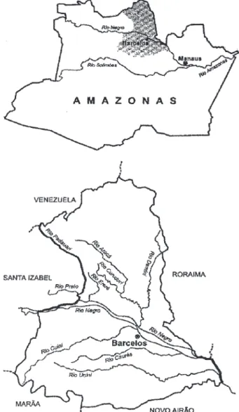
The specimens of R. brethesi were obtained from colo-nies maintained in the Parasitic Disease Laboratory, De-partment of Tropical Medicine of Instituto Oswaldo Cruz. They had been collected by means of modified Noireau traps and Shannon-type traps on piassaba palm trees in four rivers located in the left margin of Negro River, in the northern part of the state of Amazonas, Brazil: Acará River, Curuduri River, Preto River, and Padauiri River. The first two rivers are situated in the municipality of Barcelos (lati-tude 68°55’N and longi(lati-tude 0°10’W), the third in the mu-nicipality of Santa Isabel do Rio Negro (latitude 62°55’S and longitude 1°W), and the last is divisor of both mu-nicipalities (Fig. 1).
Five male and five female specimens were utilized. The insects were killed using ethyl acetate and were dissected
to remove the structures. These were mounted on metal-lic supports suitable for scanning electron microscopy, using double-sided tape. The structures analyzed were the head in dorsal and ventral views, eyes, antennae, buc-cula, labrum, rostrum, stridulatory sulcus and the feet, to view the apex of the tibia, spongy fossette and ctenidium. Measurements were made in the ocular and inter-ocellar regions.
These structures were covered with gold using an evaporation system known as “sputtering”, in which the gold is removed by means of bombardment in high vacuum (Hayat 1970), utilizing Balzers apparatus. The samples were observed at 15-20 kV using the Jeol 5310 scanning elec-tron microscope (Akishima, Tokyo, Japan) belonging to the Carlos Chagas Filho Biophysics Institute of the Fed-eral University of Rio de Janeiro (UFRJ). The images ob-tained were captured directly onto the computer by utiliz-ing the SemAfore software.
917 917 917 917 917 Mem Inst Oswaldo Cruz, Rio de Janeiro, Vol. 100(8), D ecem ber 2005
RESULTS
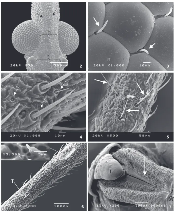
The head of R. brethesi presented rugose cuticular features in its dorsal region, with 1+1 longitudinal smooth areas delimiting a central area in which uniformly arranged bristles were seen (Fig. 2). The specimens presented com-posite eyes that were totally covered with ommatidia, in-cluding in the posterior-inferior area of this structure. Between every two or three ommatidia, there were bristles set in protruding buttons that were sometimes visible. These were corrugated and short or long, with apices that were rounded or slightly dilated (Fig. 3). The mean inter-ocular distance measured was 470 µm on the females and 464 µm on the males, while the mean inter-ocellar distance was 703.4 µm on the females and 660.3 µm on the males.
The antennae had four segments and presented cor-rugated integument and sensilla of varied shapes and sizes (Fig. 4). At the base of the second segment of the an-tenna, there were three trichobotria distributed on the ex-ternal lateral face (Fig. 5) and two on the dorsal face, and also another four located on the remainder of the seg-ment, thus totaling nine of these structures. At the base of each trichobotrium, the cuticular area was differenti-ated by presenting lamellar structures and fingerlike pro-longations (Fig. 6).
The pyriform labrum that rested on the first segment of the rostrum presented smooth integument with slight depressions and coarse bristles that were generally curv-ing downwards. Under the labrum, there was a triangular depression with regularly distributed granulation that ex-tended laterally and symmetrically as far as the apex of the first segment of the rostrum (Fig. 7).
The third segment of the rostrum presented sensilla of different shapes and sizes (Fig. 8). At the ventral apex, there was an elliptical hairless depression with two perfo-rations located in the basal third. The mouth styli were surrounded by translucent sheaths located in the ros-trum. When the rostrum was distended, this made it pos-sible to view the buccula, which was located ventrally between the apex of the head and the anterior-ventral re-gion of the first segment of the rostrum. It was an oval structure, with medial constriction, resembling a figure-of-eight. The basal part had raised borders covered with dispersed tubercles, and in the medial region a slight de-pression was observed, with corrugated integument. The apical portion had two lateral depressions with longitudi-nal and transversal striae (Fig. 9).
The stridulatory sulcus was presented in the shape of an amphora (Fig. 10), with transversal striae of appear-ance varying according to the region observed. In the basal region, they were poorly defined (Fig. 11), becom-ing delineated from the area of the constriction onwards as far as the apex, which had a rugose appearance (Fig. 12). In the lateral portion of the sulcus, a reticulated area was seen, with granular appearance and close silky tu-bercles covering its whole extent (Fig. 13)
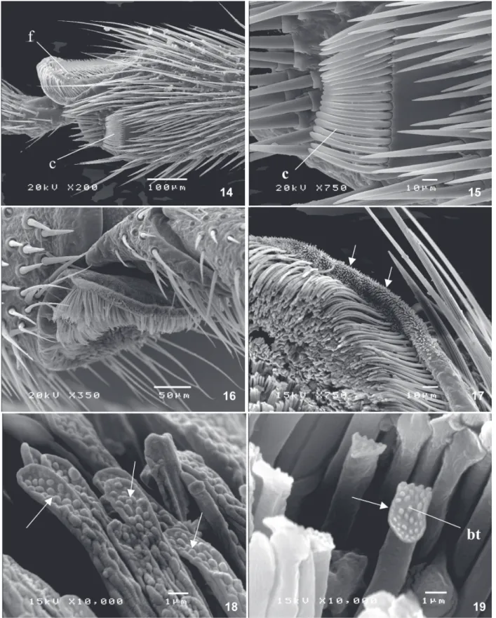
On the feet, two cuticular structures were prominent at the apex of the tibia: the ctenidium and the spongy fossette (Fig. 14). The ctenidium was found only on the first pair of feet, in both sexes (Fig. 15). The spongy fos-sette (Fig. 16) was present on the first and second pairs of
feet, in both males and females, and had a pyriform ap-pearance with an external margin covered with short fin-gerlike projections (Fig. 17). Around the internal margin, bristles were seen, turning inwards; this margin had a lat-erally flattened appearance and its surface was covered with button-shaped structures (Fig. 18). In the medial re-gion, the bristles were straight, and there was a straight and flattened apex covered with buttons (Fig. 19).
DISCUSSION
Of the species of triatomine found in Amazon Brazil, ten are infected with T. cruzi (Almeida 1971, Lent & Wygodzinsky 1979, Miles et al. 1981, 1983, Brazil et al. 1985, Barrett & Guerreiro 1991), among them R. brethesi
(Coura et al. 1999).
At a recent international meeting on the surveillance and prevention of Chagas disease in the Amazon region, one of the points of consensus was that American trypa-nosomiasis has high prevalence and wide dispersion as a wild enzootic disease throughout the Amazon region, pre-senting characteristics favorable towards its expansion as a human endemic disease. Among the particular trans-mission situations, it was reported that it occurred in ar-eas with extraction activities consisting of the gathering of piassaba palm tree fibers, where the vector species R. brethesi has been found (Technical Report 2005).
As stated earlier, few studies have been conducted on this species. These have mainly been restricted to its biology, and no morphological approaches at the ultra-structural level comparable with what has been done for other triatomine species are known of. The present study therefore expands the information available on this vec-tor species coming from the Upper and Middle Negro River, microregion of Negro River. In this first analysis using adult specimens, the presence of some structures already described in the literature for other species of triatomines has been confirmed, and some other, previously unre-ported structures have been described. These may have taxonomic value, in accordance with the continuation of the comparative study.
Observations on the presence of bristles between the ommatidia in the genera Alberprozenia, Belminus, Microtriatoma, Cavernicola, Rhodnius,Psammolestes, Dipetalogaster, Eratyrus, Panstrongylus, Paratriatoma,
and Triatoma, by means of scanning electron microscopy, were made by Galíndez Girón et al. (1994), thus providing confirmation for the results obtained in the present study for the genus Rhodnius.
918 918 918 918
918 Scanning electron microscopy of R. brethesi • Jacenir Reis dos Santos-M allet et al.
Fig. 2: dorsal region of Rhodnius brethesi head with 1+1 longitudinal smooth areas (arrows) showing the inter-ocular (a) and inter-ocellar (b) regions where the measurements were made. Fig. 3: bristles between ommatidia (arrow). Fig. 4: sensilla in the antennae with varied shapes and sizes. Fig. 5: three trichobotria on the external lateral face of antennae (arrows). Fig. 6: cuticular area at the base of trichobotrium (T) with lamellar structures and fingerlike prolongations. Fig. 7: the labrum with smooth integument; triangular depression (arrow); first segment of the rostrum (*).
However, Catalá and Schofield (1994) wrote that, in adult specimens of the genus Rhodnius, the total number of trichobotria varied between five and nine, and that this
919 919 919 919 919 Mem Inst Oswaldo Cruz, Rio de Janeiro, Vol. 100(8), D ecem ber 2005
Detailed studies (Andrade et al. 2002) on the principal sensilla found on the antennae of R. brethesi (bristle types I, II, and III) are being conducted with the aim of
elucidat-ing their taxonomic value, and also to evaluate the recep-tor pattern in relation to habitat adaptations. These as-pects were also observed by Catalá (1994, 1998) in others
920 920 920 920
920 Scanning electron microscopy of R. brethesi • Jacenir Reis dos Santos-M allet et al.
species of Rhodnius genus. Recently, Catalá et al. (2004) demonstrated that specimens kept in a laboratory for long periods of time can present modifications to the sensilla,
thus suggesting that the behavioral and physiological results obtained with insects from laboratories might be compromised.
921 921 921 921 921 Mem Inst Oswaldo Cruz, Rio de Janeiro, Vol. 100(8), D ecem ber 2005
In the present study, the third segment of the rostrum presented sensilla of different shapes and sizes. The im-portance of the rostrum for characterizing the genus as
Rhodnius was made by Pinto (1931) using light micros-copy. Likewise, Catalá (1996) emphasized this in an analy-sis by scanning electron microscopy of the rostrum of eight species of the genus Triatoma. This latter author concluded that the numbers and distribution of the sen-silla did not differ between nymph and adult forms, but did differ between the species of triatomines.
The buccula is a structure that has been demonstrated to have taxonomic value, and in the present study on R. brethesi it was found to have the format of a figure-of-eight. In this, it differs from the species Triatoma williami
and Triatoma gerstaeckeri (Ferro et al. 1997) and also from Triatoma guazu and Triatoma jurbergi (Silva et al. 2003), which have a U-shaped format.
The stridulatory sulcus is another structure present-ing features of taxonomic value, in relation to shape, num-ber of striae and lateral ornamentation of the integument. In the present study on R. brethesi, it was found to have the shape of an amphora. In this, it differs from what has been observed for other genera, and for other species of the same genus, as demonstrated by Lent and Wy-godzinsky (1979) and Silva et al. (2003).
The ctenidium, which has the function of removing impurities from the insect’s cuticle, was found only on the first pair of feet in the adult form. The spongy fossette has an adhesive function that allows adult specimens to move across smooth surfaces and allows the male to seize the female during copulation (Lent & Wygodzinsky 1979). It was only found on the first and second pairs of feet of both the male and female adults, compatible with species of the genus Rhodnius. These authors found the spongy fossette in all the nymph stages of Parabelminus and at least in the fifth nymph stage of Microtriatoma. Cam-pannuci et al. (1997) stated that the presence of the spongy fossette on the first and second pairs of feet of Triatoma infestans served to keep the female in a position that en-abled successful copulation. They also suggested that some type of secretion might be released in relation to sexual interactions and/or the spongy fossette might fur-nish sensory information associated with sexual behav-ior. In the present study, the differences in the bristles found in the spongy fossette suggest the need for com-parative studies within and between species.
The present work has expanded the morphological knowledge of R. brethesi, since previous descriptions of specimens were basically produced from optical micros-copy. Among the structures analyzed, it is suggested that detailed features of the buccula and the stridulatory sul-cus could be included as diagnostic characteristics to be utilized in new dichotomous keys, because of the notable differences in these structures that are presented in com-parisons with species in different genera and also with different species. In addition to these two characteristics, ultrastructural study of the antennae, rostrum and spongy fossette of adults could be included in these new keys, to complement the information existing in the literature.
The results obtained from this work, allied with the observations made by various authors on other species
utilizing scanning electron microscopy, furnish data that will contribute towards establishing specific diagnostic characteristics, especially with regard to differentiating between cryptic species, and also in the analysis of speci-mens from different localities, either with or without mak-ing associations with their habitats.
ACKNOWLEDGEMENTS
To Hertha Meyer Cell Laboratory of the Carlos Chagas Filho Biophysics Institute, Federal University of Rio de Janeiro, for allowing the use of the scanning electron microscope; to Samuel Ferreira de Deus and Maria José da Silva de Souza from Parasitic Disease Laboratory, Department of Tropical Medi-cine of Oswaldo Cruz Institute, and Adalberto José da Silva from Nucleus of Morphology and Ultrastructure of Arthro-poda, Department of Entomology of Oswaldo Cruz Institute, for technical support.
REFERENCES
Almeida FB 1971. Triatomíneos da Amazônia. Encontro de três espécies naturalmente infectada por Trypanosoma semelhante ao cruzi, no Estado do Amazonas (Hemiptera, Reduviidae). Acta Amazonica 1: 89-93.
Andrade ZP, Gonçalves TCM, Carvalho-Moreira CJ, Junqueira ACV, Spata CM, Santos-Mallet JR 2002. Morphological and morphometric analysis of antennal sensilla of Rhodnius brethesi Matta, 1919 (Hemiptera: Reduviidae) by scanning electron microscopy. Simpósio de Metodologias Integradas no Estudo da Biologia/Evento de Microscopia e Mi-croanálise do Mercosul, Curitiba, PR.
Barata JMS 1981. Aspectos morfológicos de ovos de Tria-tominae. II Características macroscópicas e exocoriais de dez espécies do gênero Rhodnius Stal, 1859 (Hemiptera Reduviidae). Rev Saúde Públ S Paulo 15: 490-542.
Barata JMS 1998. Macroscopic and exochorial structures of Triatominae eggs (Hemiptera, Reduviidae): In RU Carcavallo, I Galíndez Girón, J Jurberg, H Lent (eds), Atlas of Chagas Disease Vectors in the Americas,Vol. II, Fiocruz, Rio de Janeiro, p. 409-448.
Barrett TV, Guerreiro JCH 1991. Os triatomíneos (Hemiptera, Reduviidae) em relação a doença de Chagas na Amazônia. In AL Val (eds), Bases Científicas para Estratégia de Preservação e Desenvolvimento da Amazônia: Fatos e Perspectivas, Instituto Nacional de Pesquisa da Amazônia, Manaus, p. 119-130.
Brazil RP, Silva AF, Albarelli A, Valle JF 1985. Distribuição e infecção de triatomíneos por Trypanosoma cruzi na Ilha de São Luiz, Maranhão. Rev Soc Bras Med Trop 18: 257-260.
Campannuci V, Insausti TC, Lazzari CR 1997. The functional morphology of triatominae legs: I. The distal tibia. Mem Inst Oswaldo Cruz 92 (Suppl. I): 487.
Carcavallo RU, Curto de Casas SI, Sherlock IA, Galíndez-Girón I, Jurberg J, Galvão C, Mena Segura CA 1999. Geographical distribution and altilatitudinal dispersion. In R Carcavallo, I Galíndez Girón, J Jurberg, H Lent (eds), Atlas of Chagas Disease Vectors in the Americas,Vol. III, Fiocruz, Rio de Janeiro, p. 747-792.
922 922 922 922
922 Scanning electron microscopy of R. brethesi • Jacenir Reis dos Santos-M allet et al.
Catalá S 1994. The cave organ of Triatominae bugs. Mem Inst Oswaldo Cruz 89: 275-277.
Catalá S 1996. Sensilla associated with the rostrum of eight species of Triatominae. J Morphol 228: 195-201.
Catalá S 1998. Antennae and rostrum. In R Carcavallo, I Galíndez Girón, J Jurberg, H Lent (eds), Atlas of Chagas Disease Vectors in the Americas,Vol. II, Fiocruz, Rio de Janeiro, p. 409-448.
Catalá S, Schofield C 1994. Antennal sensilla of Rhodnius. J Morphol 219: 193-203.
Catalá S, Maida DM, Caro-Riaño H, Jaramillo N, Moreno J 2004. Changes associated with laboratory rearing in anten-nal sensilla patterns of Triatoma infestans, Rhodnius prolixus, and Rhodnius pallescens (Hemiptera, Reduviidae, Triatominae). Mem Inst Oswaldo Cruz 99: 25-30.
Costa J, Barth OM, Marchon-Silva V, Almeida CE, Freitas-Sibajev MGR, Panzera F 1997. Morphological studies on the Triatoma brasiliensis Neiva, 1911 (Hemiptera, Reduvi-idae, Triatominae) genital structures and eggs of different chromatic forms. Mem Inst Oswaldo Cruz 92: 493-498.
Costa J, Jurberg J, Barth OM 1991. Estudos morfológicos de Cavernicola lenti Barrett & Arias, 1985 (Hemiptera: Redu-viidae: Triatominae). Mem Inst Oswaldo Cruz 86: 247-263.
Coura JR, Arboleda Naranjo M, Willcox HPF 1995. Chagas’ disease in the Brazilian Amazon. II. A serological survey. Rev Inst Med Trop São Paulo 37: 103-107.
Coura JR, Barrett TV, Naranjo MA 1994. Ataque de populações humanas por triatomíneos silvestres no Amazonas: uma nova forma de transmissão chagásica? Rev Soc Bras Med Trop 27: 251-253.
Coura JR Junqueira ACV, Boia MN, Fernandes O 1999. Chagas disease: from bush to huts and house. Is it the case of the Brazilian Amazon? Mem Inst Oswaldo Cruz 94 (Suppl. I): 379-384.
Coura JR, Junqueira ACV, Boia MN, Fernandes O, Bonfante C, Campos JE, Santos L, Devera R 2002a. Chagas disease in the Brazilian Amazon. IV. A new cross-sectional study. Rev Inst Med Trop São Paulo44: 159-165.
Coura JR, Junqueira ACV, Fernandes O, Valente SAS, Miles M 2002b. Emerging Chagas disease in Amazonian Brazil. Trends Parasitol 18: 171-176.
D’Alessandro A, Barreto P, Duarte CA 1971. Distribuition of Triatominae transmited trypanosomiase in Colombia and new records of the bugs and infections. J Med Entomol 8: 159-172.
Dias JCP, Prata A, Schofield CJ 2002. Chagas disease in the Amazon: an overview of the current situation and perspec-tive for prevention. Rev Soc Bras Med Trop 35: 669-678.
Ferro ZPA, Barbosa HS, Jurberg J, Carcavallo RU 1997. The buccula and gula of Triatominae nymphs by scanning elec-tron microscopy (Hemiptera: Reduviidae). Acta Microsc 6: 572-573.
Ferro ZPA, Carvalho-Moreira CJ, Junqueira ACV, Spata CM, Silva LM, Gonçalves TCM 1999. Morphological studies of Rhodnius brethesi Matta, 1919 (Hemiptera, Reduviidae, Triatominae) by scanning electron microscope and confo-cal. Mem Inst Oswaldo Cruz 94 (Suppl. II): 242.
Ferro ZPA, Junqueira ACV, Moreira CJC, Spata CM, Gonçalves
TCM 1998. Preliminary analysis of external morphology of Rhodnius brethesi Matta, 1919 by scanning microscopy (Hemiptera, Reduviidae, Triatominae). Mem Inst Oswaldo Cruz 93 (Suppl. II): 342.
Fraiha Neto H, Valente SAS, Valente VC, Pinto AYN 1995. Doença de Chagas – Endêmica na Amazônia? An Acad Méd Pará Belém 6: 53-57.
Galvão C, Carcavallo RU, Rocha DS, Jurberg J 2003. A check-list of the current valid species of the subfamily Triatominae Jeannel, 1919 (Hemiptera, Reduviidae) and their geographi-cal distribution, with nomenclatural and taxonomic notes. Zootaxa 202: 1-36.
Galíndez Girón I, Carcavallo RU, Valderrama A 1994. Erwinilas o cerdas interomatidiales em la subfamília Triatominae (Hemiptera, Reduviidae). Entomol Vect 1: 94-96.
Gonçalves TCM, Jurberg J, Costa JM, Souza W 1985. Estudo morfológico comparativo de ovos e ninfas de Triatoma maculata (Erichson, 1848) e Triatoma pseudomaculata. Corrêa & Espínola 1964 (Hemiptera, Reduviidae, Tria-tominae). Mem Inst Oswaldo Cruz 80: 263-276.
Hayat MA 1970. Principles and Techniques of Electron Mi-croscopy. Biological Applications, Vol. 1, Van Nostrand Reinhold Company, New York.
Lent H 1948. O gênero Stal, 1859 (Hemiptera, Reduviidae). Rev Bras Biol 8: 319.
Lent H, Jurberg J 1969. O gênero Rhodnius com um estudo sobre a genitália das espécies (Hemiptera, Reduviidae, Triatominae). Rev Brasil Biol 29: 487-560.
Lent H, Wygodzinsky P 1979. Revision of the Triatominae (Hemiptera, Reduviidae), and their significance as vectors of Chagas’ disease. Bull Am Mus Nat Hist 163:123-520.
Mascarenhas B M 1982. Triatomíneos da Amazônia: mor-fometria do ovo de Rhodnius brethesi Matta, 1919 (Hemi-ptera: Triatominae). Acta Amazonica 12: 661-664.
Mascarenhas B M 1987. Descrição dos estádios imaturos de Rhodnius brethesi Matta, 1919 (Hemiptera, Reduviidae). Bol Mus Paran Emílio Goeldi, sér Zool 3: 183-194.
Mascarenhas BM 1990. Triatomíneos da Amazônia: sobre o ciclo evolutivo de Rhodnius brethesi Matta, 1919 (Hemi-ptera: Reduviidae: Triatominae). Bol Mus Paran Emilio Goeldi, sér Zool 6: 191-202.
Mascarenhas BM 1991. Triatomíneos da Amazônia: sobre o habitat e algumas considerações comportamentais de Rhodnius brethesi Matta, 1919 (Hemiptera: Reduviidae: Triatominae) na região do médio Rio Negro, Amazonas. Bol Mus Paran Emilio Goeldi, sér Zool 7: 107-116.
Matta A 1919. Um novo reduvídeo do Amazonas, Rhodnius brethesi n. sp. Amazonas Med 2: 93-94.
Matta A 1922. Sobre o gênero Rhodnius do Amazonas. Ama-zonas Med 5: 161-162.
Miles MA, Arias JR, Souza AA 1983. Chagas disease in the Amazon Basin. V. Periurban palms as habitats of Rhodnius robustus and Rhodnius pictipes, triatomíneos vectors of Chagas disease. Mem Inst Oswaldo Cruz 78: 391-398.
923 923 923 923 923 Mem Inst Oswaldo Cruz, Rio de Janeiro, Vol. 100(8), D ecem ber 2005
Pinto C 1931. Valor do rostrum e das antenas na caracterização dos triatomíneos. Boletim Biológico 19: 45-137.
Rebelo JMM, Barros VLL, Mendes WA 1998. Espécies de Triatominae (Hemiptera: Reduviidae) do Estado do Maranhão, Brasil. Cad Saúde Públ 14: 187-192.
Rosa J A, Barata, J M S, Cilense M, Neto F M B 1999. Head morphology of 1st and 5th instar nymphs of Triatoma circummaculata and Triatoma rubrovaria (Hemiptera, Reduviidae). Int J Insect Morphol Embriol 28: 363-375.
Silva MBA, Barbosa HS, Galvão C, Jurberg J, Carcavallo RU 2003. Comparative study of the stridulatory sulcus, buc-cula and rostrum of the nymphs of Triatoma guazu Lent &
Wygodzinky, 1979 and Triatoma jurbergi Carcavallo, Galvão & Lent 1998 by scanning electron microscopy (Hemi-ptera, Reduviidae). Mem Inst Oswaldo Cruz 98: 335-344.
Technical Report/Relatório Técnico 2005. Reunião Internacional sobre Vigilância e Prevenção da Doença de Chagas na Amazônia. Implementação da Iniciativa Intergovernamental deVigilância e Prevenção da doença de Chagas na Amazônia. Rev Soc Med Trop 38:82-89.