Alexandre Castanho Barata Leitão
Dissertation presented to obtain the Ph.D degree in Biology
Instituto de Tecnologia Química e Biológica António Xavier | Universidade Nova de Lisboa
Oeiras,
July, 2014
Alexandre Castanho Barata Leitão
Dissertation presented to obtain the Ph.D degree in Biology
Instituto de Tecnologia Química e Biológica | Universidade Nova de Lisboa
Oeiras, July, 2014
Alexandre Castanho Barata Leitão
Dissertation presented to obtain the Ph.D degree in Biology
Instituto de Tecnologia Química e Biológica | Universidade Nova de Lisboa
Oeiras, July, 2014
Esta dissertação é o resultado do meu próprio trabalho desenvolvido entre Março de 2010 e Julho de 2014 no laboratório do Dr. Élio Sucena, Instituto Gulbenkian de Ciência em Oeiras, Portugal, no âmbito do Programa Gulbenkian de Doutoramento (edição 2009-2010). Parte deste trabalho encontra-se em preparação para ser publicado.
This dissertation is the result of my own research, carried out between March 2010 and July 2014 in the laboratory of Dr. Élio Sucena, Instituto Gulbenkian de Ciência in Oeiras, Portugal, under the Gulbenkian Doctoral Programme (2009-2010 edition). Part of this work in being prepared for publication.
Apoio Financeiro/Financial Support
Apoio financeiro da FCT e do FSE no âmbito do Quadro Comunitário de Apoio, bolsa de doutoramento #SFRH / BD / 51175 / 2010
i
Acknowledgments
I would like to thank the IGC for giving me the opportunity to do my Ph.D. Certainly I got more from the IGC than the opposite.
To my thesis committee, Luís Teixeira and Rui Martinho, and to my Ph.D courses director, Thiago Carvalho. These three people, with small words, changed a lot my route.
To all the member of Evolution and Development Lab, Variation: Development and Selection Lab and Development, Evolution and the Environment lab. Thank you for all the discussions and ideas that certainly are impregnated in this thesis.
For all those of you that reside outside the IGC perimeter but, nevertheless, contributed to my happiness and to the joy of continuing my science career, my sincere gratitude.
ii
Summary
All coelomate animals possess a population of cells that do not make part of an organ and instead freely flow inside the body cavity. These cells, termed hemocytes (in invertebrates) or blood cells (in vertebrates), are involved in varied functions including immune response, clearance of apoptotic cells and distribution of nutrient and gases (Grigorian & Hartenstein 2013). To perform all these functions correctly, blood cells need to achieve an optimal concentration and proportion between cell types (Almeida et al. 2005). This balance is achieved by several mechanisms of cell fate choices and proliferation in a process collectively termed hematopoiesis.
In vertebrates, immune cells differentiate from Hematopoietic Stem Cells (HSC) residing in hematopoietic organs. Several layers of genetic control dependent on cytokines and cell contacts ensure the correct proportion of produced cell types. Invertebrates produce fewer blood cell types but almost all species possess a hematopoietic organ (Hartenstein 2006). In Drosophila melanogaster hematopoiesis is
iii The same mature cell types can be found in circulation. Importantly, both plasmatocytes and crystal cells found in circulation are not derived from the lymph gland because without an immune challenge this organ does not release hemocytes until pupariation (Holz et al. 2003).
Also, tough hemocytes can be found in circulation, up to two thirds of them are sessile and reside in the epithelium underneath the larval cuticle (Lanot et al. 2001). These sessile cells are not randomly dispersed but aggregated in clusters in every segment of the larva. Recently, it has been shown that the peripheral nervous system supports hemocyte homing to these sites and influences their proliferation/survival (Makhijani et al. 2011). It has been suggested that these sessile hemocytes clusters function as hematopoietic compartments because cells taken from them can differentiate into lamellocytes (Márkus et al. 2009). However, to define the hemocyte clusters as hematopoietic compartments one needs evidence to show that the clustering of cells is necessary to induce cell fate choices.
iv
responsible for inducing crystal cells differentiation are themselves hemocytes. Moreover, we show that the clusters are necessary to achieve a correct proportion between plasmatocytes and crystal cells. Nevertheless, a striking difference exists between crystal cell development in hemocyte clusters and in the lymph gland. Whereas, in the lymph gland crystal cells differentiate from prohemocytes, in the hemocyte clusters they differentiate from plasmatocytes.
With the confirmation that hemocyte clusters function as hematopoietic tissue we conducted a Genome Wide Association Study (GWAS) to search for genetic players involved in the regulation of hemocyte numbers/proportions. Our results show that the G protein-coupled receptor kinase 1 (Gprk1) is essential to achieve the
correct number of hemocytes. Although the function of Gprk1is poorly
studied in Drosophila the vertebrate ortholog (GRK2) plays several
roles in hematopoiesis and is essential for the correct number of monocytes and neutrophils (Otten et al. 2013). This indicates that
Gprk1 is another example of the evolutionary conservation in genetic
control of hematopoiesis between insects and vertebrates.
Together, our results show that the regulation of hemocyte numbers/proportions in the Drosophila larva is achieved by a
v
Sumário
Todos os animais celomados têm uma população de células que não fazem parte de um órgão mas, ao invés, estão em livre circulação dentro da cavidade do corpo. Estas células, denominadas células sanguíneas (em vertebrados) ou hemócitos (em invertebrados), estão envolvidas em diferentes funções incluindo: resposta imunitária, fagocitose de corpos apoptóticos e distribuição de nutrientes e gases (Grigorian & Hartenstein 2013). Para que estas funções sejam efetuadas corretamente pelas células sanguíneas é necessário que estas atinjam a concentração e proporção ótima entre diferentes tipos celulares (Almeida et al. 2005). Este balanço é conseguido através de diferentes mecanismos de determinação de diferenciação celular e proliferação, um processo denominado hematopoiese.
Em vertebrados, as células imunitárias diferenciam-se a partir de células estaminais hematopoiéticas. A produção de diferentes tipos celulares nas proporções corretas é conseguida através de diferentes níveis de controlo genético dependente de citoquinas e contactos celulares. Os animais invertebrados produzem menos tipos celulares mas quase todas as espécies têm um órgão hematopoiético (Hartenstein 2006). Em Drosophila melanogater, o
vi
sinalizador posterior para manterem o seu estado indiferenciado ou para migrarem para a zona cortical e começarem a sua diferenciação.
Os mesmos tipos de hemócitos encontram-se em circulação. Um aspeto importante a notar é que tanto os plasmatócitos como as células cristal em circulação não derivam da glândula linfática porque, sem um infeção, este órgão não liberta os hemócitos produzidos até se formar a pupa (Holz et al. 2003).
Fora da glândula linfática os hemócitos encontram-se em circulação mas dois terços dos hemócitos estão num estado séssil no epitélio da cutícula larvar (Lanot et al. 2001). Estas células sésseis não estão distribuídas aleatoriamente mas sim agregadas em conjuntos nos vários segmentos da larva. Recentemente, foi demonstrado que o sistema nervoso periférico influencia a atracão dos hemócitos para estes locais e influencia a sua proliferação/sobrevivência (Makhijani et al. 2011). Já foi sugerido que os agregados de hemócitos sésseis funcionam como tecido hematopoiético porque células retiradas destes agregados conseguem diferenciar-se num terceiro tipo de hemócito, o lamelócito (Márkus et al. 2009). No entanto, para definir os agregados de hemócitos como tecido hematopoiético é necessário mostrar que a estrutura destes agregados é necessária para induzir escolhas de destinos celulares.
vii questão de desenvolvimento. A sinalização Notch foi uma das primeiras cascatas genéticas demonstrada ser essencial na diferenciação de células cristal (Duvic et al. 2002; Lebestky et al. 2003). Inspirados pelo facto que os hemócitos dentro dos agregados estão em contacto por longos períodos de tempo, nós delineámos uma série de experiências para testar se os agregados funcionam como tecido hematopoiético onde as células cristal seriam diferenciadas sob a influência de outros hemócitos. Os nossos resultados mostram que as células cristal são formadas nestes agregados e que, tal como na glândula linfática, as células que induzem a sinalização Notch são hemócitos. Também demonstramos aqui que os agregados são necessários para manter a proporção correta entre plasmatócitos e células cristal. Com a confirmação de que os agregados funcionam como tecido hematopoiético conduzimos um estudo de associação genómica para procurar genes envolvidos na regulação dos números/proporção de hemócitos. Os nossos resultados mostram que o gene G protein-coupled receptor kinase 1 (Gprk1) é essencial para desenvolver o número correto de
hemócitos. Apesar deste gene ser pouco estudado em Drosophila, o
ortólogo em vertebrados (GRK2) desempenha importantes funções
em hematopoiese e é essencial para o estabelecimento do número correto de monócitos e neutrófilos (Otten et al. 2013). Isto é uma indicação que Gprk1 é outro exemplo de conservação evolutiva no
controlo genético da hematopoiese entre insetos e vertebrados.
Em conjunto, os nossos resultados mostram que a regulação dos números/proporções de hemócitos na larva de Drosophila é
viii
ix
Table of contents
Acknowledgments ... i
Summary ... ii
Sumário ... v
Table of contents ... ix
1 Introduction ... 1
1.1 Hemocytes in Drosophila ... 3
1.1.1 Prohemocyte ... 4
1.1.2 Plasmatocyte ... 6
1.1.3 Crystal Cell... 7
1.1.4 Lamellocyte ... 9
1.2 Embryonic hematopoiesis ... 11
1.3 Larval hematopoiesis ... 16
1.3.1 Lymph gland development and structure ... 16
1.3.2 Hematopoiesis within the lymph gland ... 18
1.3.3 Hematopoiesis outside the lymph gland ... 21
1.4 Objectives of this work ... 23
2 Hemocyte clusters function as a hematopoietic tissue ... 25
2.1 Summary ... 26
2.2 Introduction ... 27
x
2.3.1 Fly stocks and parasitoids maintenance ... 30
2.3.2 Larva staging ... 31
2.3.3 Flow cytometry analysis and cell viability assay ... 31
2.3.4 Cell cycle analysis ... 32
2.3.5 Movie analysis ... 32
2.3.6 Total hemocyte loads counts and crystal cells counts 33 2.3.7 Hemocyte immunohistochemistry ... 34
2.3.8 Imaging ... 34
2.3.9 Phagocytosis assay ... 35
2.3.10 Cluster disruption assay ... 35
2.3.11 Statistical analysis ... 35
2.4 Results ... 36
2.4.1 The number of crystal cell precursors increase during larval development without cell proliferation ... 36
2.4.2 Serrate expression in Hml+Lz- cells is essential to induce crystal cell differentiation ... 39
2.4.3 Plasmatocyte/crystal cell ratio in clusters is influenced by the number of contacting hemocytes ... 43
2.4.4 Differentiation of Hml+Lz+ cells occurs in clusters .... 45
2.4.5 Hemocyte clusters are necessary for the normal development of different hemocyte types ... 47
xi 2.4.7 lozenge expression induction can occur in G1 phase
of the cell cycle ... 53
2.4.8 Notch promoter reporters are expressed in crystal cells and in a subset of plasmatocytes ... 55
2.5 Discussion ... 58
2.6 Author’s Contributions ... 64
2.7 Acknowledgments... 64
2.8 Supplemental Material ... 65
3 Gprk1 is involved in the control of hemocyte loads ... 70
3.1 Summary ... 71
3.2 Introduction ... 72
3.3 Material and Methods ... 74
3.3.1 Fly stocks maintenance and egglays ... 74
3.3.2 Parasitoids maintenance and infection ... 75
3.3.3 Total hemocyte loads counts and crystal cells counts 75 3.3.4 Phenoloxidase activity ... 76
3.3.5 Hemocyte immunohistochemistry ... 76
3.3.6 Statistical analysis ... 77
3.4 Results ... 78
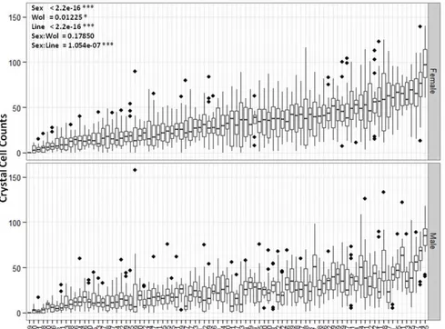
3.4.1 Crystal cell numbers vary across DGRP lines ... 78
3.4.2 No correlation is found between parasitoid resistance and crystal cell numbers ... 80
xii
3.4.4 GWAS for crystal cell numbers ... 85
3.4.5 Gprk1 is essential to control hemocyte loads ... 88
3.5 Discussion ... 89
3.6 Author’s Contributions ... 96
3.7 Acknowledgments... 96
3.8 Supplemental Material ... 97
4 General discussion ... 98
4.1 Haemocoelic hemocytes compartments ... 99
4.2 Hematopoietic cell fate decision in hemocyte clusters 101 4.3 Control of hemocyte numbers ... 106
4.4 Open questions for future enquire ... 107
4.5 Concluding remarks ... 108
1
1
Introduction
The term “hematopoiesis” results from the conjugation of two Greek words: haimat (blood) and poiesis (to make). Understanding
how animals make blood presupposes the study of two intertwined
2
that evolved in animals and alludes to the fact that this may be a very plastic process.
In invertebrates, blood cells are more commonly termed hemocytes. The number of different hemocyte types in invertebrates is smaller than blood cell types in vertebrates. This reflects the fact that a great part of vertebrate immune cells are dedicated to the adaptive immune response, a branch of the immune system that is not present in invertebrates. Nevertheless, it is possible to distinguish different hemocyte types and hematopoietic organs in most of coelomate invertebrate species studied so far (Grigorian & Hartenstein 2013). The number, location and structure of these hematopoietic organs changes greatly between species (Grigorian & Hartenstein 2013). They range from very simple structures where hemocyte proliferation is enhanced, such as the hematopoietic centers of polychaete annelids, to more complex gland like structures, exemplified by the Drosophila lymph gland (Hartenstein 2006).
Hematopoiesis in the Drosophila’s larval lymph gland produces the
3
In this introduction to the theme of hematopoiesis in
Drosophila melanogaster we will start to define the different types of
hemocytes found at the different life stages and follow with the current knowledge of hematopoiesis during embryogenesis and larval stages.
1.1
Hemocytes in Drosophila
In insects it is common to classify hemocytes into four morphologically distinct types: prohemocytes, granulocytes, plasmatocytes and oenocytoids (Lavine & Strand 2002). Confusingly, the terminology for functional equivalent cells in Drosophila is
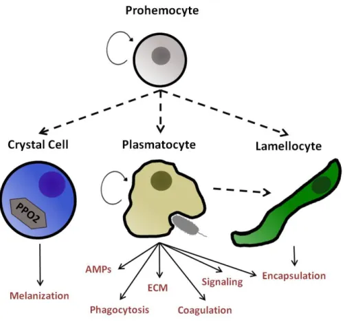
different (Ribeiro & Brehélin 2006). Prohemocytes are cells that can differentiate into the other three types of hemocytes. Granulocytes, termed plasmatocytes in Drosophila, are phagocytic cells. Large cells dedicated to encapsulation of entities too large to be phagocytized are termed plasmatocytes in most insect species and lamellocytes in Drosophila. Finally, oenocytoids are cells that produce Phenoloxidase precursors necessary for the melanization response and in
Drosophila they are called crystal cells (Ribeiro & Brehélin 2006).
Throughout this thesis we will use the Drosophila terminology when
referring to hemocyte types, described as: prohemocytes, plasmatocytes, lamellocytes and crystal cells (Rizki 1957; Roshana & Gateff 1982; Ribeiro & Brehélin 2006). This hemocyte classification into four types was validated recently using molecular markers (Kurucz, Váczi, et al. 2007).
Some variations on this theme can be found at different levels. For example, there are references to another hemocyte type in
Drosophila termed podocyte but they are often considered
4
(Rizki 1957; Rizki 1962). Also, it has been reported that the obscura group flies do not produce lamellocytes (Havard et al. 2009). A fusiform cell termed nematocyte has been recently proposed to play a role against parasitoid egg infections in Zaprionus indianus, a
phylogenetic close relative of Drosophila melanogaster (Kacsoh et al.
2014). At a larger scale, it is also possible to distinguish another hemocyte type termed spherule cell in Lepidoptera for which the function remains unknown (Lavine & Strand 2002). Life-stage also determines the blood cell composition of Drosophila. Hemocytes can
be found in every developing stage of Drosophila melanogaster but
only during larval stages may we find all four types of hemocytes. Embryo, pupa and adult stages only contain a subset of hemocyte types.
A summary of hemocyte types and their functions is represented in Figure 1.1.
1.1.1 Prohemocyte
Prohemocytes are quiescent cells that can differentiate into the other three hemocyte types. They are found in the medullary zone of the larva hematopoietic organ, the lymph gland. Prohemocytes are characterized by their lack of maturation markers (Jung et al. 2005). In addition, they express genes that are down-regulated once they start to differentiate into mature hemocytes. Examples of these genes are DE-cadherin (DE-Cad), domeless (dome), unpaired3 (upd-3), and patched (ptc) (Jung et al. 2005; Mandal et al. 2007). Some of these
genes are essential to maintain the prohemocyte state. For example, prohemocytes will start to upregulate differentiation markers if
hedgehog, the ligand for the receptor Patched, is mutated (Mandal et
pro-5
hemocytes during embryogenesis when the first wave of hematopoiesis occurs. During hemocyte development in the embryo the cells pass through a stage when they only express early hemocyte markers and still are bipotent cells that may differentiate into plasmatocytes or crystal cells (Waltzer et al. 2010). Nevertheless, this is only a transient state that lasts few hours during embryogenesis. After that, all cells have either plasmatocyte or crystal cell markers (Waltzer et al. 2010).
Some reports suggest the presence of prohemocytes in circulation (Lanot et al. 2001; Sinenko et al. 2010). For example Lanot and colleagues report that hemocytes dividing in circulation do not phagocyte Indian ink particles (Lanot et al. 2001). This could indicate that there are two subpopulations of plasmatocytes, one with the capacity to divide and another devoted to phagocytosis. An alternative explanation would be that plasmatocytes do not phagocyte during cell division but a recent study shows that cells phagocyte in equal proportion whether they are synthesizing DNA or not (Makhijani et al. 2011). All larval hemocytes outside the lymph gland expresses
Hemolectin and Peroxidasin GAL4 drivers (Goto et al. 2003; Stramer
6
1.1.2 Plasmatocyte
Plasmatocytes are present in all developmental stages of
Drosophila and are always the most abundant hemocyte type. They
7
(Shia et al. 2009; Charroux & Royet 2009) (Figure 1.1). In contrast to their larval function, plasmatocytes are dispensable for the activation of humoral response in the adult fat body (Defaye et al. 2009). Later, when entering pupariation, an ecdysone peak increases the mobility of plasmatocytes and their chemo-attraction towards damaged epithelia (Regan et al. 2013). Also, concomitantly with this increase in phagocytic activity, plasmatocytes become bigger and increase the number of granules (Regan et al. 2013). During this period they change their expression profile by down-regulating Hemese
expression and becoming positive for the Ad1 antigen (Honti et al. 2014). In flies where hemocytes are depleted, by over-expressing a apoptotic gene, increased mortality rate is observed during pupal stages (Defaye et al. 2009; Charroux & Royet 2009). However, this phenotype is rescued by raising the larvae under sterile conditions, demonstrating the necessity for hemocytes for a immune response in the pupa but excluding a need for plasmatocytes in pupal development (Defaye et al. 2009; Charroux & Royet 2009). Embryonic and larval derived plasmatocytes persist into the adulthood in a post-mitotic state (Holz et al. 2003). Plasmatocyte is virtually the only hemocyte type found in adult and when they are depleted, the flies are more susceptible to bacterial challenge (Defaye et al. 2009; Charroux & Royet 2009).
1.1.3 Crystal Cell
8
Drosophila melanogaster genome encondes three pro-Phenoloxidase
(PPO) genes. A recent report demonstrates that crystal cells are the sole source of PPO1 and PPO2 but only PPO2 is present in the crystals (Binggeli et al. 2014). The melanization cascade is used as an immune response in several invertebrate groups whereby the production of toxic intermediates in the cascade kills bacteria and/or melanin deposition sequesters microbes and hardens capsules formed around parasitoid eggs (Lemaitre & Hoffmann 2007; Jiravanichpaisal et al. 2006). Since crystal cells are non-phagocytic cells and produce pro-Phenoloxidase crystals it is most likely that they are dedicated to the melanization response. This is evident when larvae are bled and crystal cells rupture right away releasing the crystal contents. The rupture of crystal cells in vivo depends on JNK
pathway and on Eiger, a gene with high homology to Tumor Necrosis
Factor (TNF) family members (Bidla et al. 2007).
9
are not present but some rare cells do express the crystal cell marker C1 (Kurucz, Váczi, et al. 2007).
1.1.4 Lamellocyte
10
Figure 1.1 – Hemocyte types and their functions in Drosophila melanogaster. Prohemocytes are cells with the capacity to proliferate and
11
1.2
Embryonic hematopoiesis
In Drosophila, hematopoiesis occurs in two distinct phases of
development. During embryogenesis hemocytes differentiate from cells in the head mesoderm and during larval stages they are produced in the lymph gland (Tepass et al. 1994; Roshana & Gateff 1982). In impressive single cell transplantation experiments, Holz and colleagues have shown that, as early as the blastoderm stage, a small anlage is already cell-autonomously committed to hemocyte fate (Holz et al. 2003). This commitment corresponds to the domain and time of expression of serpent, the first transcription factor known
to be essential for hemocyte differentiation (Rehorn et al. 1996). Serpent is one of the five GATA transcription factors found in
Drosophila, so called because they bind to the consensus DNA
sequence WGATAR (Waltzer et al. 2010). Nonetheless, it is worth noting that this commitment to hemocyte fate cannot be explained solely on serpent expression since it is expressed in other
mesodermal tissues such as fat body (Waltzer et al. 2010).
Two other zinc finger transcription factors are expressed in all pro-hemocytes at embryonic stage 5: Glial cells missing (Gcm) and Glial cells missing 2 (Gcm2) (Bataillé et al. 2005; Alfonso & Jones
2002; Bernardoni et al. 1997). This expression of Gcm and Gcm2 is
dependent on serpent (Bernardoni et al. 1997). Overexpression of Gcm is sufficient to induce expression of plasmatocyte markers and
in Gcm/Gcm2 mutants the number of plasmatocytes is strongly
reduced (Bernardoni et al. 1997; Alfonso & Jones 2002). This indicates that Gcm has instructive roles in plasmatocyte differentiation. Shortly after, at stage 6 of embryogenesis, Gcm is
down-regulated in the most anterior row of serpent expressing cells,
12
(Bataillé et al. 2005) (Figure 1.2). In 40% of these lozenge positive
cells, expression is discontinued and these cells differentiate into plasmatocytes. The other 60% maintain lozenge expression and
differentiate into crystal cells (Figure 1.2). In Gcm/Gcm2 mutants all lozenge positive cells differentiate into crystal cells (Bataillé et al.
2005). Moreover, forced expression of Gcm in lozenge positive cells
is sufficient to provoke a full differentiation into plasmatocytes (Bataillé et al. 2005). Together these results show that Gcm plays a
repressive role in crystal cell development and at the same time it promotes plasmatocyte differentiation.
How the expression of lozenge is initiated and limited to the
first rows of cells of the embryonic anlage is not determined. Although Notch signaling is sufficient and required in hemocytes for lozenge
expression in larval lymph gland cells (Duvic et al. 2002; Lebestky et al. 2003), this is not the case during embryonic development (Bataillé et al. 2005).Nevertheless, Notch signaling plays a role in determining the number of embryonic crystal cells (Lebestky et al. 2003; Bataillé et al. 2005). lozenge expression is also dependent on Serpent and its
maintenance can be assured by a regulatory loop through an enhancer activated by the Lozenge/Serpent complex (Ferjoux et al. 2007). Moreover, other crystal cell specific loci such as
pro-Phenoloxidases contain such Lozenge/Serpent binding sites (Ferjoux et al. 2007). However, lozenge expression is maintained in crystal
cells during larval stages while serpent is no longer detectable in
these cells.
13
GATA (FOG) family gene u-shaped (Ush) (Waltzer et al. 2010). By
stage 10 all prohemocytes express Ush (Fossett et al. 2001). Later in
development, by embryonic stage 13, Lozenge positive cells start to reduced Ush expression while in plasmatocytes Ush continues to be
maintained at high expression levels. In Ush mutants the number of
crystal cells increases significantly (Fossett et al. 2001). Moreover, the interaction of Serpent N-Zinc finger with Ush modulates the expression of Crq in plasmatocytes (Waltzer et al. 2010). Hence, Ush
adds another layer in the gene regulatory network that governs the cross-talk between specification of crystal cells and plasmatocytes.
At early stage 12, plasmatocytes start to migrate throughout the embryo along stereotypical routes (Tepass et al. 1994) and eventually colonize the germ band in a process that requires the dissolution of epithelial cell-cell junctions (Siekhaus et al. 2010). Pvf2 and Pvf3 are two ligands of the PDGF-and VEGF-receptor related (Pvr), that are expressed in epithelial cells along the migration routes of embryonic hemocytes and are proposed to guide this process (Cho et al. 2002). Later work has shown that the Pvf ligands are used as trophic factors that are essential for germband invasion (Brückner et al. 2004; Parsons & Foley 2013). During migration, plasmatocytes start to phagocyte dead cells and are essential for the proper development of the central nervous system (Defaye et al. 2009). In contrast, crystal cells do not migrate and remain in a cluster close to the anterior midgut. No role has been attributed to crystal cells during embryogenesis. It is possible that they form a pool of cells ready to act after larval hatching. This is the first observed behavioral difference seen between plasmatocytes and crystal cells.
In late embryogenesis, plasmatocytes stop serpent and Gcm
14
such as Hemolectin (Goto et al. 2003). Once the larva hatches the
plasmatocytes populate the body cavity and continue to proliferate. Crystal cells maintain lozenge expression and similarly enter the body
15
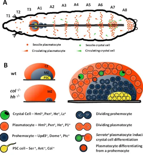
Figure 1.2. – Embryonic hematopoiesis. At embryonic stage 5 an anlage of cells start to express serpent (srp) in the head mesoderm. These srp+
cells are at this stage committed to differentiate into hemocytes. Following serpent expression all prohemocytes induce glial cell missing (Gcm)
expression. At stage 7 all prohemocytes initiate u-shaped (ush) expression.
At the same time the anterior-most rows of prohemocytes are lozenge (lz)
positive. 40% of Lz+ prohemocytes discontinue lozenge expression, maintain high ush expression and differentiate into plasmatocytes as the majority of
16
1.3
Larval hematopoiesis
1.3.1 Lymph gland development and structure
In the Drosophila larva, hematopoiesis proceeds within the
lymph gland. It consists of three to four paired lobes surrounding the anterior part of the dorsal vessel (Jung et al. 2005). The primary lobes of the lymph gland are bigger and, in homeostatic conditions, contain prohemocytes as well as differentiating and mature plasmatocytes and crystal cells. However, the lymph gland is an immune responsive organ and if a parasitoid wasp female punctures the cuticle of the larva to deposit an egg, the lymph gland will trigger the production of lamellocytes at the expense of prohemocytes (Makki et al. 2010). The lymph gland is differentiated in the embryo from thoracic lateral mesoderm cells (Holz et al. 2003). At stage 11 the transcription factor
collier (col) is expressed in two clusters of cells in T2 and T3
segments that eventually coalesce to form the paired lobes of the lymph gland (Crozatier et al. 2004). Looking at the expression pattern of another transcription factor, odd-skiped (Odd), one can see the
same T2 and T3 clusters plus some cells in T1 that will also make part of the lymph gland (Krzemien, Crozatier, et al. 2010). Similarl to embryonic hemocytes, serpent is expressed in lymph gland
hemocytes but only after collier (Crozatier et al. 2004). Unlike the
embryonic hemocytes, that lose serpent expression after hatching,
lymph gland hemocytes will maintain serpent expression throughout
larval stages. In late embryogenesis the lymph gland precursor consists of a pair of lobes, around 20 cells each, flanking the dorsal vessel. Only the 3 to 4 most posterior cells of each lobe maintain high
collier expression (Crozatier et al. 2004). This differential expression
17
third instar larva, when one can distinguish three regions: the Posterior Signaling Center (PSC) formed by the cells maintaining
collier expression, the medullary zone composed of prohemocytes
and the cortical zone with differentiated hemocytes (Jung et al. 2005). These three zones can be distinguished by the expression of different markers (Jung et al. 2005).
The PSC in third instar larva is composed of 30 to 45cells and is defined by the expression of Antennapedia, collier and Serrate
(Mandal et al. 2007; Lebestky et al. 2003). Both Antennapedia and collier mutants lose the PSC (Figure 1.2). Without a functional PSC,
prohemocytes of the medullar zone abandon their characteristic quiescence and begin differentiating into plasmatocytes and crystal cells (Mandal et al. 2007). Moreover, in the absence of PSC lymph glands cannot produce lamellocytes upon challenge, probably by the lack of prohemocytes (Krzemień et al. 2007). This set of evidences show that the PSC is necessary to maintain the hematopoietic precursors in the lymph gland medullary zone (Mandal et al. 2007). This role of the PSC involves also the Hedgehog pathway. hedghog
is expressed in PSC cells and the receptor, patched (ptc) in medullary
zone cells. Mutants for hedgehog still have the specification of the
PSC but similarly to collier and Antennapedia mutants lose the
18
JAK/STAT activity in the medullary zone is dependent on Latran, a protein that forms a heterodimer with Domeless, the receptor for JAK/STAT cytokines, and antagonizes its activity (Makki et al. 2010).
1.3.2 Hematopoiesis within the lymph gland
Hemocytes inside the medullary zone, where they lack differentiation markers, migrate into the cortical zone and they start to express early differentiation markers such as Hemolectin and Peroxidasin in second larval instar (Jung et al. 2005). Mature
molecular markers for plasmatocytes (P1) and crystal cells (pro-Phenoloxidase) are expressed only in the late second instar larvae. However, cell lineage tracing experiments show that in second instar prohemocytes in the medullary zone are already restricted to plasmatocyte or crystal cell fate (Krzemien, Oyallon, et al. 2010). Activation of lozenge expression requires Notch signaling with
Serrate acting as ligand (Lebestky et al. 2003). Although PSC cells express Serrate they are not the source of the ligand for crystal cell
formation (Crozatier et al. 2004). Instead, Serrate expressing cells
scattered inside the lymph gland may be the ones inducing Notch signaling (Crozatier et al. 2004; Lebestky et al. 2003) (Figure 1.2). Notch activated cells will induce lozenge expression and both
proteins will cooperate in the transcription of crystal cell specific genes (Terriente-Felix et al. 2013). After maturation, crystal cells maintain a vital high Notch expression, since without Notch crystal
cells burst (Mukherjee et al. 2011). The maintenance of Notch signaling in mature crystal cells in no longer dependent on Serrate but dependent on similar (sima), a hypoxia-inducible factor
19
to explain how crystal cells survive in circulation, where they do not have cells in contact.
20
Figure 1.2 – Hemocytes during larval stages (A) The majority of
plasmatocytes (orange dots) and crystal cells (green dots) during larval stages are clustered in patches underneath the epidermis. The number of clustered hemocytes is higher in the posterior segments of the larva. The lymph gland is located in the anterior part of the dorsal vessel. (B) The
21
1.3.3 Hematopoiesis outside the lymph gland
After hatching embryonic hemocytes persist in the larva and can be found in circulation or immotile in direct contact with the epidermis forming hemocyte sessile patches/clusters (Lanot et al. 2001) (Figure 1.2). Hemocyte load increases drastically throughout larval development from less than 200 cells in early first instar up to 6000 cells just before pupariation (Lanot et al. 2001; Makhijani et al. 2011). In homeostatic conditions, the number of sessile hemocytes is always higher than circulating ones, and constitute up to two thirds of the total cell count up until mid third instar larva (Lanot et al. 2001). In first instar larvae, the majority of hemocytes accumulate in the last segment and in lateral patches surrounding the oenocytes (Makhijani et al. 2011). Throughout development, with the increase in hemocyte load, they start to form dorsal patches in the epidermis and accumulate along the dorsal vessel. A discrete population of hemocytes also populates the proventriculus (Zaidman-Rémy et al. 2012) and a considerable number is attached to imaginal discs (Lanot et al. 2001).
22
As stated above, lymph gland hemocytes do not enter circulation in homeostatic conditions until pupariation (Holz et al. 2003; Honti et al. 2010). The haemocoelic hemocyte load increases throughout development because hemocytes are mitotically active. However, only plasmatocytes have been shown to divide, mature crystal cells do not (Lanot et al. 2001; Rizki 1957). This suggests that crystal cells are differentiated in circulating and/or sessile hemocytes. The formation of crystal cells in the larva is dependent on Notch signaling (Duvic et al. 2002; Lebestky et al. 2003). This is evident when Notch signaling is disrupted in larvae carrying a Notch thermo
sensitive allele raised at restrictive temperature (Duvic et al. 2002). In this experiment crystal cell numbers decreases in hemocyte clusters and in the lymph gland.
Notch receptor is activated by two different ligands in Drosophila: Serrate and Delta. Only Serrate plays a role in crystal cell differentiation both in the lymph gland and in sessile hemocytes (Duvic et al. 2002; Lebestky et al. 2003). Since a Notch expressing
cell requires cell contact with a Serrate expressing cell for its
activation, it is tempting to propose that crystal cells are differentiated in hemocyte clusters where they make cell contacts with other hemocytes, epidermal cells and neurons (Lanot et al. 2001; Makhijani et al. 2011).
23
and suggest the patches act as hematopoietic tissue. However, the existence of bona fide lamellocyte progenitors is questionable since
later work demonstrates that lamellocytes can differentiate from mature plasmatocytes (Honti et al. 2010; Stofanko et al. 2010). During a parasitoid wasp egg infection plasmatocytes down-regulate plasmatocyte specific genes and start to express lamellocyte specific factors (Honti et al. 2010).
Overall, the evidences gathered so far indicate that hematopoietic cell fate decisions occur in hemocytes outside the lymph gland. While plasmatocytes are mitotically active, crystal cells must differentiate outside the lymph gland. Furthermore, similarly to the lymph gland case, haemocoelic hemocytes can change their normal development upon parasitoid infection and produce lamellocytes. How the number of crystal cells and lamellocytes is controlled is certainly an important topic for further investigation that should reveal how circulating cells in animals achieve the correct concentration/proportion to perform their functions at optimal efficiency.
1.4
Objectives of this work
The maintenance of the plasmatocyte/crystal cell ratio during
Drosophila melanogaster development is striking since plasmatocytes
24
25
Hemocyte clusters function as a hematopoietic
26
2.1 Summary
Blood cells can be found in virtually all species of coelomate animals. Their functions are usually compartmentalized into different cell types for which the correct establishment of proper numbers and ratios is essential for homeostasis (Almeida et al. 2005). This depends upon a regulated balance between proliferation and differentiation mostly carried out in the hematopoietic organs (Hartenstein 2006). In Drosophila melanogaster, the larval
hematopoietic organ (lymph gland) produces two types of mature hemocytes (blood cells), plasmatocytes and crystal cells. Strikingly, in homeostatic conditions, hemocytes produced in the lymph gland are not released until pupariation. Yet, as larval development proceeds, the numbers of circulating hemocytes of both types increase, an observation difficult to reconcile with the post-mitotic character of crystal cells (Rizki 1957; Lanot et al. 2001). In this light, it has been proposed that hematopoietic properties must reside elsewhere, namely within the clusters of hemocytes found in close association with epidermal and neuronal cells along the larval body axis. Here, we show that hemocyte clusters function as a bona fide
hematopoietic tissue as their structure is necessary for Notch-dependent differentiation of crystal cells. Moreover our results suggest that, in contrast to the lymph gland, crystal cells formed in clusters do not derive from prohemocytes but from the transdifferentiation of plasmatocytes. The existence of this novel hematopoietic tissue, relying on structure-dependent signaling events to promote blood homeostasis, creates a new paradigm for addressing outstanding questions in Drosophila hematopoiesis and
27
2.2 Introduction
Blood cells can be found in almost any species of coelomate animals. Their functions are varied and include: gases transport, phagocytosis, extracellular matrix deposition and antibody production. The different functions performed by blood cells are to some degree compartmentalized in different cell types. Thus, the correct establishment of different blood cell numbers/ratios is essential for their proper function (Almeida et al. 2005). Some mature blood cells retain the ability to proliferate when in circulation but the majority of blood cell proliferation and differentiation occurs in hematopoietic organs (Grigorian & Hartenstein 2013). These organs provide the correct ‘molecular environment’ for the control of cell proliferation and differentiation. Thus, the study of hematopoietic organ structure and function is essential to understand how different mature blood cells numbers are controlled.
In Drosophila melanogaster, embryonic hematopoiesis
28
1957). Plasmatocytes increase in number because they are mitotically active cells but mature crystal cells do not divide (Rizki 1957; Lanot et al. 2001; Krzemien, Oyallon, et al. 2010; Makhijani et al. 2011). This observation implies that crystal cells differentiate/mature during larval stages.
The larva possesses a hematopoietic organ, the lymph gland, where plasmatocytes and crystal cells are differentiated from prohemocytes. Prohemocytes residing in the medullary zone of the lymph gland are influenced by cells from the Posterior Signaling Center (PSC) to maintain their quiescent state or to differentiate into mature plasmatocytes and crystal cells (Crozatier et al. 2004; Mandal et al. 2007). During the differentiation process, cells migrate and occupy the most cortical zone of the lymph gland (Jung et al. 2005; Krzemien, Oyallon, et al. 2010). Similarly to what happens in haemocoelic hemocytes, in the lymph gland cortical zone, mature plasmatocytes are still capable to divide but mature crystal cells enter a post-mitotic state (Krzemien, Oyallon, et al. 2010). An essential aspect of Drosophila’s larval hematopoiesis is that hemocytes
produced in the lymph gland do not disperse from the organ until pupariation or upon immune challenge such as parasitoid wasp egg infection (Holz et al. 2003; Honti et al. 2010). Hence, in homeostatic conditions, differentiated hemocytes in the lymph gland do not contribute to haemocoelic hemocyte population. Without contribution from the lymph gland or cell division, how haemocoelic crystal cells augment in number during larval development remains an open question.
29
Lebestky et al. 2003). In the lymph gland the role of Notch signaling in crystal cell formation is cell autonomous. (Lebestky et al. 2003; Mukherjee et al. 2011; Small et al. 2014). Notch activation is sufficient in hemocytes to induce the expression of lozenge, the first known
transcription factor in crystal cell development (Lebestky et al. 2000). One requirement of Notch signaling is that it requires cell contact because the twoNotch ligands of Drosophila, Serrate and Delta, are
membrane bound proteins (Fiúza & Arias 2007). In the haemocoelic compartment hemocytes can make cell contacts because the majority of them are sessile cells that form clusters in the subepidermal layer of the body cavity (Lanot et al. 2001). Hemocytes in clusters are densely packed and linked through interdigitations. Neurons from the Peripheral Nervous System (PNS) attract hemocyte to the cluster location and provide trophic signals for their maintenance (Makhijani et al. 2011). Moreover, when they are sessile in clusters, plasmatocytes have a higher division rate than when they are in circulation.
30
necessary to control cell proliferation and/or cell fate choice decisions.
The hemocytes in clusters are in a dynamic equilibrium with circulating hemocytes (Babcock et al. 2008; Welman et al. 2010). Hemocytes can enter and leave the clusters, which gives the opportunity for hemocytes produced in clusters to contribute to the increase in circulating crystal cells. Thus, hemocyte clusters are a good candidate location to study haemocoelic crystal cell development. In this chapter we ask if crystal cell precursors differentiate/mature in haemocoelic hemocytes and if the structure of hemocyte clusters is important for this differentiation.
2.3 Material and Methods
2.3.1 Fly stocks and parasitoids maintenance
All fly stocks were maintained in standard fly food at room temperature. Experiments were performed at 25°C except for RNAi experiments that were performed at 29°C. The following stocks with stock numbers in brackets were obtained from the Bloomington Stock Center: Lz-GAL4 mCD8GFP (X) (6314); Lz-GAL4 (X); UAS-GFP (II) (6313); HmlΔ-GAL4 2xEGFP(II) (30140); UAS-StringRNAi (34831); UAS-WhiteRNAi (33613); UAS-FLP
UbiFRTSTOPStinger (28282); Trio-GAL4 (48798); GMR29F08-GAL4 (49492); GMR29H07-GAL4 (45189); GMR30A01-GAL4 (45554); GMR30C04-GAL4 (41342); GMR30C06-GAL4 (49528); GMR30D10-GAL4 (49632); Notch-GMR30G07-GAL4 (45561); Notch-GMR57F04-GAL4 (46387). The line UAS-WhiteRNAi was used as control for RNAi experiments. The
31
the Vienna Drosophila Resource Center: Cg9313RNAi (103600)
NotchRNAi (100002), SerrateRNAi (108348) DeltaRNAi (109491). The line
Cg9313RNAi was used as a control line for RNAi experiments. The line
HmlΔ-nuclearDsRed was a kind gift from Marc Dionne. The Eater-GAL4 (X) and Eater-Eater-GAL4 (II) were a kind gift from Robert A Schultz. Leptopilina boulardi strain G486 females were allowed to lay eggs in second instar Drosophila Dif mutants at room temperature. Adult
parasitoids were maintained in fly food vials with a drop of honey.
2.3.2 Larva staging
Around 20 female flies were placed in a cage with a food plate with yeast. Egglays took place at 25ºC for 6 hour. At ~72h midpoint after egglay 2nd instar larvae were selected based on spiracle
morphology and transferred into a new food plate. After 2h larvae that molted into 3rd instar were selected and transferred into a food tube.
This first time point is referred 2h after 3rd instar.
2.3.3 Flow cytometry analysis and cell viability assay
32
stock solution of Propidium Iodide (PI) was diluted in 200μl of Ringer’s solution to a final concentration of 2μg/ml. Positive events for PI were considered dead or dying cells.
2.3.4 Cell cycle analysis
Hemocytes were centrifuged for 5 minutes at 500g at 4ºC and ressuspended in 400μl Ringer’s solution. 4,6ml of ice cold ethanol was added to each sample while vortexing. Cells were incubated for 2h at 4ºC and centrifuged for 5 minutes at 500g. The pelleted cells were ressuspended in 1ml PBS and centrifuged again for 5 minutes at 500g. Cells were ressuspended in 400μl PI staining solution (10μg/ml PI + 100μg/ml RNAse A) and incubated form 30 minutes at room temperature. Samples were immediately analyzed by flow cytometry.
2.3.5 Movie analysis
33
slide chamber was maintained at 25°C and 95% relative humidity. A Z-stack of pictures raging 28μm was taken every 1m30s for the GFP and RFP channel for 3h. At the end of the movie each larva was checked if it was still alive by ascertaining a beating dorsal vessel and movement of mouth parts. Only one larva died during the process. Z-stacks were then analyzed manually in FIJI software (Schindelin et al. 2012).
2.3.6 Total hemocyte loads counts and crystal cells counts
To estimate hemocyte concentration in the hemolymph six wandering male or female larvae were selected, briefly washed in Ringer’s solution and dried in filter paper. The six larvae were pooled in a glass well and bled by rupturing the cuticle in the ventral side to avoid disturbance of sessile hemocytes in the dorsal part where they are more abundant. All the hemolymph was collected and pooled in a 0,5ml microcentrifuge tube. 1μl of hemolymph was diluted in 9μl of Ringer’s solution. 9,5 μl of hemolymph dilution was loaded into a Neubauer chamber and hemocytes counted in all squares of 1mm2
area. This way the hemocyte concentration can be estimated by the formula: [number of counted cells]*105 cells/ml. To estimate the total
34
2.3.7 Hemocyte immunohistochemistry
Hemocytes were allowed to settle on a glass slide in a humid chamber for 10min and fixed with 4% formaldehyde solution for 20 min. After fixation cells were washed three times with PBS and blocked with PBST (PBS + 0,1% TritonX, +1% normal goat serum) for 30 min. After washing the cells with PBS the primary antibody was added at the correct dilution and cells incubated overnight at 4°C. Cells were then washed 3 times with PBS for 15 min and the secondary antibody added in the correct concentration. Cells were incubated for 3h at room temperature or at 4ºC overnight with the secondary antibody. The secondary antibody was washed three times with PBS. DAPI was added and incubated for 3 min followed by 3 washes with PBS. Slides were mounted with 80% glycerol solution and kept at 4°C before image acquisition. Antibodies used:
anti-lozenge (1:100 dilution, Developmental Studies Hybridoma Bank) and
anti-Nimrod (1:100 dilution, mixture of P1a and P1b antibodies, kind
gift from Instiván Andó)
2.3.8 Imaging
To count Hml+ and Lz+ cells, each larva was immobilized
35
2.3.9 Phagocytosis assay
To test phagocytosis activity early third stage larvae were injected with 69nl of pHrodo Red E.coli BioParticles (1mg/ml;
Molecular Probes). Injected larvae were maintained in yeast for 1h hour before ~10 larvae were bled in 20μl Ringer’s solution. Hemocytes were allowed to settle for 20min at room temperature in a humid chamber and pictures were taken immediately.
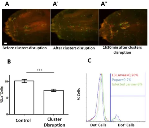
2.3.10 Cluster disruption assay
Pools of ~20 early third instar male larvae were selected and transferred to fresh yeast in a plastic petri dish maintained in a humid chamber. Every 1h30m larvae were taken from yeast, cleaned in Ringer’s solution and dried on filter paper. Groups of ~5 larvae were rolled several times by pressing a cover glass to disrupt hemocyte clusters. Control larvae were maintained on yeast. Before larva bleeding the two groups were subjected to cluster disruption to sample both circulating and sessile hemocytes.
2.3.11 Statistical analysis
36
2.4 Results
2.4.1 The number of crystal cell precursors increase during
larval development without cell proliferation
In homeostatic conditions, the larval haemocoelic hemocyte population is constituted by plasmatocytes and crystal cells. It is possible to distinguish these two cell types with several combinations of cell markers (Kurucz, Váczi, et al. 2007). To analyze hemocyte development throughout larval development we chose two live genetic drivers: HmlΔ-nuclearDsRed and Lozenge-GAL4 in combination with UAS-EGFP/UAS-mCD8GFP (Makhijani et al. 2011; Lebestky et al. 2000). With this combination of markers we can distinguish Hml+Lz- and Hml+Lz+ cells in sessile hemocytes through
the larval cuticle (Figure 2.1A-A’). Hml+Lz- cells are plasmatocytes
while Hml+Lz+ cells are fully mature crystal cells or differentiating
crystal cells (Lebestky et al. 2000). When analyzing total hemocyte counts by cell morphology it is indisputable that both plasmatocytes and crystal cells increase throughout larva development (Rizki 1957; Lanot et al. 2001). Since no crystal cell is dividing it is reasonable to assume that new crystal cells are differentiated throughout development. This can be achieved by inducing new crystal cell precursors during larva development or simply by maturation of precursors cells already present in the larva body cavity.
Lozenge is the first marker known in the genetic cascade that leads to crystal cell differentiation (Lebestky et al. 2000). Hence, we checked to see if Hml+Lz+ cells increase in number throughout the
third instar larva or if its number is fixed and crystal cell number increases due to maturation of Lz+ precursors. We counted the
37
image (Makhijani et al. 2011) (see methods). The total number of Hml+ cell increases throughout the third instar larva development in
males and females (Figure 2.1B). The number of Lz+ cells also
increases in the same time period (Figure 2.1C). In females there is no difference in the number of Lz+ in the last 16h of development.
With this, late third stage larva females have less crystal cells than males which is not common in the majority of fly stocks where females tend to have a higher number of crystal cells than males (see results below). Nevertheless, the results clearly show that Hml+Lz+
cells are increasing in number during the third instar larval development. The proportion of Hml+Lz+/Hml+Lz- cells even increases
in the first 24h of third larval instar development while Hml+Lz- cells
are also increasing (Supplementary Figure 2.1). Therefore, we can conclude that there is no fixed number of Hml+Lz+ cells throughout
development.
Although mature crystal cells do not divide it is still possible that Lz+ precursors proliferate before full crystal cell maturation. To
check if proliferation of Lz+ cells plays a role in the increase of crystal
cell loads we inhibited cell division using RNAi for string. String is an
essential phosphatase for entering mitosis (Edgar & O’Farrell 1990). When we drive string RNAi with HmlΔ-GAL4 driver there is a
significant reduction in the concentration of hemocytes, which demonstrates the necessity of string for hemocyte proliferation
(Figure 2.1E). Only females were tested in this experiment because the number of males is much reduced in this cross. Next, we asked if
stringRNAi expressed only in Lz+ cells would decrease the hemocyte
concentration and/or specifically the number of crystal cells. StringRNAi
38
treatment that blackens mature crystal cells (Rizki 1957; Neyen et al. 2014). To estimate crystal cell numbers changes we counted the number of cells in the dorsal part of the seventh abdominal segment (see methods and below). When driven by Lz-GAL4 driver the number of crystal cells is not reduced (Figure 2.1F). This result suggests that once a cell becomes Lz+ it enters a postmitotic state.
Hence, the increase in Hml+Lz+ cell numbers observed in third larval
instar is due to de novo differentiation of Hml+Lz+ cells and not their
39
Figure 2.1 (preceding page) – Hml+Lz+ cells increase during larval development without cell proliferation. (A) Dorsal view of a third instar
larva where hemocyte nuclei are marked with HmlΔ-nuclearDsRed. It is possible to see the lymph gland (arrow) sessile hemocytes along the body axis and a big cluster of cells in the A7 segment (square). (A’) In the magnification of hemocyte cluster it is possible to see that is constituted by Hml+Lz- and Hml+Lz+ cells.
(B) It is possible to see a increase throughout
third instar larval development of Hml+ cells both in females (light grey) and
males (dark grey). (C) Lz+ cells also increase during third instar larval
development. (D) string knockdown with RNAi reduces the hemocyte
concentration when driven in all hemocytes (HmlΔ-GAL4) but not when driven by Lz-GAL4 driver. (E) The number of crystal cells is not reduced in larvae when stringRNAi is driven in Lz+ cells. Error bars represent SEM n.s. = non-significant p-values *** = p-value <0.0001
2.4.2 Serrate expression in Hml
+Lz
-cells is essential to induce
crystal cell differentiation
Crystal cell numbers are reduced in larvae raised at restrictive temperature in a thermo sensitive Notch allele background (Duvic et
al. 2002).This reduction is visible in haemocoelic hemocytes and in the lymph gland (Duvic et al. 2002; Lebestky et al. 2003). In the lymph gland Notch signaling has a cell autonomous role in Hml+Lz
-precursors (Mukherjee et al. 2011). To check if the role of Notch is also cell autonomous in haemocoelic hemocytes we expressed
NotchRNAi specifically in all hemocytes making use of the HmlΔGAL4
40
cells is reduced in the same proportions in all treatments when comparing total crystal cells with the A7 dorsal cluster (Supplementary Figure 2.2A). Hence, from this point onwards the number of crystal cells was only estimated from the dorsal part of the A7 segment. Notch downregulation reduces the number of sessile
crystal cells both in males and females (Figure 2.2A-B). The knockdown of Notch does not disrupt the hemocyte clusters nor
changes the concentration of hemocytes in circulation (Figure 2.2C-D). Two other GAL4 drivers expressed in all larval hemocytes (Pxn and Trio) reduce the number of crystal cells when driving NotchRNAi
(Supplementary Figure 2.3A). When expressed in plasmatocytes only during embryogenesis, using the driver CrqGAL4 (Honti 2010), there is no difference in the number of larva crystal cells (Supplementary Figure 2.3A). In the lymph gland Notch activation is essential to
induce lozenge upregulation (Lebestky et al. 2003). To confirm that Notch knockdown inhibits the induction of Lz+ in haemocoelic cells
and not the maturation of Lz+ cells into crystal cells, we measured the
proportion of Lz+ cells with anti-Lozenge antibody while inhibiting
Notch expression in all hemocytes (Trio-GAL4>NotchRNAi). The
proportion of Lz+ cells in this case is clearly reduced (Figure 2.2E).
Altogether these results confirm that Notch activation is essential to induce lozenge expression in larval hemocytes that will mature into
crystal cells.
In Drosophila, Notch is activated by two different ligands:
Serrate and Delta. Only Serrate mutants have reduced haemocoelic
crystal cell numbers (Duvic et al. 2002; Lebestky et al. 2003). Interestingly, knocking down Serrate in hemocytes (HmlΔ
-GAL4xSerrateRNAi) reduces the number of crystal cells to similar level
41
to induce Notch signal in hemocytes is expressed itself in hemocytes. Similarly to what has been described before (Duvic et al. 2002), the downregulation of Delta does not lead to a reduction in crystal cell
numbers (Figure 2.2A-B). Since we have used the HmlΔ-GAL4 driver it is possible that Serrate is necessary in Hml+Lz- or in Hml+Lz+ cells.
To distinguish between these two hypotheses we performed knockdown of Serrate with Lz-GAL4 driver. In this case there is no
reduction in the number of crystal cells (Figure 2.2F). Hence Hml+Lz
-cells (plasmatocytes) are responsible for Serrate signaling to activate Notch. Unexpectedly, Notch knockdown in Lz+ cell does not lead to
reduction in the total number of crystal cells (Supplementary Figure 2.4A). In the lymph gland it is reported that Lz-Gal4 driving NotchRNAi
43
Figure 2.2 (preceding page) – Serrate downregulation in hemocytes leads to crystal cell numbers reduction. (A) NotchRNAi driven in all
hemocytes reduces the number of crystal cells in all body (Blue) and specifically in the dorsal part of A7 segment (Red). A similarly level of reduction is seen with SerrateRNAi but not with DeltaRNAi. (B) The results in males are similarly than in males with the exception that males have fewer crystal cells than females. (C) Notch pathway manipulation in hemocytes
does not disrupt hemocyte clusters. (D) Notch pathway manipulation does
not alter hemocyte concentrations. (E) NotchRNAi driven in all hemocytes
reduces the proportion lozenge positive cells measured with antibody
staining. (F) SerrateRNAi driven only in Lz+ cells does not reduce the number of crystal cells. Error bars represent SEM n.s. = non-significant p-values ** = p-value <0.001 *** = p-value <0.0001
2.4.3 Plasmatocyte/crystal cell ratio in clusters is influenced by
the number of contacting hemocytes
For Notch pathway activation, cells need to be in contact because the ligand Serrate is membrane bound (Guruharsha et al. 2012). The fact that Serrate expression in Hml+Lz- hemocytes is
necessary for crystal cell development suggests that Lz+ cells are
induced when hemocytes are in contact in hemocyte clusters. If this is the case, we can predict that it is more probable to find Hml+Lz+
44
in Hml+Lz- and Hml+Lz+ sessile hemocytes and considered how many
nuclei are less than 23μm away. This distance was used as a proxy for the number of contacting cells. The probability of a cell to be Hml+Lz+ increases with the number of cells it is in contact with (Figure
2.3B). Because we used the distance between nuclei to estimate the number of cells that are in contact there is a clear overestimation of cells in contact. However, this caveat does not affect our interpretation because it only turns our cell contacts estimation more conservative. The fact that we see a positive correlation between cells that are in contact and the percentage of those cells that are Lz+
goes in line with the fact that induction of Notch pathway is dependent on other hemocytes. The dependence on other hemocytes to induce
Notch signaling influences the proportion of induced Hml+Lz+ and
thus cluster structure.
Figure 2.3 – The number of clustered hemocytes influences their constitution. (A) Histogram representing the distribution of distances
between nuclei in contacting cells. Cytoplasm was visible by GFP driven by HmlΔ-GAL4 and nuclei visible by HmlΔ-nuclearDsRed (B) Probability of a
45
2.4.4 Differentiation of Hml
+Lz
+cells occurs in clusters
All the descriptions before lead us to propose that Hml+Lz+
cells are induced from Hml+Lz- cells in clusters of sessile hemocytes.
A way to test this hypothesis is to directly visualize the transformation of Hml+Lz- cells into Hml+Lz+ cells with live time-lapse imaging. To
achieve this we developed a method for imaging hemocytes in clusters in live larvae for periods of three hours. HmlΔ-nuclearDsRed; LzGAL4>EGFP/mCD8GFP early L3 male larvae (<12h after L3 ecdysis) were selected and prepared for imaging (see methods). Larvae had pressure from a cover slip in the dorsal part, which affects A7 dorsal hemocyte cluster most probably because this alters hemolymph circulation. Hence, we imaged more anterior clusters that were not so affected. As expected, it was possible to see the division of Hml+Lz- cells nuclei (Arrows in Figure 2.4A). The proportion of
dividing cells in these movies was ~7% of Hml+Lz- cells present in the
beginning of the movie (n=13 movies with mean 50 Hml+Lz- and 6 Hml+Lz+ cells per movie). No case of Hml+Lz+ cell division was seen
in any movie, once again confirming that Lz+ cells are postmitotic
cells while plasmatocytes are in proliferation in sessile clusters. Notably, it was possible to see induction of GFP in GFP- cells,
demonstrating that an Hml+Lz- cell is turning into an Hml+Lz+ cell
(Arrowhead in Figure 2.4A). The new Hml+Lz+ cells can be in contact
with other Hml+Lz+ cells or only in contact with Hml+Lz- cells. This
supports the idea that Serrate+ inducing cells are Hml+Lz- cells. In our
movies cells that are LzGFP+ never become GFP- and cells that start
with low GFP tend to increase it with time (Supplementary Figure 2.5A).
When we bleed larvae and analyze Hml+Lz+ cells we see a