ISJ 12: 5-12, 2015
ISSN 1824-307X
RESEARCH REPORT
Do snails
Lymnaea stagnalis
have phenoloxidase activity in hemolymph?
YL Vorontsova
a,#, IA Slepneva
b,#, NI Yurlova
a, VV Glupov
aa
Institute of Systematics and Ecology of Animals, Siberian Branch of Russian Academy of Sciences, Novosibirsk, Frunze Str., 11, 630091, Russia
b
Voevodsky Institute of Chemical Kinetics and Combustion, Siberian Branch of Russian Academy of Sciences, Institutskaya Str., 3, Novosibirsk, 630090, Russia
#
Equal contribution
Accepted December 11, 2014
Abstract
Hemocytes and hemolymph of snail
Lymnaea stagnalis
were analyzed to detect phenoloxidase
(PO) activity. No PO activity was found in the hemocytes of snails. A low level of PO activity (by DOPA
oxidation) was registered in the hemolymph without cells. Addition of a specific PO inhibitor revealed
the lack of effect on enzyme activity in the hemolymph, whereas hydrogen peroxide has increased it.
Studying the electron paramagnetic resonance (EPR) spectrum of DOPA- and dopamine-semiquinone
indicates the peroxide-dependent dopamine oxidation. Our results suggest that peroxidase, rather
than phenoloxidase, plays a key role in the oxidation of DOPA and dopamine in the hemolymph of
L.
stagnalis
. It is just peroxidase activity that may be important in the formation of cytotoxic molecules,
such as
o
-semiquinones, during snail defense immune reactions.
Key Words: phenoloxidase; peroxidase; Lymnaea stagnalis; immunity
Introduction
The enzyme phenoloxidase (PO) oxidizes phenols to form melanin which plays an important role in various physiological processes, such as egg production in gastropods (Bai et al., 1997), sclerotization of a new postmolt exoskeleton (Terwilliger, 1999) and immunity of invertebrates (Söderhäll and Cerenius, 1998). Highly reactive quinoid intermediates are generated during melanization (Johansson and Söderhäll, 1995; Slepneva et al., 1999). These can be involved in cytotoxic reactions in defense mechanism (Nappi and Ottaviani, 2000). Phenoloxidases occur as inactive precursors, termed the prophenoloxidases (proPO). ProPO are activated by a proteolytic cascade system. The cascade is activated by microbial cell wall components (Johansson and Söderhäll, 1995; Söderhäll and Cerenius, 1998) or some chemical compounds (Fisher and Brady, 1983). PO activity has been described for many invertebrates, including crustaceans, insects, and
___________________________________________________________________________
Corresponding author: Yana L. Vorontsova
Laboratory of Insect Pathology
Institute of Systematics and Ecology of Animals Siberian Branch of Russian Academy of Sciences Frunze Str., 11, 630091 Novosibirsk, Russia [email protected]
molluscs (Ashida and Söderhäll, 1984; Ashida and Yamazaki, 1990; Aspán and Söderhäll, 1991; Nellaiappan and Sugumaran, 1996).
Lymnaea stagnalis is a freshwater gastropod snail, which is used as a model organism to investigate immunological defense mechanisms (Sminia et al., 1973; van der Knaap et al., 1993; Plows et al., 2006). Surprisingly, despite this, no study has been carried out to strongly investigate the presence of PO activity in the hemolymph of L. stagnalis. Only Seppälä and co-authors reported the phenoloxidase-like activity determined in the hemolymph of L. stagnalis (Leicht et al., 2013; Seppälä and Leicht, 2013). They consider the PO-like activity as an important parameter of snail defense against some immune elicitors (Seppälä and Leicht, 2013). Unfortunately, the authors didn’t study whether the enzyme activity is present in the hemolymph or hemocytes.
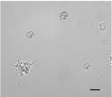
Fig. 1 Light microscopy of hemocytes from Lymnaea stagnalis stained for phenoloxidase activity. Scale bar = 10 µm.
data on PO activity in the hemocytes of L. stagnalis are lacking. Therefore, the presence of the PO activity in the hemolymph of L. stagnalis is still a problem.
The aim of our study was to determine the PO activity in the hemolymph and in the hemocytes of L. stagnalis.
Materials and Methods
Snails
Lymnaea stagnalis (Gastropoda, Pulmonata, Basommatophora) were collected at the littoral zone of Lake Chany, Russia (54°30′-55°09′ N, 76°48′ -78°12′ E) and maintained under laboratory conditions (14/10 light/dark daily cycles, 20 ± 1 °C) in 5 L aquaria containing dechlorinated tap water supplemented with mussel shell as calcium source. The snails were fed with pesticide-free lettuce daily ad libitum, and the water was replaced once a week.
Chemicals
4-aminoantipyrine, catalase, 3,3′ -diaminobenzidine tetrahydrochloride hydrate (DAB), diethylenetriaminepentaacetic acid (DTPA), dihydroxy-L-phenethylamine (dopamine), 3,4-dihydroxy-L-phenilalanine (DOPA), ethylene-diaminetetraacetic acid (EDTA), formaldehyde, glucose, glutaraldehyde, hydrogen peroxide, phenylthiourea (PTU), potassium phosphate, sodium azide, sodium chloride, trishydroxymethylaminomethane (Tris), were purchased from Sigma-Aldrich (USA). All solutions were prepared with bidistilled deionized water.
Hemolymph collection
Three adult snails (shell height: 25 mm) were rinsed with distilled water. The hemolymph was
collected by stimulation of the foot sole, as described by Sminia (1972). When the snail retracts into its shell, a drop of hemolymph is extruded through the hemal pore and collected with a micropipette. The collected hemolymph was mixed with either antiaggregant buffer (for hemocytes analysis) or phosphate-buffered saline (PBS: 50 mM phosphate buffer, 150 mM NaCl), pH 7.2 (for peroxidase and phenoloxidase assays in hemolymph) (2 parts hemolymph: 1 part buffer) and was kept on the ice to prevent hemocyte clumping.
Hemocyte monolayers preparation
The hemolymph with antiaggregant buffer (AB: 62 mM NaCl, 100 mM glucose, 10 mM EDTA, 30 mM sodium citrate, 26 mM citric acid, pH 4.6) (1 part hemolymph : 2 parts AB) was centrifuged at 500 g for 5 min at 4 °C. The supernatant was removed and the hemocytes were rinsed twice in AB. Hemocytes were then resuspended in PBS and 10 µl of hemocyte suspension (104 cells) were placed on a microscope slide. The slides were kept in a moist chamber at 22 °C for 15 min to allow hemocytes to adhere and spread. Then hemocytes were fixed with either 1G4F fixative (1 % glutaraldehyde : 4 % formaldehyde) for peroxidase assay or acetone for phenoloxidase assay. Acetone was used not only as fixative, but also as the activator of proPO (Fisher and Brady, 1983).
Peroxidase assay
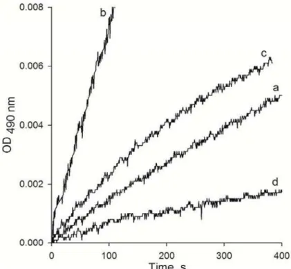
Fig. 2 Kinetics of DOPA (a) and dopamine (b) oxidation in hemolymph of L. stagnalis, effect of adding of H2O2 (c) and catalase (d) on the rate of DOPA oxidation.
water. Hemocytes were observed for the presence of brown deposits using a light microscope (Zeiss; Axioscope 40). Hemocytes with brown coloration were described a peroxidase-positive cells, i.e., the hemocytes with peroxidase activity. The percentage ratio of the peroxidase-positive hemocytes was calculated. Cells on control slides were incubated in media lacking DAB or hydrogen peroxide. The effect of peroxidase inhibitor was checked by adding 0.8 mM sodium azide to the hemocyte suspension. The peroxidase activity in the hemolymph was assayed spectrophotometrically using 4-aminoantipyrine as a substrate (Nicell and Wright, 1997). Each sample of hemolymph with PBS was then centrifuged at 500g for 5 min at 4 °C to remove the hemocytes. The hemolymph without cells (40 µl) was mixed with 0.85 mM hydrogen peroxide, 1.17 mM 4-aminoantipyrine with 80 mM phenol and 0.2 M potassium phosphate buffer; pH 7.0, up to a final volume of 250 µl. The mixture was placed in a cuvette with optical path length of 1 mm and the absorption at 510 nm for 5 min was recorded using a UV-2401 (PC) CE spectrophotometer (Shimadzu, Japan).
The effect of peroxidase inhibitor was checked by adding 0.1 mM KCN to the reaction mixture.
Determination of PO activity
After fixation with acetone, hemocytes were rinsed three times in PBS. Thereafter, hemocytes monolayers were incubated with 100 µl of DOPA (4mg/ml in PBS) at 22 °C for 15, 40, 60, 120 and 150 min in the dark moist chamber. Then, the hemocyte monolayers were rinsed with distilled
water and checked for the presence of dark-grey deposits (indicative of melanin formation) using a light microscope (Zeiss; Axioscope 40). PO activity in the hemolymph was assayed spectrophotometrically by mixing 150 µl of hemolymph without cells with either 125 µl of DOPA or dopamine (both 10 mM in PBS). The mixture was placed in the cuvette with optical path length of 1 mm and the absorption at 490 nm was detected. To study the effect of H2O2 and catalase on PO activity, either 3 mM H2O2 or 470 U/ml catalase were added to the reaction mixture. The effect of the specific PO inhibitor was checked by adding 18 µM PTU to the reaction mixture.
Determination of DOPA- and dopamine-semiquinone production
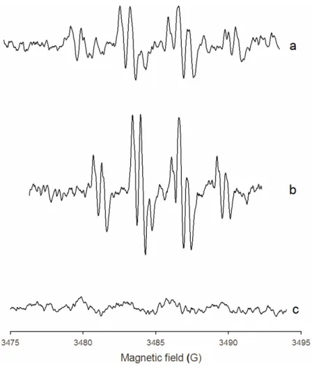
Fig. 3 EPR spectra of semiquinones of 10 mM dopamine (a) and 10 mM DOPA (c) obtained in hemolymph of L. stagnalis. Effect of 2.5 mM H2O2 added in hemolymph on intensity of dopamine-semiquinone spectrum (b). Amplitude of modulation was 0.16 G for spectra a and b, and 1.6 G for spectrum c.
DOPA or dopamine (100 µl) and 3.5 M MgCl2 (30 µl). Formation of the o-semiquinones in the mixtures was detected at room temperature by EPR method using an ER 200-D SRC X-band ESR spectrometer (Bruker). To study the effect of H2O2 on dopamine-semiquinone formation, 2.5 mM H2O2 was added to the reaction mixture. The EPR conditions were the following: field center, 3480 G; fields weep, 20 G; time constant, 1 s; microwave power, 2 mW; magnetic field modulation, 100 kHz; and modulation amplitude, 0.16 G.
Statistical analysis
The data were analyzed using the software SigmaPlot for Windows, version 9.0 (Systant Software, Inc.). When necessary, a statistical analysis was used and the data were expressed as means ± SE (n ≥ 5). Significant differences between treatments were analyzed by Student's t-test (p < 0.05) using the Origin 6.0 program.
Results
The hemocytes of L. stagnalis were analyzed to detect the PO activity. No PO-positive hemocytes
were revealed after incubation of cells with DOPA during 2.5 h (Fig. 1). This indicates the lack of the PO activity in the hemocytes of L. stagnalis.
We have spectrophotometrically registered the low level of DOPA oxidation in the hemolymph without cells. At the same time, the rate of dopamine oxidation was 5 times as high as that of DOPA (Figs 2a, b). Adding 18 µM PTU to the hemolymph had no effect on the rates of DOPA and dopamine oxidation (data not shown). The addition of hydrogen peroxide to the hemolymph increased the rate of DOPA oxidation (Fig. 2c). In contrast, the DOPA oxidation rate was observed to decrease upon addition of catalase to the hemolymph (Fig. 2d).
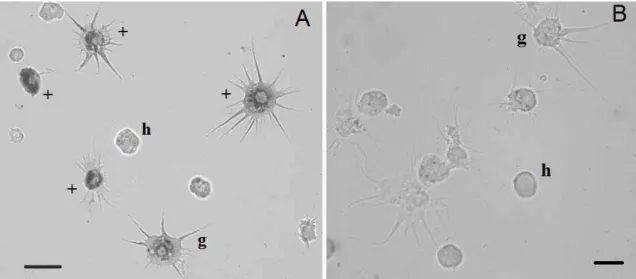
Fig. 4 (A) Light microscopy of hemocytes from L. stagnalis with peroxidase activity. (B) Effect of sodium azide on peroxidase activity in hemocytes from L. stagnalis. Abbreviations: g, granulocyte; h, hyalinocyte; “+” show peroxidase positive staining hemocytes. Scale bars = 10 µm.
the EPR spectrum was registered in the presence of the specific inhibitor of phenoloxidase, PTU (data not shown). The dopamine-semiquinone spectrum intensity increased with adding H2O2 to the hemolymph (Fig. 3b).
We have analyzed the hemocytes of L. stagnalis to detect the peroxidase activity. It was registered in granulocytes and hyalinocytes (Fig. 4a). The quantity of peroxidase-positive hemocytes was 45 ± 2 %. Adding sodium azide to a suspension of hemocytes inhibited the peroxidase activity in the hemocytes of L. stagnalis (Fig. 4b).
The peroxidase activity in the hemolymph of L. stagnalis was determined from the oxidation of 4-aminoantipyrine. We have registered the high rate of 4-aminoantipyrine oxidation (Fig. 5a). Adding 0.1 mM KCN to the hemolymph reduced the rate of this reaction (Fig. 5b).
Discussion
Phenoloxidase plays a key role in the immunity of invertebrates. This enzyme is known to be involved in encapsulation and phagocytosis of insects (Carton et al., 2008) and some species of snails (Aladaileh et al., 2007; Scheil et al., 2013). Only one group of researches reports that PO or PO-like activity of hemolymph is involved in the immune defense of the L. stagnalis (Seppälä and Jokela, 2010, 2011; Leicht et al., 2013; Seppälä and Leicht, 2013). Moreover, the authors carried out unusually long time incubation of hemolymph with DOPA (6 h) to detect the PO activity, named PO-like activity (Leicht et al., 2013; Seppälä and Leicht, 2013). This fact allows us to have some doubts on the real presence of PO activity in the hemolymph of the snails because it is known that DOPA can be oxidized not only by PO, but by peroxidase too (Kalyanaraman et al., 1984; Puiu et al., 2010) and the question of the presence of phenoloxidase in the
L. stagnalis hemolymph and its role in the immune defense is still ambiguous. Our histochemical experiments revealed no PO activity in the hemocytes of L. stagnalis. The level of DOPA oxidation in the snail hemolymph was very low as compared with that in the insect hemolymph (Lee and Anstee, 1995; Kryukova et al., 2011). The specific PO inhibitor, PTU, was employed to verify the participation of PO in DOPA oxidation. The concentration of PTU we used (18 µM) should inhibit the PO-dependent reaction completely (Ryazanova et al., 2012). In our experiments, PTU has failed to inhibit the DOPA oxidation in the hemolymph of snail which indicates that the DOPA oxidation occurs under the action of other enzyme. Moreover, we have detected a significantly higher level of dopamine oxidation in the L. stagnalis hemolymph which was not inhibited by PTU. These data allow us to suggest that the observed DOPA and dopamine oxidation in the hemolymph is provided by the activity of peroxidase rather than PO. This assumption is in fair agreement with the previous data testifying that the peroxidase is able to oxidize dopamine more effectively than DOPA (Kalyanaraman et al., 1984). Actually, in our experiments, the addition of hydrogen peroxide to the reaction mixture has increased the DOPA and dopamine oxidation rate, while the addition of catalase to the reaction mixture decreased it. Based on spectrophotometrical results, we can conclude that PO is not involved in the oxidation of DOPA and dopamine in the hemolymph of L. stagnalis.
Fig. 5 Detection of peroxidase activity by oxidation of 4-aminoantipyrin in hemolymph of L. stagnalis (a), inhibitory effect of KCN (0.1 mM) (b).
al., 2003; Komarov et al., 2005). Based on the results from these assays, we used the EPR method to detect the o-semiquinone radical in the L. stagnalis hemolymph. The specific spectrum of very low intensity with DOPA was observed as compared with dopamine (Figs 3a, c). This result demonstrates the lack of the PO activity in the hemolymph of snails. In order to identify the enzyme involved in dopamine oxidation, we added hydrogen peroxide and PTU to the hemolymph. The increasing intensity of the dopamine-semiquinone EPR spectrum upon addition of H2O2 and the lack of PTU effect on the spectrum indicate to the peroxide-dependent dopamine oxidation.
As demonstrated earlier, peroxidase is present in the hemolymph of L. stagnalis and is involved in snail’s immunity (Dikkeboom et al., 1984; Adema et al., 1992; Mohandas et al., 1992). Sminia and Barendsen (1980) registered the peroxidase activity in the lysosomal system of the spreading hemocytes of L. stagnalis. Peroxidase histochemical reaction products are present in the Golgi apparatus and in the lysosomes. We have also registered the activity of peroxidase in the L. stagnalis snail hemocytes of two types (Fig. 4). Furthermore, we have observed the activity of peroxidase in the hemolymph of snails using a typical substrate for peroxidase assay. Enzyme activity decreased due to the addition of peroxidase inhibitor, KCN, to the hemolymph (Fig. 5) which confirms the presence of the peroxidase activity in the hemolymph of L. stagnalis. Gornowicz and co-authors (2013) have also detected the peroxidase activity in these snails. Moreover, they revealed a significant increase of enzyme activity in the hemolymph of L. stagnalis naturally infected with digenean trematodes (Gornowicz et al., 2013). Thus, the peroxidase activity in the hemolymph is likely to play a key role in the host defense function.
Taken together, the results of this study show that peroxidase rather than PO plays a key role in the DOPA and dopamine oxidation in the hemolymph of L. stagnalis. It is only peroxidase activity that may be important in the formation of cytotoxic molecules such as o-semiquinones during snail’s defense immune reactions.
Acknowledgements
We thank anonymous referees, who gave fruitful comments on the manuscript. The work was supported by the Federal Fundamental Scientific Research Programme for 2013-2020 (VI.51.1.5) and the Russian Foundation for Basic Research, grants 13-04-02075 and 12-04-01057.
References
Adema CM, Harris RA, van Deutekom-Mulder EC. A comparative study of hemocytes from six different snails: morphology and functional aspects. J. Invertebr. Pathol. 59: 24-32, 1992. Aladaileh S, Nair SV, Raftos DA. Induction of
phenoloxidase and other immunological activities in Sydney rock oysters challenged with microbial pathogen-associate molecular patterns. Fish Shellfish Immunol. 23: 1196-1208, 2007.
Ashida M, Söderhäll K. The prophenoloxidase activating system in crayfish. Comp. Biochem. Physiol. 77B: 21-26, 1984.
Ashida M, Yamazaki HI. Biochemistry of the phenoloxidase system in insects: with special reference to its activation. In: Ohnishi E, Ishizaki H (eds), Molting and metamorphosis, Springer-Verlag, Berlin,pp 239-265, 1990.
Phenoloxidase activity in the reproductive system of Biomphalaria glabrata: Role in egg production and effect of schistosome infection. J. Parasitol. 83: 852-858, 1997.
Carton Y, Poirié M, Nappi AJ. Insect immune resistance to parasitoids. Insect Science 15: 67-87, 2008.
Dikkeboom R, van der Knaap WPW, Mueleman EA, Sminia T. Differences between blood cells of juvenile and adult specimens of the pond snail Lymnaea stagnalis. Cell Tissue Res. 238: 43-47, 1984.
Eaton DR. Complexing of metal ions with semiquinones. An electron spin resonance study. Inorganic Chem. 3: 1268-1271, 1964. Fisher CW, Brady UE. Activation, properties and
collection of haemolymph phenoloxidase of the American cockroach, Periplaneta americana. Comp. Biochem. Physiol. 75C: 111-114, 1983.
Gornowicz D, Dmochowska K, Zbikowska E, Zolotowska K. Total antioxidative status and the activity of peroxidase and superoxide dismutase in the haemolymph of Lymnaea stagnalis (L.) naturally infected with digenean trematodes. J. Mollusc. Stud. 79: 225-229, 2013.
Johansson MW, Söderhäll K. The prophenoloxidase activating system and associated proteins in invertebrates. In: Rinkevich B, Muller WEG (eds), Invertebrate Immunology, Springer, Berlin, pp, 46-66, 1995.
Kalyanaraman B. Characterization of o-semiquinone radicals in biological systems. Methods Enzymol. 186: 333-343, 1990.
Kalyanaraman B, Felix CC, Sealy RC. Peroxidatic oxidation of catecholamines. A kinetic electron spin resonance investigation using the spin stabilization approach. J. Biol. Chem. 259: 7584-7589, 1984.
Komarov DA., Slepneva IA, Glupov VV, Khramtsov VV. Detection of free radical formation in haemolymph of insects by EPR spectroscopy. Appl. Magn. Reson. 28: 411-419, 2005.
Kryukova NA, Dubovskiy IM, Chertkova EA, Vorontsova YL, Slepneva IA, Glupov VV. The effect of Habrobracon hebetor venom on the activity of the prophenoloxidase system and the generation of reactive oxygen species and encapsulation in the haemolymph of Galleria mellonella larvae. J. Insect Physiol. 57: 796-800, 2011.
Lee MJ, Anstee JH. Phenoloxidase and its zymogen from the haemolymph of larvae of the lepidopteran Spodoptera littoralis (Lepidoptera: Noctuidae). Comp. Biochem. Physiol. 11B: 379-384, 1995.
Leicht K, Jokela J, Seppälä O. An experimental heat wave changes immune defense ad life history traits in a freshwater snail. Ecol. Evol. 3: 4861-4871, 2013.
Mohandas A, Adema CM, van der Knaap WPW, Sminia T. The effect of haemolymph extraction on distribution of lysosomal enzymes in Lymnaea stagnalis haemocytes: a cytochemical study. Comp. Hematol. Int. 2: 61-67, 1992.
molecules in invertebrates. BioEssays 22: 469-480, 2000.
Nellaiappan K, Sugumaran M. On the presence of prophenoloxidase in the haemolymph of the horseshoe crab, Limulus. Comp. Biochem. Physiol. 113B: 163-168. 1996.
Nicell JA, Wright A. A model of peroxidase activity with inhibition by hydrogen peroxide. Enzym. Microb. Technol. 21: 302-310, 1997.
Plows LD, Cook RT, Davies AJ, Walker AJ. Phagocytosis by Lymnaea stagnalis haemocytes: a potential role for phosphatidylinositol 3-kinase but not protein kinase A. J. Invertebr. Pathol. 91: 74-77, 2006. Puiu M, Babaligea M, Oimazu C, Raducan A,
Oancea D. Peroxidase-mediated oxidation of L-dopa: a kinetic approach. Biochem. Engineer. J. 52: 248-254, 2010.
Ryazanova AD, Alekseev AA, Slepneva IA. The phenylthiourea is a competitive inhibitor of the enzymatic oxidation of DOPA by phenoloxidase. J. Enzym. Inhibit. Med. Chem. 27: 78-83, 2012.
Scheil AE, Hilsmann S, Triebskorn R, Köhler H-R. Shell colour polymorphism, injuries and immune defense in three helicid snail species, Cepaea hortensis, Theba pisana and Cornu aspersum maximum. Results Immunol. 3: 73-78, 2013. Seppälä O, Jokela J. Maintenance of genetic
variation in immune defense of a freshwater snail: role of environmental heterogeneity. Evolution 64: 2397-2407, 2010.
Seppälä O, Jokela J. Immune defence under extreme ambient temperature. Biol. Lett. 7: 119-122, 2011.
Seppälä O, Leicht K. Activation of the immune defence of the freshwater snail Lymnaea stagnalis by different immune elicitors. J. Exp. Biol. 216: 2902-2907, 2013.
Slepneva IA, Glupov VV, Sergeeva SV, Khramtsov VV. EPR detection of reactive oxygen species in haemolymph of Galleria mellonella and Dendrolimus superans sibiricus (Lepidoptera) larvae. Biochem. Biophys. Res. Commun. 264: 212-215, 1999.
Slepneva IA, Komarov DA, Glupov VV, Serebrov VV, Khramtsov VV. Influence of fungal infection on the DOPA-semiquinone and DOPA-quinone production in haemolymph of Galleria mellonella larvae. Biochem. Biophys. Res. Commun. 300: 188-191, 2003.
Sminia T. Structure and function of blood and connective tissue cells of the fresh water pulmonate Lymnaea stagnalis studied by electron microscopy and enzyme histochemistry. Z. Zellforsch. Mikrosk. Anat. 130: 497-526, 1972.
Sminia T, Barendsen LA. A comparative morphological and enzyme histochemical study on blood cells of the freshwater snails Lymnaea stagnalis, Biomphalaria glabrata, and Bulinus truncates. J. Morphol.165: 31-39, 1980.
prophenoloxidase-activating system in invertebrate immunity. Curr. Opin. Immunol. 10: 23-28, 1998.
Terwillinger NB.Hemolymph proteins and molting in crustaceans and insects. Americ. Zool. 39: 589-599. 1999.
van der Knaap W, Boerrigter-Barendsen LH, van Den Hoeven DSP, Sminia T.
humoral defence factor in blood cells (Amoebocytes) of the pond snail, Lymnaea stagnalis. Cell Tissue Res. 219: 291-296, 1981. van der Knaap W, Adema C, Sminia T. Invertebrate