*e-mail: [email protected]
Extraction of Alginate Biopolymer Present in Marine Alga
Sargassum filipendula
and Bioadsorption of Metallic Ions
Sirlei Jaiana Kleinübing*, Frederico Gaia, Caroline Bertagnolli, Meuris Gurgel Carlos da Silva
School of Chemical Engineering, University of Campinas – UNICAMP, Cidade Universitária Zeferino Vaz, CEP 13081-970, Campinas, SP, Brazil
Received: August 1, 2012; Revised: September 19, 2012
This paper studies the bioadsorption of Pb2+, Cu2+, Cd2+ and Zn2+ ions by marine alga
Sargassum filipendula and by the alginate biopolymer extracted from this alga. The objective is to evaluate the importance of this biopolymer in removing different metallic ions by the marine alga S. filipendula. In the equilibrium study, the same affinity order was observed for both bioadsorbents: Pb2+ > Cu2+ > Zn2+ > Cd2+. For Pb2+ and Cu2+ ions when the alginate is isolated and acting as
bioadsorbents, adsorption capacities greater than those found for the alga were observed, indicating that it is the main component responsible for the removal of metallic ions. For Zn2+ and Cd2+ ions,
greater bioadsorption capacities were observed for the alga, indicating that other functional groups of the alga, such as sulfates and amino, are also important in the bioadsorption of these ions.
Keywords: Sargassum filipendula, extraction, alginate, heavy metals, bioadsorption
1. Introduction
Seaweeds are one of the types of macroscopic biomass known for their metal-sorbing potential. In several ocean locations, marine macroalgae are abundant and of very fast growth, which allows them to be easily collected in large quantities1.
In brown seaweeds, the capacity of removing several metallic species is attributed to the biochemical constitution of their cellular wall, which is basically composed by three types of biopolymers: alginate, fucoidan and cellulose, which might provide several functional groups as binding sites (amino, carboxyl, sulfates). Alginate is its main polysaccharide, and the presence of mannuronic (M) and guluronic (G) acids in this biopolymer is directly related to the bioadsorption capacity of metallic ions.
Bioadsorption studies with marine alga Sargassum sp. have shown that carboxylic and sulfate groups (present in alginic acid and fucoidan) are active in ionic exchange. The carboxylic groups presented in the biomass are mostly constituted (typically approximately 70%) in sites in dry alga and play an important role in bioadsorption, being responsible for approximately 90% metal immobilization in the process2.
The alga bioadsorption capacity is directly proportional to the presence of these sites in the alginate polymer.
The alginate biopolymer represents up to 40% dry matter in brown marine algae. It is a linear polymer with high molecular weight constituted by two uronic acids – β -D-mannuronic (M) and α-L-guluronic (G)3. Differences in
molecular conformation between M and G blocks are the main responsible for the variation in alginate affinity for metallic ions4,5.
Several studies point out the high efficiency of different species of the marine alga Sargassum in the bioadsorption of different metallic ions, many of these using
Brazilian Sargassum sp.6-9 No studies were found using
alginate extracted from Brazilian algae for application as bioadsorbent. The isolated study of this biopolymer as a bioadsorbent might aid the understanding of the mechanisms involved in the bioadsorption process of different metallic ions by the marine alga.
The alginate biopolymer is extracted as sodium alginate and can be characterized by determining the M/G ratio using nuclear magnetic resonance spectroscopy (NRS). The addition of calcium ions to a solution of sodium alginate promotes instantaneous jellification of the contact region, which happens when the calcium ions are diffused through the gel-membrane interface10 and the gel formed can no
longer be managed.
The mechanism forming alginate gel can be explained by the ‘egg box’ model for interactions of G segments with calcium ions. Block G regions are aligned side by side, resulting in the formation of a cavity, with calcium ions forming the link between the chains, similar to an egg in its package, making a tri-dimensional network.
This study used the jellification process for the preparation of calcium alginate used as the bioadsorbent, where the bioadsorption of Pb2+, Cu2+, Cd2+ and Zn2+ ions
by marine alga Sargassum filipendula and by the alginate biopolymer extracted from this alga in the form of calcium alginate were evaluated.
2. Material and Methods
2.1.
Metal solutions
In order to perform this study, Pb2+, Cd2+, Cu2+ and
Zn2+ synthetic solutions were prepared from lead nitrate
salts. Concentrations are expressed in mmol.L–1. The solution
pH was adjusted to 4.5 using nitric acid HNO3 (0.5 M). Heavy metal concentration was determined by Atomic Absorption Spectrophotometer – AA 100 – Perkin Elmer from the Environmental Engineering Laboratory – Unicamp.
2.2.
Marine alga Sargassum filipendula
The marine alga S. filipendula was collected on the North Coast of São Paulo State, in the city of São Sebastião, Brazil. The alga was rinsed with distilled water and dried at 60 °C for 24 hours prior to being stored. Biomass was grinded and separated by dynamic sieving with a mean diameter of 0.59 mm.
2.3.
Extraction of alginate present in the alga
The method described by McHugh11 was used for
extracting the alginate. The dry alga was soaked in formaldehyde 0.4% for 30 minutes, rinsed with water and dropped into a HCl solution (0.1 mol.L–1) for 2 hours.
Then, the samples were rinsed again in distilled water before extraction in 2% sodium carbonate solution. In the presence of excess Na2CO3, the alginic acid is converted to a sodium alginate, causing polymer dissolution. The alginate extraction was carried out at 60 °C by soaking for 5 hours. The sample was filtered to separate the alga residue and the precipitated sodium alginate with ethanol.
2.4.
Preparation of alginate for application as
bioadsorbent
In order to be used as adsorbent, calcium alginate beads were prepared. A solution of sodium alginate was prepared by dissolving 5 g alginate in 100 mL deionized water under high agitation. The solution was then dripped into an aqueous solution of calcium chloride at 4% (CaCl2.2H2O, Nuclear brand). The alginate beads were kept in contact with calcium chloride for 24 hours. Following this period, the beads were rinsed in deionized water and left to dry at room temperature.
The morphology of the calcium alginate beads was evaluated by scanning electronic microscopy. This technique allowed to obtain images with high amplification and high resolution, enabling the evaluation of particle forms by means of interaction between an incident ray of electrons scanning the surface of the sample. The samples received carbon coating, and micrographs were obtained in a LEO, model LEO 440i scanning electronic microscope, with increases of 50-, 150- and 3000-fold.
Alginate bead average diameter and sphericity before and after drying were determined by microphotographs obtained in optical microscope coupled to an image acquisition system, using SigmaScan Pro 4 software.
2.5.
Fourier Transform Infrared Spectroscopy
(FT-IR)
FT-IR was used to identify the functional groups in the alga and alginate extracted from alga samples before and after the bioadsorption of metallic ions. Infrared spectra were recorded in the 4000-650 cm–1 region using a Thermo
Nicolet instrument, model IR-200. The ATR (attenuated total reflection) device allowed the obtaining of information about the surface.
2.6.
Bioadsorption kinetics
The behavior of metallic ion bioadsorption kinetics was studied using the marine alga S. filipendula and the alginate biopolymer extracted from that alga, with the objective of evaluating the rate of bioadsorption in relation to time. The tests were performed with alginate and alga beads (1 g) added to 500-mL metallic solution (pH 4.5 and 250 rpm agitation) in a metallic ion initial concentration of approximately 4.0 mmol.L–1.
The metal adsorption capacity by the adsorbents was calculated according to the mass equilibrium, equation 1.
0
( j f)
j s C C V q
m
−
= (1)
where: qj = metallic ion j adsorption capacity (mmol.g–1);
0 j
C = metallic ion j initial concentration (mmol.L–1);
f
C = metallic ion final concentration after equilibrium has been reached (mmol.L–1); V = solution volume (L);
ms = adsorbent dry mass (g).
2.7.
Equilibrium bioadsorption
In order to obtain equilibrium bioadsorption data for metallic ions, the static method was used, where a series of solutions (100 mL) with different metallic ion concentrations were placed in contact with 0.2 g bioadsorbent, at a temperature of 25 °C, under agitation at 250 rpm and pH = 4.5, adjusted with nitric acid at 0.05 M. The Langmuir Isotherm model was used to adjust the equilibrium data. The adjustment was performed by non-linear regression using the software Origin 6.0.
3. Results and Discussions
3.1.
Calcium alginate beads obtained before and
after the drying process
Figure 1a presents the beads, in gel form, obtained from the jellification of sodium alginate (extracted from the alga) in calcium chloride, before the drying process.
Micrographs for the dry calcium alginate beads are presented in Figure 1b-d with 50-, 150- and 3000-fold magnification. It can be observed that the surface is very rugged and presents depressions, resulting from the shrinking that takes place during the drying process. The rugosity can increase the superficial area, easing the diffusion of metal ions in the particle. Lagoa & Rodrigues12 observed a similar
appearance for dry beads with 2% sodium alginate, although they were not as spherical as the ones presented in Figure 1. The diameter of moist beads, calculated by the SigmaScan Pro 4 software, was 3.14 mm ± 0.18, while the sphericity was 0.83 ± 0.02. The dry spheres presented diameter of 1.02 mm ± 0.75 and sphericity of 0.82 ± 0.07.
3.2.
Evaluation of functional groups presented
in marine alga Sargassum filipendula and
extracted alginate biopolymer
Fourier Transform Infrared (FT-IR) analysis has been frequently used to detect how vibrational frequencies (bands) vary in marine algae13-17. This technique offers
cellular surface. Band shifting provides an indication of the functional group interaction with metallic ions.
3.3.
Identification of functional groups present in
Sargassum filipendula alga
By analyzing the spectra obtained for the marine alga S. filipendula, the following bands and corresponding related functional groups were identified:
The 1414- and 1616-cm–1 bands are attributed to
carboxylic (COOH) groups present in the alginate biopolymer18,16. The band at 1229 cm–1 is attributed to sulfate
(SO3) groups present in the fucoidan biopolymer16. The
band at 1537 cm–1 is attributed to amino groups present in
proteins19,15. The band at 1029 cm–1 is attributed to alcohol
groups1. Also, according to Mathlouthi and Koenig18, the
band frequency between 3200 and 3500 cm–1 corresponds
to amino (–NH2) and alcohol (–OH) groups.
3.4.
Identification of functional groups present in
the alginate biopolymer extracted from the
Sargassum filipendula alga
By analyzing the spectra obtained for the alginate extracted from marine alga S. filipendula, the following bands and corresponding related functional groups
were identified: 1418- and 1620-cm–1 bands present in
mannuronic (M) and guluronic (G) acids in the alginate biopolymer18,16; 1320-, 1123- and 1086-cm–1 bands
correspond to CC, CO and CCH groups14, present in acids
forming the alginate biopolymer. 1026- and 3443-cm–1 bands
are attributed to alcohol groups16. The bands identified for
mannuronic and guluronic groups were at 815 cm–1 and
778 cm–1, respectively.
3.5.
Efficiency of the extraction process
Initially, a comparison between the spectra obtained for marine alga S. filipendula and the alginate biopolymer extracted from this alga was performed. The alginate extraction yield from the marine alga S. filipendula was 17%. According to Percival and McDowell20, the amount of
alginate present in brown algae ranges from 10 to 40%. It can be observed that the band attributed to the sulfate group in the fucoidan biopolymer and the bands corresponding to protein amino groups present in the marine alga disappear in the spectrum obtained for the extracted alginate, indicating efficiency in the extraction. For alginate extracted from the alga, the identified bands corresponded to carboxylic, alcohol groups, as well as mannuronic and guluronic acids forming the biopolymer. It can also be noticed, in the
spectrum obtained for the extracted alginate, the appearance of 1320-, 1123- and 1086-cm–1 bands corresponding to CC,
CO and CCH groups present in mannuronic and guluronic acids forming the alginate biopolymer.
3.6.
Changes after the bioadsorption of different
metallic ions by marine alga Sargassum
filipendula
Figure 2 presents the spectra obtained for the marine alga S. filipendula before and after the bioadsorption of different metallic ions. Table 1 presents the changes suffered by the bands after the bioadsorption of different metallic ions by marine alga S. filipendula.
Shifts in the 1416- and 1614-cm–1 bands can be seen,
which represent the carboxylic groups in the alginate biopolymer. According to Sheng et al.16, the 1640-cm–1 band
is due to COO-M carboxylic groups, where M can be Na+,
K+, Ca+ or Mg+, naturally present in brown marine algae.
Since these species are competition elements, this result indicates that the ionic exchange process is taking place.
By analyzing the shift in 1537-cm–1 band, its
disappearance after the bioadsorption of Cu2+ and
displacement to 1531 cm–1 after the bioadsorption of the
remaining ions can be observed, indicating that the amino groups, present in proteins, are involved in the bioadsorption process of these ions.
The spectra corresponding to 1240- and 1034-cm–1
bands represent the SO3 groups in the fucoidan biopolymer and the alcohol group in the alginate biopolymer. It can be observed that these groups are participating in the bioadsorption process of all metallic ions, since the ions cause the shifting of this band. It is also noticed that the alcohol group suffered a more significant shift when Pb2+
ion was present in the solution.
The bands corresponding to mannuronic and guluronic acids, which form the alginate biopolymer, disappear after the ion bioadsorption process, indicating the strong participation of these groups in that process.
Kleinübing et al.13 studied the functional groups
present in marine alga S. filipendula, before and after the bioadsorption of Cu2+ and Ni2+ metallic ions using FT-IR
technique. The authors noticed that the carboxylic (COOH) and alcohol groups present in the alginate biopolymer, the sulfate (SO3) groups present in the fucoidan biopolymer and the amino groups present in the proteins participate in the bioadsorption process of metallic ions.
3.7.
Changes after the bioadsorption of different
metallic ions by the alginate extracted from
the alga
Figure 3 presents the spectra obtained for the alginate extracted from the marine alga S. filipendula, before and after the bioadsorption of different metallic ions. Table 2 presents the shifts suffered by the bands after the bioadsorption of different metallic ions by the alginate biopolymer extracted from the alga.
Shifts can be observed in the 1618-cm–1 band, which
represents the carboxylic groups, COO-M, where M stands for calcium ions used in the jellification process for the formation of calcium alginate beads, indicating that the ionic exchange mechanism between the metallic ions and calcium ions is working.
For all ions, shifts in 1320-, 1123- and 1086-cm–1 bands
correspond to the CC, CO and CCH groups, present in
Table 1. Shifting observed in the bands.
S. filipendula (cm–1) Pb2+ (cm–1) Cu2+ (cm–1) Cd2+ (cm–1) Zn2+ (cm–1)
1614 1632 1617 1632 1632
1537 1531 - 1531 1531
1416 1411 1411 1411 1418
1229 1226 1232 1233 1226
1029 1009 1027 1028 1028
819 - - -
-Figure 2. Fourier transform infrared (FT-IR) spectra of S. filipendula
alga and saturated with Cu2+, Pb2+, Zn2+ and Cd2+.
acids forming the alginate biopolymer. The 1320-cm–1 band
disappeared in the presence of Pb2+ and Cu2+ ions, and was
shifted to Cd2+, indicating the importance of the CC group
in capturing these ions. The 1086-cm–1 band presented a
more significant shifting in the presence of Pb2+. The band
at 1123 presented a significant shifting in the presence of Pb2+ and Cu2+ ions, and disappeared in the presence of Cd2+
and Zn2+ ions, indicating the importance of the CO group
in removing metallic ions.
The band at 1026 cm–1 attributed to alcohol groups was
shifted in the presence of ions, emphasizing the Pb2+ ion
with very significant results. The bands corresponding to
the mannuronic and guluronic groups forming the alginate biopolymer were shifted after the ion bioadsorption process, showing the effective participation of these acids in the bioadsorption process.
3.8.
Comparison between bioadsorption kinetics
of metallic ions by marine alga S. filipendula
and alginate extracted from the alga
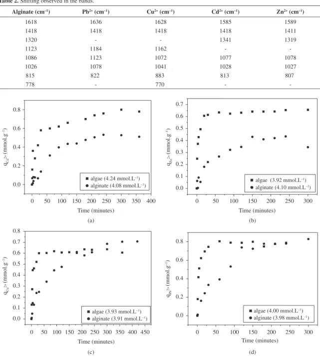
Figure 4 presents the bioadsorption kinetic curves of different metallic ions by the marine alga S. filipendula and by the alginate biopolymer extracted from the alga. It can be observed that for the marine alga S. filipendula, the kinetic
Table 2. Shifting observed in the bands.
Alginate (cm–1) Pb2+ (cm–1) Cu2+ (cm–1) Cd2+ (cm–1) Zn2+ (cm–1)
1618 1636 1628 1585 1589
1418 1418 1418 1418 1411
1320 - - 1341 1319
1123 1184 1162 -
-1086 1123 1072 1077 1078
1026 1078 1041 1028 1027
815 822 883 813 807
778 - 770 -
is very fast at the beginning, tending towards an equilibrium state before 100 minutes, except for the Cd2+ ion, which took
approximately 250 minutes to reach equilibrium. Different behaviors in the bioadsorption kinetic curves of metallic ions by alginate biopolymer extracted from the alga were observed, with changes in the shape of curves and increase in the time needed to reach equilibrium. The curve becomes longer, indicating that resistance to the transfer of mass in the calcium alginate particles is taking place.
The alginate is extracted from the alga in the form of sodium alginate, and in order to be used as bioadsorbent, it is jellified in the form of calcium alginate. Since the ionic exchange is the main mechanism responsible for removing metallic ions in the alginate biopolymer, the greater resistance to mass transfer observed for the alginate in comparison to the alga can be related to the exclusive presence of calcium ion in the alginate, whereas in the alga, different ions such as Mg2+, Na2+, Al2+, Si2+, Ca2+ are
naturally presented21. These ions present in the biomass are
more easily exchanged when compared to the calcium ion in the calcium alginate. Consequently, a greater resistance to mass transfer and an increase in the time needed to reach adsorption equilibrium could be noticed for the alginate biopolymer.
3.9.
Comparative study of the equilibrium of
different metallic ions by the marine alga
Sargassum filipendula and by the extracted
alginate
Figure 5 shows the bioadsorption isotherms for Pb2+,
Cu2+, Cd2+ and Zn2+ in marine alga S. filipendula and in
calcium alginate beads, with adjustment by Langmuir model. Table 3 presents the parameters obtained from these adjustments.
The following affinity order was observed for the marine alga S. filipendula and for the alginate biopolymer extracted from the alga by the metallic ions: Pb2+ > Cu2+ >
Zn2+ > Cd2+. It is important to emphasize the role of this
biopolymer in the removal of Pb2+ and Cu2+ ions. For these
ions, when the alginate is isolated, it acts as a bioadsorbent, and adsorption capacities higher than those found for marine alga S. filipendula were found, indicating that this is the main group responsible for the removal of metallic ions.
Haug22 studied the release of ions present in the alginic
acid extracted from the Laminaria digitata alga with the presence of different heavy metals in solution. In the paper, the authors could observe the exchange between ions existing in the alginate and the heavy metals present in the solution and, also, that the amount of exchanged protons
varied according to the heavy metal in the following order: Pb2+ > Cu2+ > Cd2+ > Ba2+ > Sr2+ > Ca2+ > Co2+ > Ni2+ >
Mn2+ > Mg2+.
However, for Zn2+ and Cd2+ ions, the opposite was
observed. Greater bioadsorption capacities were observed for the marine alga in comparison to the alginate biopolymer, indicating that the other functional groups present in the alga, such as sulfate and amino, are important in the bioadsorption of these ions.
Moreover, although the link between biomass and metallic ions can be seen as an ionic exchange process between H+ ions and metallic ions present in acid functional
groups, the high degree of selectivity presented by biomass from certain ions suggest that some of them are directly coordinated with the functional groups via the formation of internal sphere complexes. This type of adsorption is particularly important for Cu2+ and Pb2+ ions.
The greater importance of covalent binding for Cu compared to Ni is due to the fact that while electrostatic attraction is equally strong for both metals, covalent binding constants are higher for Cu than for Ni23. The higher
electronegativity to Cu leads to more equal (covalent) sharing of electrons with a ligand atom such as oxygen as it occurs in carboxyl groups24. Correspondingly, covalent
binding was more important for Cu than for Ni, which was bound predominantly by electrostatic attraction.
4. Conclusions
This paper focused on the bioadsorption of Pb2+, Cu2+,
Cd2+ and Zn2+ ions by the marine alga Sargassum filipendula
and by the alginate biopolymer extracted from this alga. With the results obtained, it was possible to conclude that the marine alga S. filipendula adsorbs the metallic ions in a different manner. The same ion affinity order was found for both bioadsorbents (Pb2+ > Cu2+ > Zn2+ > Cd2+). However,
Pb2+ and Cu2+ ions presented high affinity both by the marine
alga S. filipendula and by the alginate extracted from the alga. The Zn2+ and Cd2+ ions presented reduced removal
capacities in the extracted alginate when compared to the alga, indicating that functional groups such as sulfates and amino present in the alga are important in the removal of these ions. By analyzing the FT-IR, it can be concluded that the extraction process for the biopolymer was efficient and that the functional groups present in the alga and in the alginate extracted from the alga are participating in the bioadsorption process of all metallic ions studied herein.
Acknowledgements
The authors would like to acknowledge CAPES/PNPD and FAPESP for the financial support and CEBIMar (USP) for collection and identification of the algal material.
Table 3. Langmuir parameters estimated in the removal of ions in marine alga S. filipendula and alginate extracted from the alga.
Metal
Langmuir parameters
S. filipendula Extracted alginate
qm(mmol.g–1) b
j(L.mmol
–1) R2 q
m(mmol.g
–1) b
j(L.mmol
–1) R2
Pb2+ 0.95 ± 0.07 4.94 ± 1.11 0.97 1.16 ± 0.042 2.22 ± 0.294 0.99
Cu2+ 0.81 ± 0.042 4.92 ± 1.02 0.97 1.04 ± 0.075 0.65 ± 0.116 0.98
Zn2+ 0.80 ± 0.020 4.45 ± 0.56 0.99 0.71 ± 0.037 0.44 ± 0.061 0.99
Cd2+ 0.71 ± 0.062 7.02 ± 2.66 0.94 0.44 ± 0.037 0.83 ± 0.190 0.97
References
1. Volesky B. Detoxification of metal-bearing effluents: biosorption for the next century. Hydrometallurgy. 2001; 59:203-216. http:// dx.doi.org/10.1016/S0304-386X(00)00160-2
2. Kratochvil D and Volesky B. Advances in the Biosorption of Heavy Metals. Trends Biotechnology. 1998; 16:291-300. http:// dx.doi.org/10.1016/S0167-7799(98)01218-9
3. Haug A. The affinity of some divalent metals to different types of alginates. Acta Chemica Scandinavica. 1961; 15:1794-1795. http://dx.doi.org/10.3891/acta.chem.scand.15-1794
4. Haug A and Smidsrod O. Selectivity of some anionic polymers for divalent metal ions. Acta Chemica Scandinavica. 1970; 24:843-54. http://dx.doi.org/10.3891/acta.chem.scand.24-0843
5. Papageorgiou SK, Katsaros FK, Kouvelos EP, Nolan JW, Le Deit H and Kanellopoulos NK. Heavy metal sorption by calcium alginate beads from Laminaria digitata. Journal of Hazardous Materials. 2006; 137(3):1765-1772. PMid:16797834. http:// dx.doi.org/10.1016/j.jhazmat.2006.05.017
6. Kleinübing SJ, Guibal E, Da Silva EA and Da Silva MGC. Copper and nickel competitive biosorption simulation from single and binary systems by Sargassum filipendula.
Chemical Engineering Journal. 2012; 184:16-22. http://dx.doi. org/10.1016/j.cej.2011.11.023
7. Vieira MGA, Oisiovici RM, Gimenes ML and Da Silva MGC. Biosorption of Chromium (VI) using a Sargassum sp Packed-Bed Column. Bioresource Technology. 2008; 99(8): 3094-3099. PMid:17689245. http://dx.doi.org/10.1016/j.biortech.2007.05.071
8. Fagundes-Klen MR, Ferri P, Martins TD, Tavares CRG and Silva EA. Equilibrium study of the binary mixture of cadmium-zinc ions biosorption by the Sargassum filipendula
species using adsorption isotherms models and neural network.
Biochemical Engineering Journal. 2007; 34:136-146. http:// dx.doi.org/10.1016/j.bej.2006.11.023
9. Da Silva EA, Cossich ES, Tavares, CRG, Cardozo Filho L and Guirardello R. Modeling of Copper(II) Biosorption by Marine Alga Sargassum sp. in Fixed-Bed Column. Process Biochemistry. 2002; 38:791-799. http://dx.doi.org/10.1016/ S0032-9592(02)00231-5
10. Glicksman M. Red seaweed extracts. In: Glicksman M, editor.
Food Hydrocolloids. Boca Raton: CRC Press, Inc.; 1983.
12. Lagoa R and Rodrigues JR. Kinetic analysis of metal uptake by dry and gel alginate particles. Biochemical Engineering Journal. 2009; 46(3):320-326. http://dx.doi.org/10.1016/j. bej.2009.06.007
13. Kleinübing SJ, Vieira RS, Beppu M, Guibal E and Da Silva MGC. Characterization and Evaluation of Copper and Nickel Biosorption on Acidic Algae Sargassum Filipendula. Materials Research. 2010; 13:1-10. http://dx.doi.org/10.1590/S1516-14392010000400018
14. Fuks L, Filipiuk D and Majdan M. Transition metal complexes with alginate biosorbent. Journal of Molecular Structure. 2006; 792-793:104-109. http://dx.doi.org/10.1016/j. molstruc.2005.12.053
15. Chen JP and Yang L. Study of a heavy metal biosorption onto raw and chemically modified Sargassum sp. via spectroscopic and modeling analysis. Langmuir. 2006; 22:8906-8914. PMid:17014134. http://dx.doi.org/10.1021/la060770+
16. Sheng PX, Ting Y-P, Chen JP and Hong L. Sorption of lead, copper, cadmium, zinc, and nickel by marine algal biomass: characterization of biosorptive capacity and investigation of mechanism. Journal Colloid and Interface Science. 2004; 275:131-141. PMid:15158390. http://dx.doi. org/10.1016/j.jcis.2004.01.036
17. Figueira MM, Volesky B and Matheus HJ. Instrumental analysis study of iron species biosorption by Sargassum biomass.
Environmental Science and Technology. 1999; 33:1840-1846. http://dx.doi.org/10.1021/es981111p
18. Mathlouthi M and Koenig JL. Vibrational Spectra of Carbohydrates. Advances in Carbohydrate Chemistry and Biochemistry. 1986; 44:7-66. http://dx.doi.org/10.1016/S0065-2318(08)60077-3
19. Fourest E and Volesky B. Contribution of sulphonate groups and alginate to heavy metal biosorption by the dry biomass of Sargassum fluitans. Environmental Science and Technology. 1996; 30(1):277-282. http://dx.doi.org/10.1021/ es950315s
20. Percival EGV and McDowell RH. Chemistry and Enzymology of Marine Algal Polysaccharides. London: Academic Press; 1967. p. 219.
21. Kleinübing SJ, Da Silva EA, Da Silva MGC and Guibal E. Equilibrium of Cu(II) and Ni(II) biosorption by marine alga Sargassum filipendula in a dynamic system: Competitiveness and selectivity. Bioresource Technology. 2011; 102(7):4610-4617. P M i d : 2 1 2 9 5 9 7 2 . h t t p : / / d x . d o i . o r g / 1 0 . 1 0 1 6 / j . biortech.2010.12.049
22. Haug A, Larsen B and Smidsrod O. Uronic acid sequence in alginate from different sources. Carbohydrate Research. 1974; 32:217-25. http://dx.doi.org/10.1016/S0008-6215(00)82100-X
23. Schiewer S and Wong MH. Ionic strength effects in biosorption of metals by marine algae. Chemosphere. 2000; 41:271-282. http://dx.doi.org/10.1016/S0045-6535(99)00421-X