ABSTRACT
Multiple endocrine neoplasia type 2 (MEN2) is an autosomal dominant inherited tumor syndrome caused by RET proto-oncogene germline muta-tions (RET). Here we tested the Conformation Sensitive Gel Electrophoresis (CSGE) as a screening method for RET hot-spot mutations. Seven MEN2 families were studied by direct sequencing analysis, CSGE and Single Strand Conformational Polymorphism (SSCP). Using CSGE/SSCP, we were able to detect four out of five types of RETmutations verified by sequencing analysis: Cys620Arg, Cys634Arg, Cys634Tyr, and Met918Thr, furthermore a missense substitution at codon 648 (Val648Ile). RET polymorphisms 691 and 769 were also verified. Data obtained using CSGE/SSCP were fully concor-dant. We conclude that CSGE showed to be a sensitive, fast, low-cost, and simple procedure to detect RET mutations in codons which are reported as the most prevalent RET variants (~ 95%) in large MEN2 series. As to the Val804Met mutation, this method still needs to be optimized. (Arq Bras Endocrinol Metab 2007;51/9:1468-1476)
Keywords:Genetic screening; CSGE; SSCP; Genetic sequencing; MEN-2; RET proto-oncogene
RESUMO
Rastreamento de Mutações do Gene RETna Neoplasia Endócrina Múltipla Tipo 2 por Eletroforese em Gel Sensível à Conformação (CSGE).
A neoplasia endócrina múltipla tipo 2 (NEM2) é uma síndrome tumoral herdada por mutações germinativas no proto-oncogene RET (RET). Ana-lisamos a aplicação do método Eletroforese em Gel Sensível à Confor-mação (CSGE) no rastreamento de mutações hot spots do RET. Sete famílias com NEM2 foram rastreadas pelo seqüenciamento gênico, CSGE e análise do Polimorfismo Conformacional de Cadeia Simples (SSCP). Usando ambas as metodologias de rastreamento, identificamos quatro dos cinco tipos de mutações verificadas pelo seqüenciamento: Cys620Arg, Cys634Arg, Cys634Tyr e Met918Thr, além da variação gênica Val648Ile. Das análises englobando mutações hot spotsdo RET, 90,6% concordaram com o seqüenciamento genético (incluindo a variação gênica Val648Ile). Polimorfismos nos códons 691 e 769 foram documentados. Os dados obti-dos por CSGE/SSCP foram totalmente concordantes. Concluímos que o CSGE revelou ser metodologia sensível, rápida, de fácil execução e baixo custo no rastreamento de mutações nos códons associados à grande maioria (~ 95%) dos pacientes com NEM2. (Arq Bras Endocrinol Metab 2007;51/9:1468-1476)
Descritores:Rastreamento gênico; CSGE; SSCP; Seqüenciamento gêni-co; NEM2; Proto-oncogene RET
artigo original
Gel Electrophoresis (CSGE)
MARCELO A. C. G. DOS SANTOS ELISANGELAPEREIRA DES. QUEDAS RODRIGO DEALMEIDATOLEDO DELMAR M. LOURENÇO-JÚNIOR SERGIOPEREIRA DEA. TOLEDO
Endocrine Genetics Unit (LIM-25), Endocrinology, Department of Internal Medicine, Hospital das Clínicas, and University of São Paulo School of Medicine, SP.
M
ULTIPLE ENDOCRINE NEOPLASIA type 2 (MEN2) is an inherited tumor syndrome characterized by the presence of medullary thyroid carcinoma (MTC), primary hyperparathyroidism (HPT), and pheochro-mocytoma (PHEO). MEN2 is classified as MEN2A (MTC, PHEO, and HPT); MEN2B (MTC, PHEO, and mucosal neuromas); and familial MTC (FMTC) (1-5). The genetic basis of MEN2 is associated with germline activating mutations in the RET proto-onco-gene (RET). RET gene contains 21 exons, which encode a tyrosine kinase receptor protein with an extra-cellular domain rich in cysteine residues and an intra-cellular domain enriched in tyrosine residues (1,6).Several Brazilian studies on MEN2 have been recently published, in which clinical and genetic aspects of MEN2 were extensively reviewed (2-5,7-14). All clinical variants of MEN2 have a high pene-trance rate for MTC and most (90%) RETmutation carriers will exhibit evidence for MTC during their lifetime (15). Briefly, MEN2A comprises 75% of MEN2 cases and most cases (98%) harbor a RET mutation in exons 10 or 11 (codons 609, 611, 618, 620, 630, and 634). RET variants in codon 634 cause 85% of MEN2A cases, mostly with the Cys634Arg and Cys634Tyr mutations (1,7). Furthermore, FMTC cases comprise 20% of MEN2 patients and most of them (85%) have RET mutations in codons 10 or 11 (1,8,15). Less frequent mutations in codons 609, 611, 768, and 804 are usually associated with FMTC, but rarely to MEN2A (1,7). Met918Thr (exon 16) muta-tion has been reported in 95% of MEN2B cases, which comprise 5% of MEN2 cases (1,15). Rarely, two RET variants have been reported as co-segregating in MEN2 cases (3,8). Thus, the vast majority (95%) of RET disease-causing mutations are associated to codons 620, 634, and 918 (15).
Molecular diagnosis of RET mutations has become a crucial tool for the management of MEN2 as it may (a) identify 1–7% of inherited MTC cases among MTC patients previously considered as “spo-radic” cases; (b) identify RET mutation carriers in at-risk family members and eliminate the need for annu-al follow-up in relatives who do not carry RET muta-tions; and finally (c) it is the rational basis for indicat-ing preventive total thyroidectomy, usually under 5 years of age, in all RET mutation carriers (4-6,15). Direct DNA sequencing is the gold-standard method for genetic studies in the detection of disease-causing mutations. However, several genetic screening tech-niques, such as denaturating gradient gel elec-trophoresis (DGGE), restriction enzymes, single strand conformational polymorphism (SSCP) and
denaturing high performance liquid chromatography (DHPLC) have been applied to genetic screening of at-risk relatives in families with several inherited dis-eases (1-8,15,16).
Conformation Sensitive Gel Electrophoresis (CSGE) has been shown to be a useful method for mutation detection. CSGE detects the differences in electrophoresis migration patterns of homo- and het-eroduplex formations. These diverse bands are caused by structural alterations in DNA double helix, which are favored by mild denaturing solvents (16-18). Interestingly, this method presents important advan-tages when compared to restriction enzymes, SSCP, DGGE (19), and DHPLC methodologies. CSGE may present higher sensitivity for 200–800bp PCR frag-ment sizes, when compared to SSCP sensitivity (16,20). Comparing to DGGE, CSGE is a feasible method to standardize and does not need a 50bp GC-clamp coupled to one of the sequences (16,20). More-over, CSGE does not require an expensive wave DNA fragment analysis system, such as in DHPLC. Although CSGE has been applied to several inherited conditions, limited information is available on CSGE to identify RET mutations (19,20). As there is an increasing demand for RET genetic diagnosis in the endocrine practice, the optimization of RET muta-tions is a worthwhile effort. Thus, this study aimed at validating CSGE as genetic screening approach for RET hot-spot mutations in patients with classical MEN2 phenotype presentations.
MATERIALS AND METHODS
Patients
as healthy individuals were used as controls. All individuals have been followed at the Outpatient Service of the Division of Endocrinology, Hospital das Clínicas, University of Sao Paulo School of Medicine. Index cases were diagnosed by classical clinical, biochemical, and genetic parameters (2,3,8-12,15).
Methods
Genomic DNA extraction and PCR
Genomic DNA was extracted using the salting-out method (21). DNA concentration was measured by spectrophotom-etry (Pharmacia Biotech, Sweden). Exons 10, 11, 13, 14, 15, and 16 were amplified by PCR (Minicycler — MJ Research, Waterstone, MA, USA) (22), using previously reported primers (7,13,23). Other primers were designed specifically for CSGE and SSCP analyses, as shown in table 1. PCR protocols were optimized as follows: 10 mM Tris HCl pH 8.4, 50 mM KCl pH 8.4, 0.4 mM of dNTPs, 2.0U of Taq-polimerase, 5% of Dimethyl sulfoxide – DMSO. The MgCl2and primer concentrations were: 1.76 mM; 20 pmol
for exon 10; 2.40 mM; 14 pmol for exon 11; 2.40 mM; 20 pmol for exons 13 and 15; 3 mM; 10 pmol for exon 14; and 3 mM; 20 pmol for exon 16. Genomic DNA concentration ranged from 100–200 ng. The optimized cycling programs used were: single cycle at 94ºC for 10 min. (exons 10, primer setting 11A, 11B, 15, and 16) and 3 min. (exons 13, primers settings 14A and 14B); 38 cycles at 94ºC for 30 sec.
(all exons), annealing for 30 sec. (exons 10 and 15) and 1 min. (primers settings 11A, 11B, 13, 14A, 14B, and 16), as indicated in table 1, and extension at 72ºC for 1 min. (all
exons), followed by an extension cycle at 72ºC for 4 min.
For exon 14, amplification with clamp primers (primer set-ting 14C) cycle temperatures were: single cycle at 95ºC for
4 min.; 35 cycles at 94ºC for 45 sec., annealing for 45 sec.
and extension at 72ºC for 10 min. PCR products were
con-firmed by electrophoresis at 70V for 1 h. (Power PAC 3000, Biorad, USA) in 2% agarose gel with TAE 1X buffer stained with ethydium bromide (0.5 mg/ml), where 5 ml of genomic amplified material were mixed to 1 ml of Loading Buffer 6 x and visualized in UV transiluminator (Foto Ana-lyst Mini Visionary, Fotodyne, USA).
Sequencing analysis
RETdirect sequencing was performed as follows: 2 ml of Big Dye terminator buffer (Applied Biosystems, Foster City, CA, USA), 6 ml of 0.25% buffer (Tris-HCl 0.5 M pH 9.0 with 25 mM of MgCl2), 1.60 pm of primer and 5–10 ng of amplified DNA, in a total volume of 20 ml. Cycle sequen-cing conditions were: single cycle at 96ºC for 2 min.; 40 cycles at 96oC for 10 sec., annealing at 60ºC for 20 sec. and
extension at 60ºC for 4 min. After sequencing, reactions were purified with isopropanol/ethanol according to the manufacturer’s instructions, and diluted in 2.5 ml of blue dextran buffer with deionized formamide (TSR, Applied Biosystems). Sequencing products were denaturated at 90ºC for 2 min. and immediately cooled before applied in an ABI 310 DNA sequencer. Electropherograms were ana-lyzed by AB Navigator software (Applied Biosystems).
CSGE
The MDE gel was prepared as follows: MDE 0.5 X (Cam-brex Bio Science, Rockland, ME, USA), 6% de TBE 10 X, 2.5% de glycerol, 220 ml of ammonium persulfate 10% and
Table 1. Primers sequences and annealing temperatures for RET hot-spot exons. [Adapted from refs. 7, 13, 23]
Exons Primers sequences Annealing Bp
(ºC)
10 10F: 5’AggCTgAgTgggCTACgTCTg 3’ 60 205
10R: 5’gTTgAgACCTCTgTggggCT 3’
11A 11F: 5’ATgAggCAgAgCATACgCAgCC 3’ 60 332
11R: 5’CTTgAAggCATCCACggAgACC 3’
11B 11F: 5’ATgAggCAgAgCATACgCAgCC 3’ 60 199
11R: 5’TTgTgggCAAACTTgTggTA 3’
13 13F: 5’AACTTgggCAAggCgATgCA 3’ 62 276
13R: 5’AgAACAgggCTgTATggAgC 3’
14A 14F: 5’CCTggCTCCTggAAgACC 3’ 62 298
14R: 5’CATATgCACgCACCTTCATC 3’
14B 14F: 5’CCTggCTCCTggAAgACC 3’ 62 241
14R: 5’CCAggCAAATgAgATgAggT 3’
14C 14F: 5’gCgCCCCCCgCCCCgCCCgCCgCggCgCCg 65 395
CCCAgggATAgggCCTgggCTTC 3’ 14R: 5’TAACCTCCACCCAAgAgAg 3’
15 15F: 5’CATggCCTgACgACTCgTgC 3’ 60 192
15R: 5’CCTgggAgCCCCgCCTCATC 3’
16 16F: 5’CTgAAAgCTCAgggATAggg 3’ 60 202
22 ml of N,N,N’,N’-Tetramethylethylenediamine. Gel was applied into plates (coated with repel-silane and gamma-methacryloxypropyltrimethoxysilane) and left to polymerize for 2 h. The electrophoresis system was pre-run (Sequencing System Model S2, Life Technologies, Gibco/BRL, Gaithersburg, USA) for 45 min. at 8W with TBE 0.6 X buffer. Temperature conditions for heteroduplex formation were: single cycle at 94ºC for 5 min. (exons 10, primer
ting 11A, 13, and 16) or 10 min. for exon 11 (primer set-ting B), followed by annealing at room temperature for 40 min. (exons 10, primers settings 11A and 11B, 13 and 16). 5 ml of sample (2:1 of amplicon and Loading Buffer 1 X) (Triple Dye Loading Buffer 6 X, Cambrex Bio Science) were loaded in gel and electrophoresed for 1 h. at 15 W (exons 11 primer setting A, and exon 13). Subsequently, exon 16 was loaded in gel, and all exons were electrophoresed at 8 W for 11 h. Amplified genomic material from exons 10 and 11 (primer setting B) were electrophoresed for 11 h. at 2 W. CSGE gel was silver stained.
SSCP
SSCP gel and electrophoresis conditions were the same as used for CSGE. After denaturation (same temperatures used in CSGE), amplicons were rested in ice before loading in gel. A loading buffer (95% formamide, 10 mM NaOH, 0.025% Bromophenol Blue, 0.025% Xylene Cyanol) was used. SSCP gel was silver stained.
RESULTS
Figure 1 and table 3 summarize RETmutation find-ings when CSGE, SSCP, and sequencing methods were applied.
Sequencing analysis
RET hot-spots exons (10, 11, 13, 14, 15, and 16) of all index cases (7 subjects) and in one genetically non-Table 2.Genotype-phenotype correlation for RET mutant carriers screened by CSGE.
Cases Mutated exon Phenotype Genotype Polymorphic exons Polymorphisms
1 11 MEN2A Cys634Arg
2 11 MEN2A Cys634Arg
3 11 MEN2A Cys634Arg 11 Gly691Ser
4 11 MEN2A Cys634Arg 11 Gly691Ser
5 11 MEN2A Cys634Arg 11 Gly691Ser
6 11 MEN2A Cys634Arg
7 11 MEN2A Cys634Arg
8 11 MEN2A Cys634Arg
9 11 MEN2A Cys634Arg
10 11 MEN2A Cys634Arg
11 11 MEN2A Cys634Arg 11 Gly691Ser
12 11 MEN2A Cys634Arg 11 Gly691Ser
13 10 MEN2A Cys620Arg 11 Gly691Ser
14 10 MEN2A Cys620Arg 11 Gly691Ser
15 10 MEN2A Cys620Arg 11 Gly691Ser
16 10 MEN2A Cys620Arg 11 Gly691Ser
17 10 MEN2A+HSCR Cys620Arg 11 Gly691Ser
18 10 MEN2A+HSCR Cys620Arg 11-13 Gly691Ser, Leu769Leu
19 10 MEN2A Cys620Arg 11 Gly691Ser
20 10 FMTC Cys620Arg
21 10 FMTC Cys620Arg
22 10 FMTC Cys620Arg
23 10 FMTC Cys620Arg
24 11 FMTC Cys634Tyr
25 11 FMTC Cys634Tyr
26 11 FMTC Cys634Tyr
27 11 FMTC Cys634Tyr
28 11 FMTC Cys634Tyr
29 11 FMTC Cys634Tyr
30 11 FMTC Cys634Tyr
31 11 FMTC Cys634Tyr
32 16 MEN2B Met918Thr
affected at-risk member of each family (7 subjects), used as healthy normal controls, were performed. Other 50 at-risk family members were submitted to RET sequenc-ing of the specific exons segregatsequenc-ing mutations and/or polymorphisms. Using direct sequencing we totally per-formed 186 amplicon analyses, and were able to identify mutations at RETcodons Cys620Arg (TGC-CGC) in 11 MEN2 cases; Cys634Arg (TGC-CGC) in 12 MEN2 cases; Cys634Tyr (TGC-TAC) in 8 MEN2 cases; Val804Met (GTG-ATG) in 3 MEN2 cases; and Met918Thr (ATG-ACG) in one MEN2 case. We also identified 2 other at-risk members from a MEN2A fam-ily, who presented Val648Ile (GTC-ATC) missense
sub-stitution, as previously described (2,8). Some of these RET mutation data had been partially published by us elsewhere (5) (figure 1). Polymorphisms at codons Gly691Ser (GGT-AGT), Leu769Leu (CTT-CTG), and Ser904Ser (TCC-TCG) were also identified. The RET Val804Met mutation and the Ser904Ser polymorphism could only be identified by direct sequencing. Of the seven MEN2 families here studied, 35 of 64 individuals (54.7%) were found to beRETmutant carriers (disease-causing mutations): 17 of them (48.6%) were MEN2A cases; 2 cases (5.7%) presented MEN2A disease associat-ed with congenital megacolon; 15 of them (42.9%) were FMTC and 1 patient (2.8%) had MEN2B.
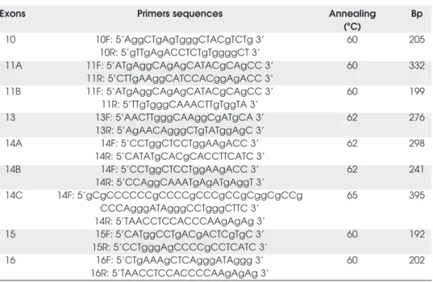
Figure 1.RETmutation analysis using direct sequencing and CSGE.
a)T to C substitution (TGC-CGC) in RETcodon 620 (exon 10) leading to a Cys620Arg mutation (5). CSGE exon 10 differentia-tion patterns: lane 1, Cys620; and lane 2, Cys620Arg.
b)(Top) T to C substitution (TGC-CGC) in RETcodon 634 (exon 11) leading to a Cys634Arg mutation (5); and, (Bottom) G to A substitution (TGC-TAC) in RETcodon 634 (exon 11) leading to a Cys634Tyr mutation. CSGE exon 11 differentiation patterns (primers 11B): lane 1, Cys634; lane 2, Cys634Arg; and lane 3, Cys634Tyr.
c)G to A substitution (GTC-ATC) in RETcodon 648 (exon 11) leading to a Val648Ile missense substitution (5). CSGE exon 11 differentiation patterns (primers 11B): lane 1, Cys648; and lane 2, Val648Ile.
d)G to A substitution (GGT-AGT) in RETcodon 691 (exon 11) leading to a Gly691Ser polymorphism. CSGE exon 11 differenti-ation patterns (primers 11A): lane 1, codons 634 and/or 648 not differentiated electrophoretic patterns; and lane 2, Gly691Ser.
e)T to G substitution (CTT-CTG) in RETcodon 769 (exon 13) leading to a Leu769Leu polymorphism. CSGE exon 13 differentia-tion patterns: lane 1, Leu769; and lane 2, Leu769Leu.
CSGE
Regarding CSGE screening mutation, the PCR products of the index cases (7 subjects) and at-risk relatives (57 family members) found to be mutated or polymorphic were screened by CSGE and compared to wild-type exons from 27 at-risk family members identified as healthy individuals. For CSGE-screened mutations, we analyzed 128 amplicons (encompassing RET hot-spot mutations and codon 648 missense substitution). 116 of 128 amplicons (90.6%) were found to be concordant with DNA sequencing (gold standard). In twelve ampli-cons (9.4%), all representing the Val804Met mutation (exon 14), the electrophoretic migration pattern could not be standardized even with alternative primer settings (14B and 14C) and electrophoresis conditions. In rela-tion to the polymorphism analysis, we screened 168 amplicons: 100 of them (59.5%) were concordant regarding CSGE and DNA sequencing. In 68 (40.5%) of the analyzed amplicons (all representing the Ser904Ser polymorphism), CSGE analysis could not be standardized. Briefly, patients presented the following genotype-phenotype correlations when examined by CSGE: (a) RETexon 10 Cys620Arg (TGC-CGC) in 5 MEN2A, 2 cases of congenital megacolon and 4 FMTC cases; (b)RETexon 11 Cys634Arg (TGC-CGC) in 12 MEN2A cases; (c) RET exon 11 Cys634Tyr (TGC-TAC) in 8 FMTC carriers; (d) RETexon 11 Val648Ile (GTC-ATC) in 2 so far clinically non-affected MEN2A carriers; and (e) RETexon 16 Met918Thr (ATG-ACG) in a case presenting MEN2B phenotype. Polymorphisms Gly691Ser (GGT-AGT) and Leu769Leu (CTT-CTG) were found to occur in RET mutation carriers, codon 648 missense substitution-carriers and non-carriers (13, 1 and 7 subjects, respectively).
SSCP
For RET hot-spot screening analysis using SSCP, we selected a subset of 37 subjects comprising all mutation, missense substitution and polymorphism genotypes here studied: 4 subjects presenting Cys620Arg + Gly691Ser +
Ser904Ser; 3 Cys634Arg; 3 Cys634Tyr; 2 Val648Ile; 3 Val804Met (one of them carrying Leu769Leu); 1 Met918Thr; 2 Leu769Leu and 19 wild-type subjects. Considering SSCP-screened mutations, 14 of 37 subjects were REThot-spot mutants carriers for codons 620, 634, 804, and 918; 2 of 37 subjects were codon 648 missense substitution carriers and nineteen subjects were healthy controls. As it was observed when applying CSGE tech-nology, the same 12 amplicons (17%) representing Val804Met mutation (exon 14), could not be standard-ized by SSCP, either. For SSCP polymorphism screening analysis, the subset group (37 subjects) comprised four subjects presenting codon 691 + 904 polymorphisms, three 769 polymorphism carriers and twelve healthy con-trol subjects. In 16 (35%) of the analyzed polymorphic amplicons (Ser904Ser), electrophoretic migration pat-terns could not be standardized. Patients presented the same genotype-phenotype correlations when screened by either SSCP or CSGE. SSCP screening method showed the same migration patterns for all exons, when examin-ing either RET mutations or polymorphisms. Data obtained by CSGE were fully concordant (100%) with those observed when SSCP was applied.
Comparing data encompassing RET hot-spot mutations and codon 648 missense substitution obtained from SSCP/CSGE assays with those from genetic sequencing, we documented that 58 analyses (83%) were concordant. Also, comparing polymorphism data obtained by SSCP/CSGE with those obtained with genetic sequencing, 30 analyses (65%) were concordant. Using CSGE and SSCP, we were able to detect four out of the five types (80%) of RET mutations verified by direct sequencing analysis in our MEN2 patients.
DISCUSSION AND CONCLUSION
Molecular diagnosis offers a specific and highly accu-rate indication for prophylactic total thyroidectomy in human RETmutation carriers, which may alter MEN2 Table 3.Screening methods: comparative study in RET hot-spot exons.
Hot-spot exons Direct sequencing CSGE SSCP Screened Screened Screened Mutations Missense Polymorphisms Substitution
10 Sensitive Sensitive Sensitive Cys620Arg
11 Sensitive Sensitive Sensitive Cys634Arg; Val648Ile Gly691Ser Cys634Tyr
13 Sensitive Sensitive Sensitive Leu769Leu
14 Sensitive Not sensitive Not sensitive Val804Met
15 Sensitive Not sensitive Not sensitive Ser904Ser
natural course of disease (15,24). In our MEN2 cases, likely due to the smallness of our sample, Val804Met RETmutation was relatively over-represented, with 3 individuals out of 35 (8.6%), compared to its less than 3% prevalence in large MEN2 series (1,15).
In the present study, we reported the CSGE use as a RETmutation screening method in typical MEN2 families. It presented high specificity for hot-spot mutations and for polymorphism analyses, as no false-positive result was detected. As for sensitivity (false negatives), Val804Met mutation could not be detect-ed by this method. Importantly, when using the CSGE method we were able to identify RET muta-tions (or polymorphisms) in exons 10, 11, 13, and 16, which have been reported to cause the majority of MEN2 cases (15). Val804Met (exon 14) is a rare genetic variation, representing less than 3% of RET disease-causing mutations reported in MEN2 cases so far (1,15,25). We have occasionally successfully screened Val804Met using DGGE (3,4,8).
Direct genetic sequencing is the “gold stan-dard” in genetic mutation analysis. However, for extended genealogies or populations, the cost of this procedure may be significantly higher than other screening methods. For instance, CSGE has been suc-cessfully used in several genetic screenings and may result in six-fold lower costs than sequencing, consid-ering only reagents. Furthermore, it has been shown that screening methods such as DHPLC (that uses a Wave system analyzer), DGGE, SSCP, and restriction enzymes are also adequate alternatives for direct DNA sequencing (4,8,16). Each of these methods has advantages and disadvantages, as follows: DHPLC uses an automatic system for reading heteroduplex bands and is a sensitive method (90–95%), although its specific wave analyzer costs must be considered (23). Restriction enzyme sensitivity and reproduction analy-sis may present some drawbacks, such as: (a) inade-quate recognition of restriction site and a consequent-ly non-defined band in agarose gel electrophoresis, and (b) incomplete discrimination of amplified sequences with similar base pair sizes (4,14,16,26). Despite these limitations, a previous study reported total correlation when comparing restriction enzymes to direct sequencing (14). SSCP sensitivity usually reaches 80% for PCR fragment sizes smaller than 300 bp, meaning that non-mutant electrophoresis migra-tion patterns do not exclude the possibility of a muta-tion carrier (4,14,16,26). According to other studies, SSCP presents an overall sensitivity of 95–100% for RET hot-spots mutation analysis (27,28). Also, DGGE is a highly sensitive method (90–100%) and is
a very useful screening approach for RET mutations, which has been used in our laboratory (3,4,8). DGGE has several advantages, such as: (a) sensitivity and reproducibility higher than SSCP; (b) a high correla-tion with direct sequencing; (c) non-radioactive pro-tocols; (d) allows little operational manipulation; and (e) does not need to recognize restriction sites. On the other hand, DGGE has some disadvantages such as: (a) requires a 50 bp GC-clamp coupled to one of the primer sequences; (b) each analyzed exon melting temperature needs to be addressed by a special com-puter software or hard standardization; (c) initial investment at specific electrophoresis system; (d) diffi-cult standardization for guanine-cytosine rich sequences; and (e) polymorphism analysis may yield false-positive results (3,4,8).
In the present study, we reported CSGE appli-cation as a RETscreening method in 7 typical MEN2 families with a sensitivity of 90.6% for hot-spot muta-tions (encompassing codon 648 missense substitution) and 59.5% for polymorphism analyses. According to the MEN Consensus, the Val804Met mutation, which was not detected in our CSGE assay, is a rare genetic variation, representing less than 3% of RET disease-causing mutations (1,15,25).
Additionally, we were interested in testing CSGE for detecting RETpolymorphisms as they may act as epigenetic factors interacting with RET muta-tions and possibly altering the clinical presentation, course, and morbidity of MEN2 disease (29-33).
In summary, our study has shown that CSGE (and SSCP) is a useful method for screening RET hot-spot mutations, considering the following points: (a) using CSGE, we have successfully screened 4 of 5 types of RET mutations among our cases; (b) these four mutations detected by CSGE have been reported as being responsible for most (95%) of the disease-causing mutations in different MEN2 samples; (c) there was a total correlation (100%) between CSGE and SSCP data; (d) CSGE is a low-cost, fast and feasi-ble to standardize method; (e) it does not use a radioactive protocol; and (f) it allows little operational manipulation, which decreases the possible chance of error. Moreover, CSGE dispenses with some require-ments, such as: (a) the recognition of restriction sites; (b) different gel concentrations for each studied exon and 50 bp GC-clamp coupled to one of the primers sequences; and finally, (c) this method does not require the use of specific software to analyze each exon melting temperature.
occurring RETmutations in MEN2. It is also worth-while to note that our results reflect a retrospective study of a small number of MEN2 families (seven fam-ilies) that need to be addressed to a prospective study from a greater number of families, where genetic sequencing and CSGE screening may be simultane-ously applied.
ACKNOWLEDGMENTS
We thank the Papaiz group and CNPq–CAPES for supporting this research, aiming at providing a faster, safe, and accurate diagnosis of multiple endocrine neo-plasia type 2. We also thank: Drs. Adriana B. Nunes, Marilza C.L. Ezabella, Neusa Abelin, Cesar Y. Hayashida, Ivone I. Mackwiack and all the staff mem-bers of the Endocrinology Genetics Unit, Endocrinol-ogy, University of Sao Paulo, School of Medicine (http://medicina.fm.usp.br/ueg). We also thank Prof. Dr. Maria Rita S.P. Bueno, Head of the Dept. of Genetics and Evolutive Biology, Institute of Bio-science IB–USP, for laboratory support.
REFERENCES
1. Hoff AO, Cote GJ, Gagel RF. Multiple endocrine neoplasias.
Annu Rev Physiol 2000;62:377-400.
2. Ezabella MCL, Hayashida CY, Abelin NMA, Toledo SPA. Neo-plasias Endocrinas Múltiplas. In: Medeiros-Neto G (ed).
Moléstias Hereditárias do Sistema Tireoideano. 1aed.
São Paulo: Roca, 1996. pp. 225-42.
3. Nunes AB, Ezabella MCL, Pereira AC, Krieger JE, Toledo SPA. A novel Val648Ile substititution in RET proto-oncogene observed in a Cys634Arg multiple endocrine neoplasia type 2A kindred presenting with an adrenocorticotropin-produc-ing pheochromocytoma. J Clin Endocrinol Metab 2002;87:5658-61.
4. Toledo SPA, Santos MA, Toledo RD, Lourenço Junior DM. Impact of RET proto-oncogene analysis on the clinical man-agement of multiple endocrine neoplasia type 2. Clinics 2006;61:59-70.
5. Santos MA, Nunes AB, Abelin N, Ezabella MC, Toledo R de A, Lourenço Júnior D, et al. Genetic screening of multiple endocrine neoplasia type 2: experience of the USP Endocrine Genetics Unit. Arq Bras Endocrinol Metab 2006;50:7-16. 6. Mulligan LM, Kwok JB, Healey CS, Elsdon MJ, Eng C,
Gard-ner E, et al. Germ-line mutations of the RET proto-oncogene in multiple endocrine neoplasia type 2A. Nature 1993;363:458-60.
7. Punales MK, Graf H, Gross JL, Maia AL. RETcodon 634 muta-tions in multiple endocrine neoplasia type 2: Variable clinical features and clinical outcome. J Clin Endocrinol Metab 2003;88:2644-9.
8. Nunes AB. Identificação de mutações do proto-oncogene RET
associadas à forma hereditária do carcinoma medular de tireóide. Tese de Doutoramento, LIM-25, HC-FMUSP,
2001.
9. Maia FFR, Júnior HJ, Araújo LR. Neoplasia endócrina múlti-pla tipo 2 — manejo diagnóstico e terapêutico. Arq Bras Endocrinol Metab 2002;46:606-10.
10. Maciel RMB. Tumorigênese molecular tiroideana: impli-cações para a prática médica. Arq Bras Endocrinol Metab 2002;46:381-90.
11. Hayashida CY, Alves VAF, Kanamura CT, Ezabella MCL, Abelin NMA, Nicolau W, et al. Immunohistochemistry of medullary thyroid carcinoma and C-cell hyperplasia by an affinity-purified anti-human calcitonin antiserum. Cancer 1993;72:1356-63.
12. Abelin NMA, Gomes S, Ivanoff MT, Ezabella MCL, Hayashida CY, Toledo SPA. Abordagem clínica e laboratorial do bócio uni-nodular sólido: vantagens da determinação da calcitoni-na sérica por métodos distintos. Arq Bras Endocrinol Metab 1999;43:104-13.
13. Silva AMA, Maciel RMB, Silva MRD, Toledo SRC, Carvalho MB, Cerutti JM. A novel germ-line mutation in RETexon 8 (Gly533Cys) in a large kindred with familial medullary thyroid carcinoma.J Clin Endocrinol Metab 2003;88:5438-43. 14. Frediani D. Rastreamento gênico através de enzimas de
restrição na forma hereditária do carcinoma medular de tireóide. Monografia, LIM-25, HC-FMUSP, 2002.
15. Brandi ML, Gagel RF, Angeli A, Bilezikian JP, Beck-Peccoz P, Bordi C, et al. CONSENSUS: Guideline for diagnosis and ther-apy of MEN type 1 and type 2. J Clin Endocrinol Metab 2001;86:5658-71.
16. Korkko J, Annunen S, Pihlajamaa T, Prockop DJ, Kokko LA. Conformation sensitive gel electrophoresis for simple and accurate detection of mutations: Comparison with denaturing gradient gel electrophoresis and nucleotide sequencing.
Proc Natl Acad Sci (USA) 1998;95:1681-5.
17. Ganguly A, Rock MJ, Prockop, DJ. Conformation-sensitive gel electrophoresis for rapid detection of single-base differ-ences in double-stranded PCR products and DNA fragments: Evidence for solvent-induced bend in DNA heteroduplexes.
Proc Natl Acad Sci USA 1993;90:10325-9.
18. Ganguly A. An update on conformation sensitive gel elec-trophoresis. Hum Mutat 2002;19:334-42.
19. Kambouris M, Jackson CE, Feldman GL. Diagnosis of multi-ple endocrine neoplasia (MEN) 2A, 2B and Familial Medullary Thyroid Cancer (FMTC) by multiplex PCR and heteroduplex analyses of RET proto-oncogene mutations. Hum Mutat 1996;8:64-70.
20. Vianello F, Lombardi AM, Bello FD, Zanon E, Cabrio L, Giro-lami A. Conformation sensitive gel electrophoresis for detec-tion of factor X gene mutadetec-tions. Thromb Res 2002 ;107:51-4.
21. Miller S, Dykes D, Poleski H. A single salting out procedures for extracting DNA from human nucleated cells. Nucleic Acids Res 1988;16:1215.
22. Saiki RK, Gelfand DH, Stoffel S, Scharf SJ, Erlich HA. Primer-directed enzymatic amplification of DNA with a thermostable DNA polymerase. Science 1988;239:487-91.
23. Solari V, Ennis S, Yoneda A, Wong L, Messineo A, Hollwarth ME, et al. Mutation analysis of the RETgene in total intestinal aganglionosis by wave DNA fragment analysis system. J Pediatr Surg 2003;38:497-501.
24. Kameyama K, Takami H. Medullary thyroid carcinoma: Nationwide Japanese survey of 634 cases in 1996 and 271 cases in 2002. Endocr J 2004;51:453-6.
25. Frohnauer MK, Decker RA. Update on the MEN 2A c804 RET
mutation: Is prophylactic thyreoidectomy indicated? Surgery 2000;128:1052-8.
26. Bugalho MJ, Domingues R, Sobrinho L. The minisequencing method: a simple strategy for genetic screening of MEN 2 families. BMG Genet 2002;3:1-5.
27. Siegelman M, Mohabeer A, Fahey TJ 3rd, Tomlinson G, Mayambala C, Jafari S, et al. Rapid, nonradioactive screening for mutations in exons 10, 11, and 16 of the RET protoonco-gene associated with inherited medullary thyroid carcinoma.
Clin Chem 1997;43:453-7.
29. Robledo M, Gil L, Pollán M, Cebrián A, Ruíz S, Azañedo M, et al. Polymorphisms G691S/S904S of RETas genetic modifiers of MEN 2A. Cancer Res 2003;63:1814-7.
30. Wiench M, Wloch J, Wygoda Z, Gubala E, Oczko M, Pawlaczek A, et al. RETpolymorphisms in codon 796 and 836 are not associated with predisposition to medullary thyroid carcinoma. Cancer Detect Prev 2004;28:231-6.
31. Baumgarter-Parzer SM, Lang R, Wagner L, Heinze G, Nieder-le B, Kaserer K, et al. Polymorphisms in exon 13 and intron 14 of the RET proto-oncogene: Genetic modifiers of medullary thyroid carcinoma? J Clin Endocrinol Metab 2005;90:6232-6.
32. Cebrian A, Llorente SR, Cascon A, Osorio A, Delgado BM, Benítez J, et al. Rapid and easy method for multiple endocrine neoplasia type 1 mutation detection using confor-mation-sensitive gel electrophoresis. J Hum Genet 2002;47:190-5.
33. Wohllk N, Soto E, Bravo M, Becker P. Polimorfismos G691S, L769L e S836S del proto-oncogene RET no se associan a mayor riesgo de cancer medular tiroideo esporádico en pacientes chilenos. Rev Med Chile 2005;133:397-402.
Address for correspondence:
Sergio P.A. Toledo
Unidade de Endocrinologia Genética, Endocrinologia, Clínica Médica Av. Dr. Arnaldo 455, 5oandar