ABSTRACT
Cushing’s syndrome due to ACTH-independent macronodular adrenal hyperplasia (AIMAH) can be associated with abnormal responses of aberrantly expressed adreno-cortical receptors. This study aimed to characterize in vitrothe pathophysiology of hypercortisolism in a β-blocker-sensitive Cushing’s syndrome due to AIMAH. Cortisol secretion profile under aberrant receptors stimulation revealed hyperresponsiveness to salbutamol (β2-adrenoceptor agonist), cisapride (5-HT4 receptor agonist), and vaso-pressin in AIMAH cultured cells, but not in normal adrenocortical cells. By RT-PCR, AIMAH tissues revealed β2-adrenoceptor overexpression rather than ectopical expres-sion. MC2R expression was similar in both AIMAH and normal adrenocortical tissues. Curiously, cortisol levels of AIMAH cells under basal condition were 15-fold higher than those of control cells and were not responsive to ACTH. Analysis of culture medi-um from AIMAH cells could detect the presence of ACTH, which was immunohisto-chemically confirmed. Finally, the present study of AIMAH cells has identified: a) cor-tisol hyperresponsiveness to catecholamines, 5-HT4 and vasopressin in vitro, in agreement with clinical screening tests; b) abnormal expression of β2-adrenoceptors in some areas of the hyperplastic adrenal tissue; c) autocrine loop of ACTH produc-tion. Altogether, the demonstration of aberrant responses to hormonal receptors and autocrine hormone production in the same tissue supports the assumption of multi-ple molecular alterations in adrenal macronodular hyperplasia. (Arq Bras Endocrinol Metab 2007;51/9:1452-1462)
Keywords:Adrenal glands/ (pathol); Hyperplasia; Hormone receptors; Cell surface; Adrenocorticotropic hormone; Adrenocortical hyperfunction; Cushing syndrome
RESUMO
Alterações Celulares e Moleculares de uma Hiperplasia Adrenal Macronodu-lar Responsável por Síndrome de Cushing Responsiva a Beta-Bloqueadores. A síndrome de Cushing secundária à hiperplasia adrenal macronodular independente de ACTH (AIMAH) pode estar associada com respostas anômalas a estímulos sobre receptores hormonais expressos de maneira aberrante no córtex adrenal. O objetivo deste trabalho foi caracterizar a fisiopatologia do hipercortisolismo in vitrona sín-drome de Cushing responsiva a β-bloqueadores decorrente de AIMAH. Em cultura de células, a secreção de cortisol apresentou resposta aumentada ao salbutamol (ago-nista β2-adrenérgico), à cisaprida (agonista de receptor 5-HT4) e à vasopressina, na AIMAH mas não no córtex adrenal normal. O estudo de receptores aberrantes por RT-PCR demonstrou que o gene do receptor β2-adrenérgico estava superexpresso (e não expresso ectopicamente) nos fragmentos da AIMAH quando comparado ao tecido normal. A expressão de MC2R foi semelhante em ambos. Curiosamente, o nível basal de secreção de cortisol pelas células da AIMAH foi 15 vezes superior às células nor-mais, não havendo resposta das células AIMAH ao estímulo com ACTH. A análise do meio de cultura das células AIMAH revelou a presença de ACTH, que foi confirmada por estudo imuno-histoquímico. Em suma, este estudo demonstrou: a) aumento dos níveis de cortisol in vitroem resposta a catecolaminas, 5-HT4 e vasopressina, corres-pondendo aos resultados dos testes clínicos para pesquisa de receptores aberrantes; b) expressão anormal de receptores β2-adrenérgicos em algumas áreas de hiperpla-sia; c) produção autócrina de ACTH. Estes resultados envolvendo ativação de recep-tores aberrantes e estímulo hormonal autócrino no mesmo tecido favorecem a hipótese da existência de alterações moleculares múltiplas na hiperplasia adrenal macronodular. (Arq Bras Endocrinol Metab 2007;51/9:1452-1462)
Descritores:Glândulas supra-renais/ (patol); Hiperplasia; Receptores hormonais da superfície celular; Hormônio adrenocorticotrópico; Hiperfunção adrenocortical; Sín-drome de Cushing
artigo original
Cushing’s Syndrome
TÂNIAL. MAZZUCO MICHAËLTHOMAS MONIQUEMARTINIE NADIA CHERRADI NATHALIESTURM JEAN-JACQUES FEIGE OLIVIERCHABRE
Unité INSERM 878, Institut de Recherches en Technologies et Sciences pour le Vivant — iRTSV, CEA-Grenoble (TLM, MT, NC, J-JF & OC); Service d’Endocrinologie, Département de Diabétologie Urologie Néphrologie Endocrinologie — DUNE (TLM, MM & OC) and Laboratoire
de Pathologie Cellulaire, Département d’Anatomie et de Cytologie Pathologiques (NS), CHU-Grenoble, France.
S
EVERAL OBSERVATIONS INDICATE that Cushing’s syndrome caused by corticotrophin (ACTH)-inde-pendent tumors or macronodular adrenal hyperplasia (AIMAH) can be accounted for aberrant responsive-ness of the tissue to various hormones or neurotrans-mitters, including gastric inhibitory polypeptide (GIP), epinephrine, vasopressin (AVP) or serotonin (5-HT) (1). Catecholamine-dependent Cushing’s syn-drome is a rare condition that has been clinically reported in six cases at present (2-7).Previous in vitro reports have provided some
evidence for abnormal responses to β-adrenergic
stim-uli on adrenocortical tumors. These adrenal β
-adren-ergic responses were first studied in the rat adrenocor-tical carcinoma 494 cells in which abnormal activation of adenylate cyclase (AC) by catecholamines (8) and the presence of isoproterenol binding sites (absent in membranes of normal adrenal tissue) were character-ized (9). In human adrenocortical tissues, activation of AC by norepinephrine and/or epinephrine was observed in adenomas (10), in adrenocortical carcino-mas (11), and in one case of AIMAH (2) but not in
normal tissues (11). The presence of β-adrenergic
binding sites was also shown in that same case of AIMAH (2), in cortisol-producing adenomas (12) and
in adrenocortical carcinomas (11). Prior to these in
vitro studies, a catecholamine-dependent Cushing’s syndrome was clinically characterized in only two cases (2,6). These include a case with a simultaneous expres-sion of several aberrant hormone receptors, a mild
cor-tisol response to isoproterenolin vivoand an absence
of β-adrenoceptor mRNA detection by RT-PCR (6).
We have previously described a clinical case of
bilateral AIMAH with hypercortisolism sensitive to β
-blockade (4), characterizing a catecholamine-dependent
Cushing’s syndrome that was well controlled by β
-adrenergic antagonist treatment. Clinical screening tests performed to search for cortisol elevation in response to several ligands revealed other potential abnormally
expressed receptors in adrenal cortex, such as 5HT4and
AVP receptors. Later on, the occurrence of pharmaco-logical therapy intolerance led to the surgical excision of the larger adrenal gland of this patient. Hyperplastic
tis-sue could thus be obtained for in vitro assays that
allowed us to study the abnormal response to β
-adreno-ceptor stimulation and to determine which type of β
-receptor is implicated in this response.
Here we report the cortisol responses of
AIMAH cells to diverse in vitropharmacological tests
in order to compare with previous in vivo responses
obtained in clinical screening tests searching for aber-rant adrenal receptors expression. Our findings
indi-cate that the abnormal clinical responsiveness to indi-
cate-cholamines was a result of abnormal expression of β
2-adrenoceptors in the hyperplastic tissue. Comparison
with normal tissue suggested β2-adrenoceptor
overex-pression rather than ectopic exoverex-pression as mentioned
in the literature. In vitroassays showed cellular
hyper-responsiveness to 5-HT and AVP, confirming the results of the clinical assays. Finally, basal overproduc-tion of cortisol by AIMAH cells led us to identify an autocrine loop of ACTH production.
MATERIALS AND METHODS
Clinical investigation protocol
A 64 yr-old Caucasian woman presenting ACTH-indepen-dent hypercortisolism due to AIMAH was investigated for abnormal expression of adrenal receptors (4). After her informed consent, plasma levels of steroids in response to various stimuli were investigated using the protocol described by Lacroix et al. (13). The protocol consisted of monitoring plasma cortisol, aldosterone, and ACTH con-centrations at 30–60 min intervals for 2–3 h during the tests as follow: supine-to-upright posture test, standard mixed meal, combined i.v. administration of 200 µg TRH and 100 µg LHRH (Stimu-TSH and Stimu-LH, Ferring, Gentilly, France), combined administration of 1 mg glucagon (Gluca-Gen, Novo Nordisk, Puteaux, France) i.v. and cisapride (Prepulsid, Janssen-Cilag, Issy-les-Moulineaux, France) oral-ly, 1 mg terlipressin (Glypressine, Ferring, Gentiloral-ly, France) i.v, 10 µg desmopressin (Minirin, Ferring, Gentilly, France) i.v., 6 IU regular insulin (Actrapid, Novo Nordisk, Bagsvaerd, Denmark) i.v, or 0.25 mg tetracosactide (Synac-thène, Novartis Pharma, Rueil-Malmaison, France) i.v. as reference test. Cortisol response to the hypoglycemia (insulin test) was investigated after normalization of blood glucose levels of this diabetic patient.
Tissue collection and cell culture
Indianapolis, IN) and 0.1 mg/mL deoxyribonuclease (Sigma, Saint Quentin Fallavier, France), for 30 min at 37oC under stirring. The suspension was then filtered on sterile nylon sieve (100 µm mesh), completed with culture medium up to 50 mL and centrifuged for 10 min at 400 x g. Cells prepared from different fragments were pooled because the number of cells from small nodules was insufficient to per-form individualized culture experiments. Adrenocortical cells were then seeded in Petri dishes at a density of 106 cells/dish with supplemented Ham’s F-12-DMEM (1:1) medium containing 10% horse serum (Eurobio, Les Ulis, France) and 2.5% fetal calf serum (FCS) (Life Technologies) with antibiotics, 0.5% insulin-transferrin-sodium selenite medium supplement (ITS) (Sigma), and incubated at 37oC in a 5% CO2–95% air atmosphere. The culture medium was renewed 24 h later. Normal adrenal cells were prepared from a normal gland collected from a brain-dead patient under-going nephrectomy for renal transplantation, following the same procedure as described above. All studies were per-formed in accordance to the guidelines of the institutional Human Research Ethics Committee.
In vitro cortisol production
Confluent cultured cells were trypsinized and plated in sup-plemented medium in multiwell dishes (12 wells, 2 x 10⁵ cells/well). Twenty-four hours later, the medium was replaced by serum-free Ham’s F12/DMEM (1:1). On the next day, various concentrations of the following agonists were added in fresh serum-free culture medium: ACTH1–24 (Neosystem, Strasbourg, France), isoproterenol, BRL 37344 (β3-adrenoceptor agonist), 5-hydroxytryptamine (5-HT), Arg8-vasopressin (AVP), human chorionic gonadotropin (hCG) (all purchased from Sigma), GIP (Bachem, Voisins-le-Bretonneux, France), dobutamine (Merck, Lyon, France), salbutamol (GlaxoWellcome, Parma, Italy), desmopressin (Ferring, Gentilly, France), glucagon (Novo Nordisk,
Puteaux, France), and cisapride (kindly provided by Prof. H. Lefebvre, University Hospital of Rouen, France). After an incubation period of 2 h, aliquots of the conditioned culture medium were taken and frozen at -20ºC until determination of cortisol by RIA. The elevation of cortisol in response to increased concentrations of some agonists reached a plateau, revealing the maximal effect (Emax) of each agonist. Cul-tured normal adrenocortical cells were incubated with the same agonists at concentrations providing the maximal response of AIMAH cells. The effect of the non-selective β -adrenergic antagonist propranolol (Zeneca Pharma, Cergy, France) on cortisol secretion was tested in a set of experi-ments of AIMAH cells.
Hormone assays
Plasma and urinary cortisol and plasma aldosterone concen-trations were determined by radioimmunoassay (Immuno-tech, Marseille, France). Cortisol concentrations in condi-tioned culture media were determined by radioimmunoassay (Immunotech, Marseille, France). Plasma and culture medi-um ACTH concentrations were measured by immunoradio-metric assay (Nichols Institute Diagnostics, San Clemente, CA), with detection limit of 0.5 pmol/L (2.3 pg/mL).
RT-PCR analysis
After surgical resection and tumor dissection, 4 small tissue samples (around 10 mg each) were individualized before enzy-matic digestion of all tumor fragments for cell culture. These samples were collected from the diffuse hyperplastic area (fig-ures 2A and 2B) and from the two major nodules of 1 and 1.2 cm diameter (figures 2C and 2D). These tissue samples were immediately frozen in liquid nitrogen and stored at -80oC until RNA extraction. Thawed tissues were homogenized with the MagNA Lyser Instrument and total RNA was extracted using the High Pure RNA Tissue Kit (both from Roche Applied Sci-ence, Penzberg, Germany). The integrity of RNA was con-Figure 1. Radiologic and macroscopic morphology of the adrenal hyperplasia. Plan
abdominal computed tomographic scan shows bilateral adrenal enlargement (left gland is indicated by the white arrow). The entire surgical piece of left adrenal was com-posed of a large polylobulated mass with two satellite nodules smaller than 2 cm. Four tissue fragments (A, B, C, and D) were collected for RNA extraction.
1 cm
External view
firmed on ethidium bromide-stained agarose gels and spec-tophotometrically, using the 260/280 nm absorption ratio. RNA quantitation was performed by measuring the absorption at 260 nm. Two µg of total RNA were reverse-transcribed with ImProm-II Reverse Transcriptase (Promega, Madison WI) and 0.5 µg of random hexamer DNA primers, according to the conditions recommended by the manufacturer. Aliquots of 100 ng cDNA were subjected to semi-quantitative PCR in a final volume of 25 µL containing 2.5 U Taq DNA polymerase (Qbiogene, Illkirch, France), and 14 pmol of each oligonu-cleotide primer. First, amplification of a DNA fragment of ribosomal protein L27 (RP-L27) was performed using the fol-lowing pair of primers: 5’-GAACATTGATGATGGCAC-CTC-3’ and 5’-GGGGATATCCACAGAGTACC-3’. All cDNA samples were adjusted to yield equal amplification of RP-L27 DNA fragment as internal standard and were analyzed in triplicate. The amplification parameters were 2 min at 94ºC, then 35 cycles each consisting of 1 min at 94ºC, 1 min at 55ºC, and 2 min at 72ºC. PCR reactions were also performed in non-reverse-transcripted RNA samples to exclude DNA contamina-tion. A commercial preparation of adult human adrenal cortex RNA pooled from 11 normal glands (Human Adrenal Cortex Poly A+RNA, Clontech, Palo Alto, CA) was used as a normal control. Amplified products were separated by agarose gel elec-trophoresis (2%) and scanned using Fluorimager (Molecular Dynamics) to calculate densitometry of bands and product/RPL-27 ratios. For RT-PCR positive control, RNA from other tissues (human placenta and normal adrenal cortex adjacent to adenoma) was extracted using the same procedure. All reactions followed by electrophoresis separation were per-formed in triplicates.
Primers design
To amplify the single exon of the β1-adrenoceptor (AR) gene, the primer pair 5’-TCGTGTGCACCGTGTGG
GCC-3’ and 5’-AGGAAACGGCGCTCGCAGCTGTCG-3’ was designed, corresponding to nucleotides (nt) 638 – 860 of the reference sequence NM 000684. For the single exon of the β2-AR gene, the primer pair 5’-ACC-CACCAGGAAGCCATCAACTG-3’ and 5’-AACTTCCT-TACGGATGAGGTTATCC-3’ was designed to amplify the nt 770 – 1116 fragment of the reference sequence NM 000024. For the β3-AR gene amplification, the primer pair 5’-GCTCCGTGGCCTCACGAGAA-3’ and 5’-CCCA ACGGCCAGTGGCCAGTCAGCG-3’ was designed to amplify the nt 220 – 490 fragment of exon 1 according to the reference sequence NM 0000025. For the single exon of MC2R gene, we used the primer pair 5’-GGAGTTTTG-GAGAATCTGATCG-3’ and 5’-ATGGTGATGTAGCG-GTCCCGCAG-3’ (corresponding to nt 112 – 396). Amplification of AVP receptors V1-R, V2-R, and V3-R genes was performed using previously published sequences of nucleotides (14). For 5-HT4 receptor gene, we used previously published sequence of primers that hybridize to all receptor splice variants (15).
Immunohistochemistry
Deparaffinized sections of the hyperplastic tissue were incubated with the following primary antibodies: mouse monoclonal antibody against a C-terminal peptide of ACTH (1:500; Biodesign International, Saco, ME), rabbit polyclonal antibody against β-melanocyte-stimulating hor-mone (bMSH, 1:10000; Prof. Bachelot, Grenoble, France) and rabbit polyclonal antibody for β-endorphin (1:300; Prof Bugnon, Lyon, France). Bound antibodies were detected using the labeled streptavidin-biotin-peroxidase method (DAKO Corp., Trappes, France). Negative con-trols of the immunohistochemical reactions were per-formed by replacing the primary antibody with preimmune serum.
Figure 2. Maximal cortisol responses of cultured cells to stimulation by several agonists. Hyperplastic (AIMAH) and normal human adrenocortical cells were exposed for 2 h to different concentrations of agonists and the maximal cortisol responses (Emax) were determined and shown in the figure. Histograms represent the mean + s.e.m. of cortisol concentration in
condi-tioned medium. A)Cells were incubated with 10 nM ACTH or with the following β-adrenergic agonists: 10 µM Isoprot (isopro-terenol, non-selective agonist), 10 µM Dobut (dobutamine, β1-agonist), 1 µM Salb (salbutamol, β2-agonist) or 100 nM BRL (BRL-37344, β3-agonist); B)Other agonists were tested to compare in vitrostimulation with previously described in vivoresponses: 10 µM 5-HT (non-selective serotoninergic agonist), 100 nM Cisapr (cisapride, 5HT4receptor agonist); 10 nM Glucag (glucagon), 10
nM AVP (vasopressin), 10 nM GIP (gastric inhibitory peptide), and 100 nM hCG (human chorionic gonadotrophin). Incubations were performed in triplicate wells in 2 or 3 different experiments [* P< 0.05; ** P< 0.01 vs. control].
Normal adrenocortical cells AIMAH cells
Cortisol secretion (nmol/10
6cells/h) 300
A
Basal ACTH Isoprot Dobut Salb BRL
250
200
150
100
50
0 Cortisol secretion (nmol/10
6cells/h) 300 350
A
Basal 5-HT Cisapr Glucag AVP GIP hCG
250
200
150
100
50
Statistical analysis
Descriptive data are shown as mean ± SEM. Variables show-ing normal distribution were analyzed usshow-ing a two-tailed Student’s t-test, whereas analyses of variance were used for variables with nonparametric distribution. P values less than 0.05 were considered as statistically significant, P values less than 0.01 were considered as highly significant. Emaxof each agonist on cortisol secretion was expressed in percentage of basal cortisol production (without agonist). If a dose-depen-dent stimulation was observed, the concentration that led to 50% maximal response (EC50) was calculated using Bio-DataFit 1.02 online (Chang Bioscience, Castro Valley, CA) and values were presented as a negative base 10 logarithm of the molar concentration (pEC50).
RESULTS
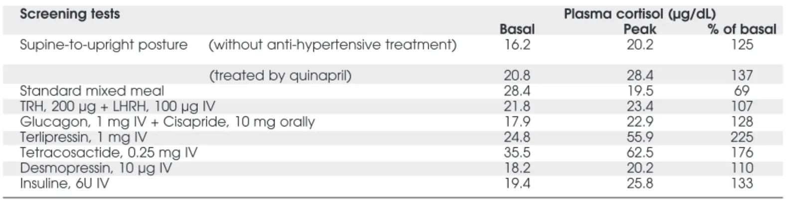
Clinical evaluation of cortisol responses Systematic search for the expression of aberrant adrenal hormone receptors according to the investi-gation protocol allowed to detect significant plasma cortisol response to the following stimulation tests: I) upright posture, II) terlipressin (a V1-vasopressin receptor agonist), III) insulin-induced hypo-glycemia, and IV) co-administration of glucagon-cisapride (table 1). Aldosterone levels remained nor-mal during all tests (data not shown). After a metic-ulous evaluation of the possible receptors involved in the orthostatism-dependent hypercortisolism, a test of cortisol secretion was performed under beta-blockade (propranolol, 320 mg/d). After 3 days, the free urinary cortisol excretion was normalized (from 434 to 36 µg/24h). Treatment discontinuation induced reappearance of hypercortisolism, which was controlled again by the reintroduction of pro-pranolol treatment.
Cortisol response of cultured cells to hormone challenge
After adrenalectomy and cell dispersion according
pre-viously described cell culture procedures, in vitro
cor-tisol synthesis was evaluated. Cells from both the AIMAH catecholamine-dependent Cushing’s syn-drome and normal adrenal cortex (control cells) were studied. The steroidogenic effect of several receptor ligands was firstly studied by incubating cultured adrenocortical cells with increasing concentrations of agonists (data not shown). The maximal steroidogenic
effect (Emax) of each agonist is shown in figure 2 and
compared to the pharmacological response of control cells to the same agonist concentration. Incubation of AIMAH cells with 10 nM ACTH elicited a
non-sig-nificant increase in cortisol levels (Emax 132 ± 53%)
whereas control cells were significantly stimulated with
a pEC50 of 10.1 ± 0.2 M (figure 2A). It should be
noted, however, that the basal level of cortisol pro-duction was 30 times higher in AIMAH cells (122
nmol/106cells/h) than in normal cells (4.1
nmol/106cells/h). Conversely to the non-selective β
-adrenoceptor agonist isoproterenol, the β2-agonist
salbutamol only induced significant cortisol
stimula-tion in AIMAH cells (pEC505.9 ± 0.1 M, Emax180 ±
58%). Other specific agonists of β-adrenoceptors
(dobutamine, β1-agonist; BRL 37344, β3-agonist)
did not induce any significant increase in cortisol pro-duction (figure 2A).
A significant stimulation of cortisol secretion was also obtained in AIMAH cell following incubation
with 5-HT and the 5-HT4 receptor agonist cisapride
(pEC506.1 ± 0.2 and 7.5 ± 0.8 M respectively) (figure
2B). Moreover, AVP elicited a dose-dependent
stimu-lation of cortisol secretion with pEC509.6 ± 0.1 M in
the same cells. Regarding 5-HT and AVP receptor
Table 1.Screening tests for abnormal cortisol response.
Screening tests Plasma cortisol (µg/dL)
Basal Peak % of basal
Supine-to-upright posture (without anti-hypertensive treatment) 16.2 20.2 125
(treated by quinapril) 20.8 28.4 137
Standard mixed meal 28.4 19.5 69
TRH, 200 µg + LHRH, 100 µg IV 21.8 23.4 107
Glucagon, 1 mg IV + Cisapride, 10 mg orally 17.9 22.9 128
Terlipressin, 1 mg IV 24.8 55.9 225
Tetracosactide, 0.25 mg IV 35.5 62.5 176
Desmopressin, 10 µg IV 18.2 20.2 110
Insuline, 6U IV 19.4 25.8 133
agonists, no significant stimulation of cortisol
secre-tion was obtained in normal cells at the Emax
concen-trations determined for AIMAH cells (figure 2B). None of the other agents (glucagon, GIP, hCG) induced any significant increase in medium cortisol levels in both cell types.
Identification of β2-adrenoceptor mRNA in adrenal tissues
These positive cortisol responses to various agonists in the AIMAH cells prompted us to examine the expres-sion of their respective receptors by semi-quantitative RT-PCR. The analysis was carried out in various tissue samples: two diffuse hyperplastic areas (B and D) and
two major nodules (A and C) of AIMAH, normal adrenal cortex (a commercial preparation of RNA) and some positive control tissues as indicated in the
leg-ends of figures 3 and 4. RT-PCR amplification of β
2-AR produced a positive signal in all AIMAH frag-ments, while no or very weak product amplification
was obtained for β1-AR and β3-AR (figure 3).
Curi-ously, normal adrenal tissue also expressed the β2-AR.
The ratios of β2-AR to RP-L27 RT-PCR signals
(mean of four fragments = 0.18) indicated a slight
increase in the expression level of β2-AR in some areas
of AIMAH tissues (figure 3) as compared to normal
cortex (β2-AR/RP-L27 ratio = 0.02). β2-AR
expres-sion in normal tissue was confirmed by RT-PCR analy-sis with the same set of primers in several human tis-sues: two adrenal cortex fragments adjacent to Conn’s adenomas, one adrenal cortex fragment adjacent to a cortisol-producing adenoma and another fragment adjacent to a silent adenoma (data not shown). The expression of the ACTH receptor MC2R was observed in both AIMAH tissue fragments (figure 3). The MC2R/RP-L27 ratio was slightly lower in AIMAH than in normal adrenal cortex or in an adrenocortical tissue fragment adjacent to a Conn’s adenoma (not shown).
Figure 3.Expression of beta-adrenoceptors genes in human adrenal hyperplasia. β1-, β2-, β3-AR, MC2R, and the control housekeeping RP-L27 mRNA were amplified by RT-PCR. Samples from AIMAH tissue fragments (A, B, C, and D — see figure 1) were used for separate RT-PCR reactions and com-pared to normal adrenal cortex mRNA (lane N, obtained from a pool of 11 human glands). Negative controls (lane-) were performed by omitting the reverse transcription step. Positive controls (lane +) were performed using human pla-cental RNA for β-AR and RP-L27 reactions; RNA from normal adrenal cortex adjacent to a Conn’s adenoma was the positive control for MC2R reaction. The PCR products were separated by agarose gel electrophoresis, revealed by ethidium bromide and photographed under UV light. DNA markers were run in parallel to determine the sizes of ampli-fied products shown on the right (indicated in base pairs, bp). The quantitation of β2-AR mRNA relative to RP-L27 mRNA signal ratio is shown in the histogram. Bars represent the mean + s.e.m. [* P< 0.05 vs. N (normal tissue)].
Figure 4.Amplification of vasopressin receptors and 5-HT4 receptor genes. V1-R, V2-R, V3-R, 5-HT4R, and the control
housekeeping RP-L27 mRNA were amplified by RT-PCR. Samples from AIMAH tissue fragments (A, B, C, D) were used for separate RT-PCR reactions and compared to normal adrenal cortex mRNA (lane N, a pool of 11 human glands). Negative controls (lane -) were performed by omitting the reverse transcription step. Positive controls (lane +) were performed using RNA from an adrenocortical tissue adja-cent to a Conn’s adenoma for V1-R, 5-HT4R and RP-L27
Analysis of AVP receptor expression showed that only the adrenal receptor V1-R was present in AIMAH tissues (figure 4). Surprisingly, a V3-R ampli-fication product was observed in normal cortex but not in AIMAH tissue. Specific signals of different sizes were detected for 5-HT4 receptor amplification from the AIMAH and the normal adrenal cortex, which could correspond to the splice variants for this gene. It must be noted that normal mRNA was originated from a pool of 11 human adrenal glands, explaining the presence of multiple bands in the lane N (figure 4).
Demonstration of ectopic autocrine ACTH secretion
Although ACTH stimulation of AIMAH cells did not elicit any significant increase in cortisol production, the expression of the ACTH receptor MC2R was well demonstrated by RT-PCR in AIMAH tissue
frag-ments. This discrepancy led us to perform several in
vitroexperiments in order to document more precise-ly the cortisol response of normal and AIMAH cul-tured cells challenged with ACTH. Under basal con-dition, a highly significant difference between basal cortisol levels of both cell types was observed (AIMAH cortisol levels were 15-fold higher than control cell
cortisol levels, P< 0.001) (figure 5A). This raised the
possibility that a ligand for a steroidogenesis-coupled receptor might be secreted by AIMAH cells. Hypoth-esis of a local production of catecholamines by conta-minating adrenomedullary cells or even ectopic adrenocortical synthesis of catecholamines was elimi-nated by the incubation of cultured medium with
pro-pranolol, a non-selective β-adrenoceptor antagonist,
which did not alter cortisol secretion (figure 5A). We then analyzed the effect of the conditioned medium from AIMAH cultured cells on cortisol pro-duction of normal cells. Addition of AIMAH condi-tioned medium to normal cells resulted in a strong increase (20 times) of cortisol production, after subtract-ing the basal concentration of cortisol in the added medium (figure 5A). The same effect was observed on primary cultures of bovine adrenocortical cells (not shown). We then assumed that ACTH could be locally produced by AIMAH cells. After 2 h of culture, samples of conditioned medium from normal and AIMAH cells were taken for ACTH assay. IRMA using an antiserum specifically directed against ACTH 1-39 revealed detectable ACTH levels in AIMAH medium (1.51 ± 0.26 pmol/L), which were significantly different from the undetectable levels in control medium (figure 5B).
Figure 5.Cortisol production and ACTH detection in cell culture medium. A) AIMAH cells were incubated for 2 h in the absence (basal) or the presence of 10 nM ACTH or 1µM of the non selective β-adrenergic antagonist propranolol (Propran). Cortisol concentrations in the conditioned media collected after 2 h of culture of AIMAH cells (white bars) were determined.
Normal human adrenocortical cells (grey bars) were incubated for 2 h in the absence (basal) or the presence of AIMAH con-ditioned medium and cortisol secretion was then measured. For determination of the steroidogenic response of normal cells to AIMAH conditioned medium, the concentrations of cortisol initially present in the conditioned medium were subtracted from those present at the end of the incubation. Histograms represent the cortisol concentration mean + s.e.m. of quadru-plicate experiments [** P< 0.01; *** P< 0.001]. B) ACTH quantitation was performed by IRMA on conditioned media from con-trol and AIMAH cells, collected after 2 h of culture (n = 4). Dashed lines represent the interval below detection limit (0.5 pmol/L).
AIMAH cells Normal cells
Cortisol secretion (nmol/10 6cells/h)
ACTH levels (pmol/L)
A
200
100
0
Basal ACTH Basal
(Limit of ACTH detection)
AIMAH conditioned
medium Propran
This prompted us to confirm this ectopic expression of ACTH in AIMAH tissue by immunohis-tochemical methods. AIMAH tissue slices containing both medulla and cortex (figure 6A) were labeled with antibodies against tyrosine hydroxylase, a marker of medullary cells (figure 6B) and against the following
pro-opiomelanocortin (POMC)-derived peptides: β
-endorphin, α-MSH, and ACTH.
Immunohistochem-istry for ACTH revealed a diffuse cellular distribution in hyperplastic adrenal cortex but not in medulla (fi-gure 6C). ACTH-positive cells had a typical aspect of spongiocytes and presented heterogeneous degrees of immunoreactivity, with some unlabelled cortical cells distributed in the tissue (figure 6D). In contrast, no immunoreactivity was observed in the adrenocortical tissue from a normal gland (data not shown).
Immunohistochemical detection of β-endorphin and
α-MSH were negative in AIMAH tissues (not shown).
DISCUSSION
This study of tissue fragments and cultured cells from an autonomously secreting adrenal macronodular
hyperpla-sia sensitive to β-blockade allowed us to identify an
abnormal expression of β2-adrenoceptors, combined
with cell hyperresponsiveness to 5-HT4 and AVP, in agreement with our previous observations from the clin-ical investigation (4). Moreover, a local ectopic ACTH
production was observed in vitro, probably contributing
to the detection of basal cortisol in AIMAH cells at high-er levels than in normal cells. Our results thus support the emergent notion that adrenal macronodular hyper-plasia can present a mosaic of alterations, as already stat-ed in few studies (3,6,16).
Under clinical investigation, abnormal stimula-tion of cortisol secrestimula-tion in both upright posture and insulin-induced hypoglycaemia tests suggested an
aberrant expression of catecholamine receptors in this case of AIMAH. Despite a slight cortisol stimulation obtained in these tests, administration of a high dose
of β-adrenergic antagonist (propranolol, 320 mg/d)
induced a strong inhibition of cortisol secretion. The
identification of the precise β-AR type implicated in
AIMAH catecholamine-dependent hypercortisolism may allow the use of specific beta-blocking treatment that will minimize the risk of side effects. Long-term propranolol treatment allowed the normalization of urinary cortisol levels during 9 months. Treatment
with the maximum dose of atenolol (a β
1-adrenocep-tor antagonist, 100 mg/d) was less efficient than pro-pranolol but it did significantly reduce cortisol levels (4). This observation could lead to the conclusion that
β1-adrenoceptors were implicated in AIMAH
steroidogenic aberrant responses. Nevertheless, in the
current work, we demonstrate in vitro cortisol
responses to both a non-selective adrenoceptor agonist
(isoproterenol) and a β2- selective (salbutamol)
ago-nist in AIMAH cultured cells, but no response to β
1-or β3-adrenoceptor agonists. These data are in
agree-ment with the results of RT-PCR analysis which
revealed mRNA expression of β2-AR but not β1-AR
in all the AIMAH fragments studied. Even though a
clinical treatment with β1-AR antagonist had some
inhibitory effect on the systemic cortisol secretion, the
present in vitrostudies rule out its involvement as an
ectopic receptor and establish the role of β2-AR
inducing the hypercortisolism. Such clinical demon-stration of cortisol levels reduction after high doses of
the β1-AR antagonist atenolol might be explained by
the loss of selectivity of beta-blockers, which is often observed at maximal therapeutic doses (17,18).
A novel aspect of aberrant β-AR expression is
raised by the detection of β2-AR mRNA in normal
adrenal cortex. This result was confirmed by perform-ing successful RT-PCR amplification of this gene
using mRNA samples from 4 specimens of normal
human adrenal cortex. It leads to conclude that β2-AR
was overexpressed rather than ectopically (1,11) expressed in the studied AIMAH tissue. Most works
reporting β-AR expression in adrenocortical tissues
used functional assays (9) and indicated no response of normal human adrenocortical cells to catecholamine stimulation. Besides the human species, cultured bovine adrenocortical cells show steroidogenic
stimu-lation through the β-adrenergic receptors according to
our unpublished results and to other authors (19).
Concerning molecular studies, the presence of β2-AR
mRNA in human adrenal gland was demonstrated by one group (20). However, the RNA from total adren-al gland used in these experiments could have been
contaminated by medullary tissue, which expresses β
-ARs (21). Furthermore, β1-, β2-, and β3-AR mRNA
were detected in human adrenal tumor-derived cells
(22). If β2-AR is present in normal adrenal cortex, it
does not seem to be coupled to steroidogenesis, as we
observed in in vitrosteroidogenic response to
salbuta-mol at pharmacological concentrations. Only isopro-terenol elicited an unexpected cortisol response in nor-mal cells. Finally, this subject is quite controversial and requires extensive functional and molecular
explo-rations to decipher the biological role of β
2-adreno-ceptor in normal adrenal cortex.
In addition to the upright posture and insulin-induced hypoglycaemia tests performed in this study,
systematic in vivosearch for the expression of aberrant
adrenal hormone receptors has been successful with other pharmacological agents: terlipressin (a V1-vaso-pressin receptor agonist) and combined glucagon-cis-apride administration (4). Further clinical explorations led to consider these responses as minor in comparison
to the β-adrenergic response. In fact, in normal
sub-jects, these tests do not increase plasma cortisol levels. The incubation of cultured AIMAH cells with 5-HT, cisapride, and AVP resulted in significant cortisol sti-mulation, but treatment with glucagon did not. As analyzed by RT-PCR, expression of AVP receptors
and 5-HT4R were not increased in AIMAH tissues. As
expected, V1-R and 5-HT4R were expressed in normal
adrenal cortex. Overexpression of the mRNAs
encod-ing the eutopic V1a and 5-HT4 receptors has been
described in tissue explants removed from AIMAHs
responsive to AVP and cisapride in vivo, respectively
(23-26). Conversely, the present AIMAH tissue pre-sented serotonin and vasopressin hyperresponsivenes
unrelated to the degree of mRNA V1-R and 5-HT4R
expression, which could be explained by differences in receptor sensitivity or in corticosteroidogenesis
cou-pling. In particular, the 5-HT4R gene is normally
expressed in adrenal cortex while the receptor stimula-tion by serotonin triggers aldosterone secrestimula-tion (27). Variants of this receptor may exhibit different coupling efficiencies (28). According to the present AIMAH,
few patients with 5-HT4-dependent AIMAH have
presented levels of mRNA expression similar to normal glands (25). In addition, in a study of vasopressin-responsive adrenocortical tumors, it was demonstrated that only a minority of tissues expressed large amounts of the V1-R (29).
In the last decade, several G protein-coupled receptors have been shown to be involved in adrenal hypercortisolism, including receptors for catheco-lamines, GIP, AVP, LH/hCG, and serotonin (1). However, it was unclear if the aberrant GPCR expres-sion was the cause or the consequence of the adrenal mass development. Recently, we have demonstrated that the enforced gene expression of the non-mutated GIP receptor (30) or LH/hCG receptor (31) can ini-tiate phenotypic alterations in adrenal cortex. In the
present work, whether the overexpression of β
2-adrenoceptor in adrenal hyperplasia is sufficient to cause both hypercortisolism and tumorigenesis is still an open question. The experimental model of xeno-transplantation using genetically modified adrenocor-tical cells (32) could contribute to better understand
the pathogenetic role of β2-adrenoceptors. Moreover,
another intriguing point of research in this field is the molecular mechanism responsible for the aberrant expression of G protein-coupled receptors. In this regard, the GIP receptor gene is the most studied, including molecular analysis of its gene promoter (33) and specific transcription factors (34). Such studies and more recently the identification of several target candidate genes (35) are contributing to clarify the causative alterations leading to aberrant expression of hormonal receptors in adrenal cortex.
According to our results, cortisol secretion of cultured AIMAH cells did not significantly increase in response to ACTH stimulation, although the MC2R was expressed in all AIMAH tissue fragments.
Similar-ly to in vitrostimulation, in vivoadministration of
syn-thetic ACTH1–24resulted in a rather moderate cortisol
response (4) as compared to the strong plasma cortisol increase (about 300–600%) published in several cases of AIMAH (2,3,36-38). In fact, AIMAH cells in cul-ture produced very elevated cortisol levels even under basal conditions. We tested the effect of propranolol on AIMAH cells and observed no modification on basal cortisol secretion, excluding an eventual
levels observed in normal cells incubated with AIMAH conditioned medium supplied further evidence that an autocrine factor/s produced by AIMAH cells was/were secreted in the culture medium and could stimulate normal cells even without any aberrant receptor expression. As no ACTH antagonist is cur-rently available, we tried to detect ACTH in culture medium by specific IRMA. We could measure ACTH concentrations about 1.5 pmol/L. Such concentration is sufficient to exert pharmacological stimulation on MC2R. Accordingly, a recent study showed intra-adrenal ACTH production of about 3–5 pmol/L in perifused adrenocortical explants (39). As ACTH plas-ma levels of these Cushing’s patients were also sup-pressed, we can conclude that the amount of local ACTH production is insufficient to be detected in the blood.
ACTH is derived by cleavage from its precursor, POMC. In order to authenticate ACTH adrenal pro-duction, we performed immunohistochemistry (IHC) analysis to detect POMC-derived peptides. It had been demonstrated that ACTH can be synthesized and released by adrenochromaffin cells (40,41), so we first identified the distribution of medullary cells within the hyperplasic adrenal mass. ACTH immunodetection in adrenocortical hyperplasia is quite common when adrenochromaffin cells are ectopically dispersed in the middle of the adrenocortical mass, being able to pro-duce ACTH (42). According to our results, ACTH immunoreactivity of AIMAH cells was unrelated to the histological localization of medullary cells (figure 6). Taken together, IRMA and IHC detections of ACTH confirm the hypothesis of a local factor able to stimulate normal adrenocortical cells, demonstrating an ectopic autocrine ACTH production by AIMAH cells. It agrees with recent evidence indicating a local production of ACTH, AVP or 5-HT in adrenocortical cortisol-pro-ducing hyperplasias that can act as ectopic ligands (16,39). This autocrine mechanism should contribute to maintain a basal adrenal cortisol production.
Finally, the present in vitro study of adrenal
cortisol autonomy due to a bilateral hyperplasia demonstrates that the so-called ACTH-independent macronodular adrenal hyperplasia (AIMAH) can actu-ally present an atypical form of ACTH-dependency by local ectopic production. Moreover, we demonstrate that abnormal clinical responsiveness to
catechola-mines is a result of abnormal expression of β
2-adreno-ceptors in this case of adrenal hyperplasia, which also
shows 5-HT4 and AVP hyperresponsiveness. This
association of mediators of adrenal response supports the recent observations that AIMAH may
simultane-ously express multiple illegitimate membrane receptors and/or present paracrine/autocrine regulatory signals.
ACKNOWLEDGEMENTS
We thank the physicians S. Favre, who referred the patient, and C. Vadot, who conducted her follow-up, and C. Guillermet for her assistance in immunostain-ing. T.L.M. was supported by a doctoral studentship from Agency for the Improvement of Graduate Train-ing of Brazil (CAPES).
REFERENCES
1. Lacroix A, Baldacchino V, Bourdeau I, Hamet P, Tremblay J. Cushing’s syndrome variants secondary to aberrant hormone receptors. Trends Endocrinol Metab 2004;15:375-82. 2. Lacroix A, Tremblay J, Rousseau G, Bouvier M, Hamet P.
Pro-pranolol therapy for ectopic beta-adrenergic receptors in adrenal Cushing’s syndrome. N Engl J Med 1997 ;337:1429-34.
3. Mircescu H, Jilwan J, N’Diaye N, Bourdeau I, Tremblay J, Hamet P, et al. Are ectopic or abnormal membrane hormone receptors frequently present in adrenal Cushing’s syndrome? J Clin Endocrinol Metab 2000;85:3531-6.
4. Mazzuco TL, Martinie M, Favre S, Bachelot I, Chabre O. ACTH-independent Cushing’s syndrome treated solely with propra-nolol therapy. The Endocrine Society’s 84th Meeting; 2002. San Francisco, CA.
5. Imohl M, Koditz R, Stachon A, Muller KM, Nicolas V, Pfeilschifter J, et al. [Catecholamine-dependent hereditary Cushing’s syndrome — follow-up after unilateral adrenalec-tomy]. Med Klin (Munich) 2002;97:747-53.
6. Miyamura N, Tsutsumi A, Senokuchi H, Nakamaru K, Kawashima J, Sakai K, et al. A case of ACTH-independent macronodular adrenal hyperplasia: simultaneous expression of several aberrant hormone receptors in the adrenal gland. Endocr J 2003;50:333-40.
7. Pignatelli D, Rodrigues E, Barbosa AP, Medina JL. Cushing syndrome due to the ectopic expression of adrenergic recep-tors in the adrenal cortex. A Case of ACTH Independent Macronodular Adrenal Hyperplasia (AIMAH). The Endocrine Society’s 86th Meeting; 2004. New Orleans, LA.
8. Schorr I, Rathnam P, Saxena BB, Ney RL. Multiple specific hormone receptors in the adenylate cyclase of an adrenocor-tical carcinoma. J Biol Chem 1971;246:5806-11.
9. Williams LT, Gore TB, Lefkowitz RJ. Ectopic beta-adrenergic receptor binding sites. Possible molecular basis of aberrant catecholamine responsiveness of an adrenocortical tumor adenylate cyclase. J Clin Invest 1977;59:319-24.
10. Matsukura S, Kakita T, Sueoka S, Yoshimi H, Hirata Y, Yoko-ta M, et al. Multiple hormone receptors in the adenylate cyclase of human adrenocortical tumors. Cancer Res 1980;40:3768-71.
11. Katz MS, Kelly TM, Dax EM, Pineyro MA, Partilla JS, Gregerman RI. Ectopic beta-adrenergic receptors coupled to adenylate cyclase in human adrenocortical carcinomas. J Clin Endocrinol Metab 1985;60:900-9.
12. Hirata Y, Uchihashi M, Sueoka S, Matsukura S, Fujita T. Pres-ence of ectopic beta-adrenergic receptors on human adreno-cortical cortisol-producing adenomas. J Clin Endocrinol Metab 1981;53:953-7.
14. Perraudin V, Delarue C, De Keyzer Y, Bertagna X, Kuhn JM, Contesse V, et al. Vasopressin-responsive adrenocortical tumor in a mild Cushing’s syndrome: in vivo and in vitro studies. J Clin Endocrinol Metab 1995;80:2661-7. 15. Lefebvre H, Cartier D, Duparc C, Lihrmann I, Contesse V,
Delarue C, et al. Characterization of serotonin(4) receptors in adrenocortical aldosterone-producing adenomas: in vivo and in vitro studies. J Clin Endocrinol Metab 2002;87:1211-6. 16. Bertherat J, Contesse V, Louiset E, Barrande G, Duparc C,
Groussin L, et al. In vivo and in vitro screening for illegitimate receptors in adrenocorticotropin-independent macronodular adrenal hyperplasia causing Cushing’s syndrome: identifica-tion of two cases of gonadotropin/gastric inhibitory polypep-tide-dependent hypercortisolism. J Clin Endocrinol Metab 2005;90:1302-10.
17. Smith C, Teitler M. Beta-blocker selectivity at cloned human beta 1- and beta 2-adrenergic receptors. Cardiovasc Drugs Ther 1999;13:123-6.
18. Wood AJ. Pharmacologic differences between beta blockers. Am Heart J 1984;108:1070-7.
19. Kawamura M, Nakamichi N, Imagawa N, Tanaka Y, Tomita C, Matsuba M. Effect of adrenaline on steroidogenesis in prima-ry cultured bovine adrenocortical cells. Jpn J Pharmacol 1984;36:35-41.
20. Thomas RF, Liggett SB. Lack of beta 3-adrenergic receptor mRNA expression in adipose and other metabolic tissues in the adult human. Mol Pharmacol 1993;43:343-8.
21. Boksa P. Studies on the uptake and release of propranolol and the effects of propranolol on catecholamines in cultures of bovine adrenal chromaffin cells. Biochem Pharmacol 1986;35:805-15.
22. Kosti O, King PJ, Hinson JP. Tumour-derived human adreno-cortical cells express beta-adrenergic receptors: steroido-genic effects of beta-adrenergic input. Endocr Res 2002;28:363-7.
23. Tatsuno I, Uchida D, Tanaka T, Koide H, Shigeta A, Ichikawa T, et al. Vasopressin responsiveness of subclinical Cushing’s syn-drome due to ACTH-independent macronodular adrenocortical hyperplasia. Clin Endocrinol (Oxf) 2004;60:192-200. 24. Mannelli M, Ferruzzi P, Luciani P, Crescioli C, Buci L, Corona
G, et al. Cushing’s syndrome in a patient with bilateral macronodular adrenal hyperplasia responding to cisapride: an in vivo and in vitro study. J Clin Endocrinol Metab 2003;88:4616-22.
25. Cartier D, Lihrmann I, Parmentier F, Bastard C, Bertherat J, Caron P, et al. Overexpression of serotonin4 receptors in cis-apride-responsive adrenocorticotropin-independent bilateral macronodular adrenal hyperplasia causing Cushing’s syn-drome. J Clin Endocrinol Metab 2003;88:248-54. 26. Mune T, Murase H, Yamakita N, Fukuda T, Murayama M,
Miura A, et al. Eutopic overexpression of vasopressin v1a receptor in adrenocorticotropin-independent macronodular adrenal hyperplasia. J Clin Endocrinol Metab 2002; 87:5706-13.
27. Lefebvre H, Contesse V, Delarue C, Soubrane C, Legrand A, Kuhn JM, et al. Effect of the serotonin-4 receptor agonist zacopride on aldosterone secretion from the human adrenal cortex: in vivo and in vitro studies. J Clin Endocrinol Metab 1993;77:1662-6.
28. Pindon A, van Hecke G, van Gompel P, Lesage AS, Leysen JE, Jurzak M. Differences in signal transduction of two 5-HT4 receptor splice variants: compound specificity and dual cou-pling with Galphas- and Galphai/o-proteins. Mol Pharmacol 2002;61:85-96.
29. Arnaldi G, Gasc JM, de Keyzer Y, Raffin-Sanson ML, Per-raudin V, Kuhn JM, et al. Variable expression of the V1 vaso-pressin receptor modulates the phenotypic response of steroid-secreting adrenocortical tumors. J Clin Endocrinol Metab 1998;83:2029-35.
30. Mazzuco TL, Chabre O, Sturm N, Feige JJ, Thomas M. Ectopic expression of the gastric inhibitory polypeptide receptor gene is a sufficient genetic event to induce benign adreno-cortical tumor in a xenotransplantation model. Endocrinolo-gy 2006;147:782-90.
31. Mazzuco TL, Chabre O, Feige JJ, Thomas M. Aberrant expres-sion of human luteinizing hormone receptor by adrenocorti-cal cells is sufficient to provoke both hyperplasia and Cush-ing’s syndrome features. J Clin Endocrinol Metab 2006;91:196-203.
32. Mazzuco TL, Chabre O, Feige JJ, Thomas M. Aberrant GPCR expression is a sufficient genetic event to trigger adrenocor-tical tumorigenesis. Mol Cell Endocrinol 2007 ;265-266:23-8.
33. Antonini SR, N’Diaye N, Baldacchino V, Hamet P, Tremblay J, Lacroix A. Analysis of the putative regulatory region of the gastric inhibitory polypeptide receptor gene in food-depen-dent Cushing’s syndrome. J Steroid Biochem Mol Biol 2004;91:171-7.
34. Baldacchino V, Oble S, Decarie PO, Bourdeau I, Hamet P, Tremblay J, et al. The Sp transcription factors are involved in the cellular expression of the human glucose-dependent insulinotropic polypeptide receptor gene and overexpressed in adrenals of patients with Cushing’s syndrome. J Mol Endocrinol 2005;35:61-71.
35. Lampron A, Bourdeau I, Hamet P, Tremblay J, Lacroix A. Whole genome expression profiling of glucose-dependent insulinotropic peptide (GIP)- and adrenocorticotropin-depen-dent adrenal hyperplasias reveals novel targets for the study of GIP-dependent Cushing’s syndrome. J Clin Endocrinol Metab 2006;91:3611-8.
36. Daidoh H, Morita H, Hanafusa J, Mune T, Murase H, Sato M, et al. In vivo and in vitro effects of AVP and V1a receptor antagonist on Cushing’s syndrome due to ACTH-independent bilateral macronodular adrenocortical hyperplasia. Clin Endocrinol (Oxf) 1998;49:403-9.
37. Mircescu H, Jilwan J, N’Diaye N, Bourdeau I, Tremblay J, Hamet P, et al. Are ectopic or abnormal membrane hormone receptors frequently present in adrenal Cushing’s syndrome? J Clin Endocrinol Metab 2000;85:3531-6.
38. Lacroix A, Tremblay J, Rousseau G, Bouvier M, Hamet P. Pro-pranolol therapy for ectopic beta-adrenergic receptors in adrenal Cushing’s syndrome. N Engl J Med 1997 ;337:1429-34.
39. Lefebvre H, Duparc C, Chartrel N, Jegou S, Pellerin A, Laque-rriere A, et al. Intraadrenal adrenocorticotropin production in a case of bilateral macronodular adrenal hyperplasia causing Cushing’s syndrome. J Clin Endocrinol Metab 2003; 88:3035-42.
40. Willenberg HS, Bornstein SR, Hiroi N, Path G, Goretzki PE, Scherbaum WA, et al. Effects of a novel corticotropin-releas-ing-hormone receptor type I antagonist on human adrenal function. Mol Psychiatry 2000;5:137-41.
41. Suda T, Tomori N, Tozawa F, Demura H, Shizume K, Mouri T, et al. Immunoreactive corticotropin and corticotropin-releas-ing factor in human hypothalamus, adrenal, lung cancer, and pheochromocytoma. J Clin Endocrinol Metab 1984; 58:919-24.
42. Pereira MA, Araújo RS, Bisi H. Síndrome de Cushing associa-da à hiperplasia macronodular associa-das adrenais: apresentação de um caso e revisão da literatura. Arq Bras Endocrinol Metab 2001;45:619-27.
Address for correspondence
Tânia Longo Mazzuco
Depto. Ciências Patológicas – CCB Universidade Estadual de Londrina Rodovia Celso Garcia Cid, Pr 445, Km 380 Caixa Postal 6001
86051-990 Londrina, PR Fax: (43) 3371-4465