cop
yr
ight
© ABE&M todos os direitos reser
v
ados
original article
RUTH CLAPAUCH
DANIEL JORGEDE CASTRO BRAGA LIZANKA PAOLA MARINHEIRO SALO BUKSMAN
YOLANDA SCHRANK
Division of Female Endocrinology and Andrology, Endocrinology Sector, Hospital da Lagoa (RC, DJCB); Instituto Fernandes Figueira, Fiocruz (LPM); Instituto Nacional de Traumato
Ortopedia (INTO), Ministry of Health (SB); Diagnósticos da América (YS); Rio de Janeiro, RJ, Brazil.
Received in 18/6/2008 Acepted in 23/10/2008
ABSTRACT
Objective: To analyze the relative risk of late-onset hypogonadism in men with osteoporosis and the usefulness of screening questionnaires. Methods: We correlated the Aging Male’s Symptoms (AMS), Androgen Defi ciency in Aging Male (ADAM) and International Index of Erectile Function (IIEF-5) ques-tionnaires and the laboratory diagnosis of hypogonadism in 216 men aged 50-84 years (110 with osteoporosis and 106 with normal bone density, paired by age and ethnicity). Results: Hypogonadism presented in 25% of the osteo-porotic and in 12.2 % of normal bone density men (OR 2.08; IC95%: 1.14-3.79) and was associated with ADAM fi rst question (low libido, p=0.013). Levels of TT below 400 ng/dl correlated with an AMS score above 26 (p=0.0278). IIEF-5 showed no correlation with testosterone levels. Conclusion: Hypogonadism was 2.08 times more prevalent in osteoporotic men. The symptom that best correlated with late-onset hypogonadism was low libido (ADAM 1 positive). (Arq Bras Endocrinol Metab 2008; 52/9:1439-1447)
Keywords: Andropause; Screening questionnaires; Testosterone; Late-onset hypogonadism; Male osteoporosis
RESUMO
Risco Relativo de Hipogonadismo Tardio (Andropausa) em Brasileiros com Mais de 50 Anos com Osteoporose e a Utilidade de Questionários de Triagem. Objetivos: Avaliar o risco relativo de hipogonadismo tardio em homens com osteoporose e a utilidade de questionários de triagem. Métodos: Correlacio-namos a pontuação dos questionários Aging Male’s Symptoms (AMS), An-drogen Defi ciency of the Aging Male (ADAM) e International Index of Erectile Function (IIEF-5) com dosagens de testosteronas em 216 homens entre 50 e 84 anos (110 com osteoporose e 106 com densidade óssea normal, pareados por idade e etnia). Resultados: Hipogonadismo ocorreu em 25% dos osteo-poróticos e em 12,2% dos com densidade óssea normal (RR 2,08; IC95%: 1,14-3,79) e esteve associado à pergunta 1 do ADAM (diminuição de libido, p = 0,013). Testosterona total < 400 ng/dL associou-se a AMS > 26 (p = 0,0278). Disfunção erétil, avaliada pelo IIEF-5, não se correlacionou com dosagens de testosteronas. Conclusão: Hipogonadismo foi 2,08 vezes mais prevalente em homens com osteoporose e esteve associado à diminuição da libido (ADAM 1 positivo). (Arq Bras Endocrinol Metab 2008; 52/9:1439-1447)
Descritores: Andropausa; Questionários de triagem; Testosterona; Hipogo-nadismo masculino tardio; Osteoporose masculina
INTRODUCTION
cop
yr
ight
© ABE&M todos os direitos reser
v
ados
SUBJECTS AND METHODS
Patients
The population analyzed in this paper was recruited from the Men’s Osteoporosis Detection Program of the Insti-tuto Nacional de Traumato-Ortopedia (INTO). The INTO program is a cross-sectional study that aims to determine the prevalence of male osteoporosis in the city of Rio de Janeiro. Up to January 2005, about 800 men voluntarily sought out the program after it was advertised in the midia (radio, newspaper and TV) that a free osteoporosis evaluation was being offered for all men over 50 years of age. Every men who spontaneou-sly presented at INTO had a lumbar spine and hip bone densitometry performed.
Men diagnosed with osteoporosis (n=132) were contacted by telephone, telegram or letter to inquire whether they would participate in a complementary evaluation of male health that addressed sexual hormo-nes, which would be performed at the Hospital da La-goa. One hundred and ten men (81%) who had a diagnosis of osteoporosis through the INTO program accepted this invitation. In order to establish the relati-ve risk of hypogonadism in osteoporotic men, a group of 106 men with normal bone densitometry from the same INTO program, matched by ethnicity and age, was also asked to participate.
Study Design
This cross-sectional study was designed to determine the prevalence of hypogonadism in men over 50 years of age who presented osteoporosis and normal bone mineral density; and to correlate laboratory and clinical data, through the responses to LOH screening questionnaires.
Data Collection
Patients were questioned individually about their medi-cal history, with emphasis on morbidities such as geni-tal surgery, drug use that could interfere with the synthesis or action of sexual hormones, depression and current use of anti-depressants. A general physical exam was performed, including the genitals, excluding pros-tate rectal exam.
Three questionnaires were given to each subject to determine the prevalence of signs and symptoms of male hypogonadism and erectile dysfunction (AMS, ADAM and IIEF-5).
young men, 54% of circulating testosterone is bound nonspecifi cally to albumin (low affi nity binding) and 44% specifi cally to sex hormone binding globulin (SHBG, high affi nity binding), while 1 to 3% is unbound, known as free testosterone (FT). FT and testosterone bound to albumin constitute the category of bioavailable testostero-ne (BT), as both have biological activity (1). As men age, there is a gradual reduction in the total testosterone se-rum concentration and a progressive increase in SHBG, so low androgen levels are best demonstrated by FT and BT dosages (2). “Gold standard” methods of assessment are equilibrium dialysis (for FT), and ammonium sulfate precipitation (for BT). Both methods are expensive, diffi -cult to perform, and largely inaccessible to most clinicians. An alternative method calculates FT and BT via a formula that uses total concentration of serum testosterone, SHBG and albumin as input variables (2,3).
Aging men with low androgen levels may experien-ce decreased libido, with or without sexual dysfunction, as well as low muscle strength, psychological changes, mainly depression and increased risk of osteoporosis (4-7). This array of psycho-somatic-sexual symptoms is referred to by many names, such as Late-onset Hypo-gonadism (LOH) or Andropause.
By defi nition, LOH is a clinical and biochemical syndrome associated with aging and characterized by a set of typical symptoms, as well as testosterone defi -ciency (8). However, these symptoms are not specifi c enough to be considered pathognomonic, which makes LOH diffi cult to clinically distinguish from aging.
Tools were recently developed to screen men exhi-biting these general symptoms for a suite of other pos-sible defi ciencies, so that the chance of making the correct clinical diagnosis could be improved. These to-ols include the Androgen Defi ciency of the Aging Male (ADAM) questionnaire (9), and the Aging Male Symp-toms (AMS) scale (10). A frequent symptom associated with hypogonadism is erectile dysfunction (ED). The International Index of Erectile Function is a well-esta-blished tool for screening men with ED (11).
cop
yr
ight
© ABE&M todos os direitos reser
v
ados
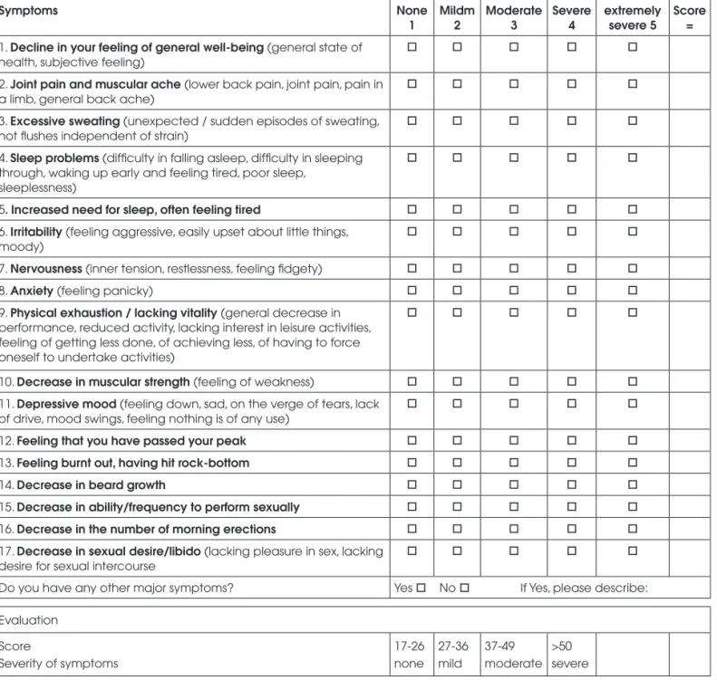
During the fi rst medical exam, the Aging Male’s Symptoms (AMS) scale questionnaire was used. This scale was created to screen for male hormone altera-tions, not to provide a defi nitive diagnosis (12). Each patient received a sheet containing the AMS question-naire (Figure 1), which a doctor read aloud to a group of four to six patients. Subjects recorded their respon-ses on a paper with a certain degree of privacy, such that each patient could not read the answers of the others.
Symptoms None
1
Mildm 2
Moderate 3
Severe 4
extremely severe 5
Score =
1. Decline in your feeling of general well-being (general state of health, subjective feeling)
2. Joint pain and muscular ache (lower back pain, joint pain, pain in a limb, general back ache)
3. Excessive sweating (unexpected / sudden episodes of sweating, hot fl ushes independent of strain)
4. Sleep problems (diffi culty in falling asleep, diffi culty in sleeping through, waking up early and feeling tired, poor sleep,
sleeplessness)
5. Increased need for sleep, often feeling tired 6. Irritability (feeling aggressive, easily upset about little things,
moody)
7. Nervousness (inner tension, restlessness, feeling fi dgety)
8. Anxiety (feeling panicky)
9. Physical exhaustion / lacking vitality (general decrease in performance, reduced activity, lacking interest in leisure activities, feeling of getting less done, of achieving less, of having to force oneself to undertake activities)
10. Decrease in muscular strength (feeling of weakness) 11. Depressive mood (feeling down, sad, on the verge of tears, lack
of drive, mood swings, feeling nothing is of any use)
12. Feeling that you have passed your peak
13. Feeling burnt out, having hit rock-bottom
14. Decrease in beard growth
15. Decrease in ability/frequency to perform sexually 16. Decrease in the number of morning erections 17. Decrease in sexual desire/libido (lacking pleasure in sex, lacking
desire for sexual intercourse
Do you have any other major symptoms? Yes No If Yes, please describe: Evaluation
Score
Severity of symptoms
17-26 none
27-36 mild
37-49 moderate
>50 severe
Figure 1. The Aging Males’ Symptoms (AMS) scale – Which of the following symptoms apply to you at this time? Please, mark the appropriate box for each symptom. For symptoms that do not apply, please mark “none” (12).
di-cop
yr
ight
© ABE&M todos os direitos reser
v
ados
vided by three sub-scales: sexual, (questions 12 to 14 and 17), psychological (questions 6 to 9 and 11) and somato-vegetative (questions 1 to 5, 10 and 13). The total score and not the sub-scales should be used, with a cut-off ≥ 27 points (13). Heinemann et al.demon-strated that the AMS scale has a sensitivity of 73.6% and specifi city of 70.4% for the improvement after andro-gen replacement therapy (14).
During the second medical exam, the St. Louis University questionnaire, also known as Androgen De-fi ciency of the Aging Male (ADAM), was given to the subjects (Figure 2) (9). Ten symptoms commonly ob-served in men with low bioavailable testosterone are assessed in the ADAM questionnaire. Affi rmative answers to questions 1 or 7 or to any other three ques-tions suggest hypogonadism. The ADAM questionnai-re demonstrated a sensitivity of 88% and a specifi city of 60% in men (9). This test has not yet been validated in Brazil. As an alternative way allowing us to use this questionnaire, the answers given by subjects from our sample, without osteoporosis or hypogonadism, were planned to be analyzed in separate. A non-statistical di-fference in testosterone values between normal subjects with positive and negative results for the ADAM ques-tionnaire could empower us to use it for the study.
The IIEF-5 questionnaire, an abbreviated version of the International Index of Erectile Function (IIEF) to evaluate erectile function (15), was also given during the second medical exam. The IIEF-5 has a maximum score of 25, and was developed by Rosen et al. (Figure 3)
(11). Scores above 21 are considered normal and wi-thout ED. Lower scores indicate ED of increasing seve-rity: mild ED (17 to 21), mild to moderate ED (12 to 16), moderate ED (8 to 11), and severe ED (5 to 7). Using a cut-off of < 22 points, the IIEF-5 demonstra-ted a sensitivity of 98% and a specifi city of 88% for the detection of the presence and severity of ED (11). This test was validated in Brazil by Rhoden et al. (16).
Laboratory measurements
Between the fi rst and second medical exams, blood samples were collected at the laboratory of the Institu-to Fernandes Figueira, between 8:30 and 10:00 in the morning, for measurement of: (i) general parameters used for detecting causes of osteoporosis; (ii) TSH for the exclusion of hypothyroidism as a clinical differential diagnosis of LOH; (iii) albumin. Part of the collected blood sample was separated and sent to the Diagnósticos 1. Do you have a decrease in libido (sex drive)?
2. Do you have a lack of energy?
3. Do you have a decrease in strength and/or endurance?
4. Have you lost height?
5. Have you noticed a decreased enjoyment of life? 6. Are you sad and/or grumpy?
7. Are your erections less strong?
8. Have you noted a recent deterioration in your ability to play sports?
9. Are you falling asleep after dinner?
10. Has there been a recent deterioration in your work performance?
Affi rmative answers to questions 1 or 7, or to any other three questions provide a positive result on the ADAM questionnaire
Figure 2. ADAM’s Questionnaire - Answer “Yes” or “No” (11).
1) How do you rate your confi dence that you could keep an erection?
(1) Very low (2) low (3) Moderate (4) High (5) Very high
(2)When you had erections with sexual stimulation, how often were your erections hard enough for
penetration (entering your partner)?
(1) Almost never or never (2) A few times (much less than half the time) (3) Most times (about half the time) (4) Much more than half the time (5) Almost always
3) During sexual intercourse, how often were you able to maintain your erection after you had penetrated (entered) your partner?
(1) Almost never or never (2) A few times (much less than half the time) (3) Most times (about half the time) (4) Much more than half the time (5) Almost always
4) During sexual intercourse, how diffi cult was it to maintain your erection to completion of intercourse?
(1) Extremely diffi cult (2) Very diffi cult (3) Diffi cult (4) Slightly diffi cult (5) Not diffi cult
5) When you attempted sexual intercourse, how often was it satisfactory for you?
(1) Almost never or never (2) A few times (much less than half the time) (3) Most times (about half the time) (4) Much more than half the time (5) Almost always Result: ≤ 21 has some degree of erectile dysfunction
cop
yr
ight
© ABE&M todos os direitos reser
v
ados
da América Laboratory, for measurement of total tes-tosterone (TT) and SHBG. Free testes-tosterone and BT were then calculated from the dosages of TT, SHBG and albumin with the formula of Vermeulen (17) through the website http://www.issam.ch/freetesto.htm.
Laboratory hypogonadism was defi ned as having cFT < 6.5 ng/dl in two samples collected at different times (5, 18, and 19).
During the second medical exam, the patient was informed about the results of his blood tests. If the cal-culated free testosterone (cFT) value was < 6.5 ng/dl, a second blood sample collection was scheduled to take place at least one month after the fi rst, in the laboratory of the Instituto Fernandes Figueira. The total testoste-rone and SHBG blood samples were once again sent to the Diagnósticos da América Laboratory for new calcu-lations of free and bioavailable testosterone. Follicle-stimulating hormone (FSH), Luteinizing hormone (LH), and prolactin were measured at the Instituto Fernandes Figueira in order to exclude secondary cau-ses of hypogonadism.
Total testosterone was measured by chemilumines-cence, using an automatic kit from Advia Centaur (Bayer Diagnostics), analytical sensitivity of 100 ng/dl, and reference values in men of 241 to 827 ng/dl. SHBG was also measured by chemiluminescence, using the Immulite 1000 kit (Siemens), which has an analyti-cal sensitivity of 0.2 nmol/l and reference values in men of 13 to 71 nmol/l. Albumin concentrations were determined by colorimetric spectrophotometry, with a kit from Targa BT Plus (Wiener Lab.), and reference
values of 3.5 to 5.5 g/dl. The FSH, LH and prolactin concentrations were measured by chemiluminescence, with the VIDAS kit (Biolab Merieux). The male refer-ence values used were: FSH (0.9-15 UI/L), LH (1.3-13 UI/L), prolactin (< 15 ng/ml).
The following factors were then analyzed: total AMS score, affi rmative responses to questions 1 or 7 or to any other three questions of the ADAM ques-tionnaire, and the total IIEF-5 score. Correlations were constructed between these scores and labora-tory hypogonadism using TT levels, as well as FT and BT levels, which were calculated by Vermeulen’s formula (17).
Statistical Analysis
The statistical analysis were done with the Student t test for continuous variables in the Graphpad Prism
version 3.00 for Windows program (Graphpad Softwa-re, San Diego, CA, USA). For categoric variables, the chi-square test by Mantel-Haenszel was performed in the Epi-info 6.04 program (CDC, Atlanta). The signi-fi cance level was set at p< 0.05.
Ethical aspects
The research objectives were explained to the men who attended the fi rst medical exam in small groups, and each subject signed an informed-consent document ap-proved under the Ethics Committee protocol.
RESULTS
Forty-one men (19% of the total sample) were diagno-sed with hypogonadism by laboratory criteria (2 sam-ples of cFT < 6.5 ng/dl): 28 in the osteoporosis group (25%) and 13 in the control group (12.2%). The OR for hypogonadism in osteoporotic men was 2.08 (IC95%:1.14-3.79). None of the subjects had hypogo-nadism secondary to hyperprolactinemia or pituitary insuffi ciency. Causes of osteoporosis that could mask the clinical diagnosis of hypogonadism were not de-monstrated by the general exams.
When the AMS and ADAM questionnaires were used as screening tools, the percentages of subjects who exhibited symptoms compatible with late-onset hypo-gonadism were determined to be 84% (179 patients) and 60% (128 patients), respectively
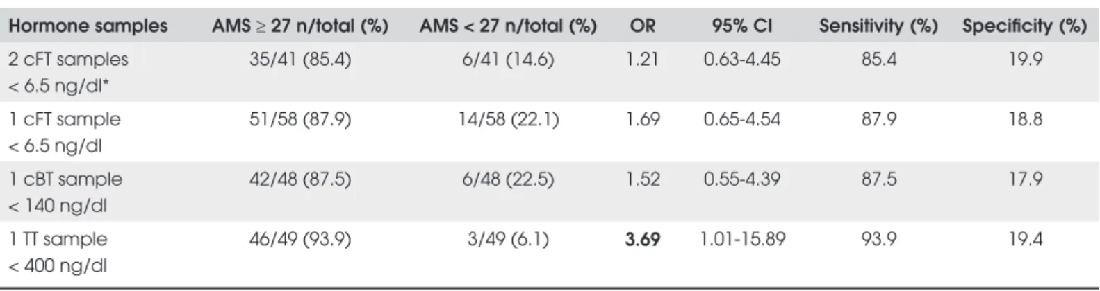
With regard to the AMS questionnaire, total scores equal to or greater than 27 showed an association only with TT levels below 400 ng/dl; no association was found with cBT or cFT (Table 1).
The answers given by 91 subjects from our sam-ple, with normal bone density and without hypogo-nadism, to the ADAM questionnaire were analyzed. The only criterion out of all those assessed that could be “validated” was the ADAM fi rst question, such that there was no statistical difference between the percentages of subjects with a positive and a negative result for this question (54,94 and 45,05% respectively, p=0.18).
cop
yr
ight
© ABE&M todos os direitos reser
v
ados
Table 1. Comparisons of AMS score ≥ 27 with measurements of calculated free, bioavailable and total testosterone.
Hormone samples AMS ≥ 27 n/total (%) AMS < 27 n/total (%) OR 95% CI Sensitivity (%) Specifi city (%)
2 cFT samples < 6.5 ng/dl*
35/41 (85.4) 6/41 (14.6) 1.21 0.63-4.45 85.4 19.9
1 cFT sample < 6.5 ng/dl
51/58 (87.9) 14/58 (22.1) 1.69 0.65-4.54 87.9 18.8
1 cBT sample < 140 ng/dl
42/48 (87.5) 6/48 (22.5) 1.52 0.55-4.39 87.5 17.9
1 TT sample < 400 ng/dl
46/49 (93.9) 3/49 (6.1) 3.69 1.01-15.89 93.9 19.4
Number of patients who answered the AMS questionnaire = 214. OR = odds ratio; CI = confi dence interval; TT = total testosterone; cFT = calculated free testosterone; cBT = calculated bioavailable testosterone; * Laboratory diagnosis of hypogonadism.
Table 2. Comparison of answer to ADAM question 1: “Do you have a decrease in libido (sex drive)?” and calculated free, bio-available and total testosterone levels.
Hormone samples Yes n/total (%) No n/total (%) OR 95% CI Sensitivity (%) Specifi city (%)
2 cFT samples < 6.5 ng/dl* 31/40 (77.5) 9/40 (22.5) 2.70 1.15-6.52 77.5 43.9 1 cFT sample < 6.5 ng/dl 43/57 (75.4) 14/57 (24.6) 2.57 1.24-5.37 75.4 45.5 1 cBT sample < 140 ng/dl 34/46 (73.9) 12/46 (26.1) 3.25 1.01-4.86 73.9 43.7 1 TT sample < 400 ng/dl 35/48 (72.9) 13/48 (27.1) 2.08 0.97-4.49 72.9 43.6
Number of patients who answered the AMS questionnaire = 213; OR = odds ratio; CI = confi dence interval; TT = total testosterone; cFT = calculated free testosterone; cBT = calculated bioavailable testosterone.
Table 3. Comparison of the prevalence of positive answers to question 1 of the ADAM questionnaire with cFT values.
ADAM 1 test
Positive Negative
cFT (ng/dl) n % n %
< 4 5 100 0 0
≥ 4 and < 5 10 76.9 3 23.1
≥ 5 and < 6.5 16 72.7 6 27.3
Total 31 77.5 9 22.5
cFT = calculated free testosterone.
men, an inverse relationship was observed between cFT values and ADAM 1 positive status (Table 3). No cor-relations were detected between question 7 or the total ADAM score and levels of TT, cFT or cBT.
The IIEF-5 questionnaire revealed no correlation between laboratory-defi ned hypogonadism and ED (p=0.269). There was no difference between TT, cFT
or cBT levels among men with IIEF-5 < 22 or ≥ 22. The correlations between IIEF-5 scores and hormone levels were very weak: TT: -0.04393, cFT: -0.05049 and cBT: -0.05077.
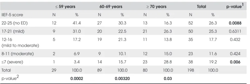
However, 147 men (74% of the subjects) complained of some degree of ED, expressed by IIEF-5 score < 22. Signifi cant differences were found between the percenta-ges of subjects without any ED seen in each age group: 41.4 % in the group of 50 to 59 years old, compared to 30.3% in the group of 60 to 69 years old and 16.3% in the group of 70 or older. Severe ED was more prevalent in men of 70 years or older (p=0.006), while in the 50 to 59 group, 31% had mild ED, 17.2% mild to moderate, 6.9% moderate and 3.4% severe. These differences were statisti-cally signifi cant (p=0.0002) (Table 4).
DISCUSSION
questio-cop
yr
ight
© ABE&M todos os direitos reser
v
ados
Table 4. Prevalence of erectile dysfunction (ED) sorted by age.
≤ 59 years 60-69 years ≥ 70 years Total p-value1
IIEF-5 score N % N % N % N %
22-25 (no ED) 12 41.4 27 30.3 13 16.3 52 26.3 0.0088
17-21 (mild) 9 31.0 20 22.5 21 26.3 50 25.3 0.6311
12-16
(mild to moderate)
5 17.2 19 21.3 11 13.8 35 17.7 0.432
8-11 (moderate) 2 6.9 9 10.1 12 15.0 23 11.6 0.424
≤7 (severe) 1 3.4 14 15.7 23 28.8 38 19.2 0.006
Total 29 100.0 89 100.0 80 100.0 198 100.0
p-value2 0.0002 0.00320 0.03
p-value1 = Refers to comparison of the degree of the percentage of erectile dysfunction in each age group; p-value2 = Refers to comparison of the distribution of the
degrees of erectile dysfunction in each age group.
nnaires, although the sensitivity and specifi city of these tests should be individualized for each culture, to be proven useful. The biochemical criteria requires two measurements of calculated free and bioavailable tes-tosterone, as demonstrated in our study.
Despite the great sensitivity for testosterone levels compatible with hypogonadism (88 to 94%), the AMS scale did not prove to be valid as an exclusive tool for screening, as shown by the low specifi city we detected (18 to 19%). Using a cutoff of score ≥ 27, the only correlation found was with TT < 400 ng/dl (OR: 3.69; 95% CI: 1.01-15.89), a level that is not diagnostic of hypogonadism, but rather indicates the need for a pre-liminary screening by which elder men have a greater chance of showing a free or bioavailable testosterone concentration below reference ranges (7). Our fi ndings agree with those in the literature (1). The AMS scale was designed to assess symptoms of aging (indepen-dent of those that are disease-related) between groups of males under different conditions, to evaluate the se-verity of symptoms over time, and to measure changes pre- and post androgen replacement therapy (10). When the AMS questionnaire was compared to ADAM for screening hypogonadism (22), a sensitivity of 83% was achieved but the specifi city was only 39%, using a very low threshold for AMS, a score of only 17 points. Regarding the ADAM scale, according to our at-tempt for an alternative validation, only ADAM 1 could be used as a screening tool for hypogonadism. In fact it was the only ADAM criterion that correlated with LOH laboratorial diagnosis in our sample. The fi rst
question in the ADAM questionnaire, “Do you have a decrease in libido (sex drive)?” was the most useful in screening for hypogonadism, with a sensitivity of 77.5% and a specifi city of 43.9%. The other criteria that responded to a positive ADAM rating, however, cor-related with neither the laboratory diagnosis of hypogonadism nor with the testosterone levels. Using ADAM 1, we observed sensitivity similar to that obtai-ned for any ADAM criteria in other populations (77.5%
versus 80-88%, respectively). The specifi city for ADAM
1 in our study (43.9%) was intermediate between that reported for any ADAM criteria in Canadian (60%), Bel-gian (21.6%) and Taiwan (20%) men (11, 23, and 24).
One way to explain the different results generally found regarding ADAM and AMS was demonstrated by Ichioka et al. (25) which examined 2211 men, 86% above the age of 40. In this study, hypogonadism bet-ter correlated with sexuality, as measured by the sexual sub-score, than with the total AMS score (25). Similar results were found in the Belgian study by T’sjoen et al. (26). Delhez et al. also described that the psychological response to decreased androgen manifests as minor de-pressive symptoms, which are not pathological (27). The AMS scale adds three different dimensions, there-fore diluting the results and generating a poor compa-rison with the ADAM questionnaire, which needs, in most cases, only one affi rmative answer to yield a posi-tive rating.
cop
yr
ight
© ABE&M todos os direitos reser
v
ados
the recommended laboratory criteria, only 41 (19%) of the subjects were judged hypogonadal after a second cFT exam, 28 in the osteoporosis group (25%) and 13 in the normal bone density group (12.2%). These num-bers show that both questionnaires lack specifi city. If applied to the general population, they would result in a large number of negative laboratory exams. Our fi n-dings support the Endocrine Society guidelines (5), in that questionnaires have not demonstrated to be a cost-effective strategy to detect LOH, and hormonal dosa-ges should rather be performed in high risk individuals, such as osteoporotic men.
If only a positive response to the fi rst ADAM ques-tion along with two samples of cFT < 6.5 ng/dl was considered, hypogonadism would be diagnosed only in 31 (15%) of the men. So, even the clinical criterion that demonstrated a better sensitivity and specifi city overall, was not able to identify all men with laboratory-defi ned hypogonadism (Table 2), because some men denied di-minished sexual desire and presented different com-plaints. This demonstrates the complexity of the clinical picture of late-onset hypogonadism and the necessity of using more than a single criterion or question for the clinical evaluation.
Sexuality combines libido and sexual function, and can be related to race, culture and religion. The IIEF-5 questionnaire, which was used to evaluate the associa-tion of sexual dysfuncassocia-tion and hypogonadism, did not demonstrate any correlation with laboratory hypogo-nadism or testosterone levels in our study. The IIEF-5 is an abridged fi ve item version of the fi fteen item In-ternational Index of Erectile Function (IIEF), an im-portant questionnaire for the evaluation of sexual erection that has an excellent sensitivity (98%) and good specifi city (88%) (11).
Although Chinese cohort studies have tried to esta-blish androgen defi ciency risks by using the IIEF-5 (28), in the majority of studies (29-31) including the Massachusetts Male Aging Study (MMAS), this corre-lation was not demonstrated (32). A possible explana-tion for the differences in these results was suggested in Mikhail’s review (33). A conclusive diagnosis of hypo-gonadism in men in these studies cannot be confi rmed, because many studies suffered from the lack of a second testosterone sample, which was collected in our study. His conclusion is that in most men, circulating levels of testosterone well below the normal range become es-sential for normal erections, and therefore only a pro-nounced hypogonadism would be evidenced by erectile
dysfunction (33). Additionally, ED becomes very common with advancing age, again as observed in our study. The great majority of hypothesized causes rela-te to vascular pathology, diaberela-tes, hyperrela-tension and the use of drugs (34), and only in some cases, hypo-gonadism (35).
CONCLUSIONS
The prevalence of late-onset hypogonadism was 25% in men with osteoporosis and 12.2% in men with normal bone density over 50 years of age (OR 2.08; IC95%: 1.14-3.79 for hypogonadism in osteoporotic men).
The symptom that best correlated with LOH was decreased sexual desire or libido (ADAM 1 positive). Among hypogonadal men, the lower the calculated free testosterone, the greater was the prevalence of a positi-ve answer to ADAM 1.
AMS scores ≥ 27 correlated only with TT levels less than 400 ng/dl.
Erectile dysfunction of any degree varied from 58 to 84%, and increased in both prevalence and se-verity with age. There were no correlations between testosterone levels or hypogonadism and erectile dysfunction.
Acknowledgements: The authors thank the Diagnósticos da América Laboratory and the Instituto Fernandes Figueira, which kindly performed the sample measurements. At the Instituto Fernandes Figueira, special thanks to Jânio Alves Cordeiro (Head of the Department of Clinical Pathology), Marcio Guil-herme de Souza (technician responsible for the hormonal analy-sis) and Rodrigo Batista dos Santos (technician responsible for the blood collection and sampling). In addition, we thank Mar-tha Suárez-Mutis, epidemiology doctor of IOC-Fiocruz, for the statistical analysis and Sergio Koifman, chief professor of the Post Graduate Program of Public Health and Environment of the National School of Public Health/Fiocruz for the sugges-tions during the study and for the review of this manuscript. There is no confl ict of interest of any author that could prejudi-ce the impartiality of the scientifi c work.
REFERENCES
1. Tsujimura A, Matsumiya K, Miyagawa Y, Takao T, Fujita K, Takada S, et al. Comparative study on evaluation methods for serum testosterone level for PADAM diagnosis. Int J Impot Res. 2005;17:259-63.
cop
yr
ight
© ABE&M todos os direitos reser
v
ados
3. Rosner W, Auchus RJ, Azziz R, Sluss PM, Raff H. Position state-ment: utility, limitations, and pitfalls in measuring testosterone: an endocrine society position statement. J Clin Endocrinol Me-tab. 2007;92:405-13.
4. Haren MT, Kim MJ, Tariq SH, Wittert GA, Morley JE. Andro-pause: a quality-of-life issue in older males. Med Clin North Am. 2006;90:1005-23.
5. Bhasin S, Cunningham GR, Hayes FJ, Matsumoto AM, Snyder PJ, Swerdloff RS, et al. Testosterone therapy in adult men with androgen defi ciency syndromes: an endocrine society clinical practice guideline. J Clin Endocrinol Metab. 2006;91:1995-2010. 6. Celec P, Ostatníková D. Testosterone: an overview; insights
into its physiology and clinical implications. Int J Endocrinol Metab. 2003;2:84-96.
7. Mooradian AD, Korenman SG. Management of the cardinal features of andropause. Am J Ther. 2006;13:145-60.
8. Nieschlag E, Swerdloff R, Behre HM, Gooren LJ, Kaufman JM, Legros JJ, et al. Investigation,treatment and monitoring of la-te-onset hypogonadism in males ISA, ISSAM, and EAU Re-commendations. Eur Urol. 2005;48:1-4.
9. Morley JE, Charlton E, Patrick P, Kaiser FE, Cadeau P, McCrea-dy D, et al. Validation of a screening questionnaire for andro-gen defi ciency in aging males. Metabolism. 2000;49:1239-42. 10. Heinemann LA, Saad F, Zimmermann T, Novak A, Myon E,
Ba-dia X, et al. The Aging Males’ Symptoms (AMS) scale: update and compilation of international versions. Health Qual Life Outcomes. 2003;1:15.
11. Rosen RC, Cappelleri JC, Smith MD, Lipsky J, Pena BM. Deve-lopment and evaluation of an abridged, 5-item version of the International Index of Erectile Function (IIEF-5) as a diagnostic
tool for erectile dysfunction. Int J Impot Res. 1999;11:319-26.
12. Heinemann LAJ, Zimmermann T, Vermeulen A, Thiel C. A new ‘Aging Males’ Symptoms’ (AMS) rating scale. Aging Male. 1999;2:105-14.
13. Moore C, Huebler D, Zimmermann T, Heinemann LA, Saad F, Thai DM. The Aging Males’ Symptoms Scale (AMS) as out-come measure for treatment of androgen defi ciency. Eur Urol. 2004;46:80-7.
14. Heinemann LA, Moore C, Dinger JC, Stoehr D. Sensitivity as outcome measure of androgen replacement: the AMS scale. Health Qual Life Outcomes. 2006;30;4:23.
15. Rosen RC, Riley A, Wagner G, Osterloh IH, Kirkpatrick J, Mish-ra A. The international index of erectile function (IIEF): a multi-dimensional scale for assessment of erectile dysfunction. Urology. 1997;49:822-30.
16. Rhoden EL, Lemos RR, Riedner CE, Telöken C, Souto CAV. Va-lidade do índice internacional de função erétil (IIEF) e IIEF-5 simplifi cado na avaliação da função eretiva. Braz J Urol. 2001;27 Suppl 1:18.
17. Vermeulen A, Verdonck L, Kaufman JM. A critical evaluation of simple methods for the estimation of free testosterone in se-rum. J Clin Endocrinol Metab. 1999;84:3666-72.
18. Kaufman JM, Vermeulen A. The decline of androgen levels in elderly men and its clinical and therapeutic implications. En-docr Rev. 2005;26:833-76.
19. Kelleher S, Conway AJ, Handelsman DJ. Blood testosterone threshold for androgen defi ciency symptoms. J Clin Endocri-nol Metab. 2004;89:3813-17.
20. Matsumoto AM. Andropause: clinical implications of the decli-ne in serum testosterodecli-ne levels with aging in men. J Gerontol A Biol Sci Med Sci. 2002;57:M76-99.
21. Morley JE. The need for a men’s health initiative. J Gerontol A Biol Sci Med Sci. 2003;58:614-7.
22. Morley JE, Perry HM, Kevorkian RT, Patrick P. Comparison of screening questionnaires for the diagnosis of hypogonadism. Maturitas. 2006;53:424-9.
23. Tancredi A, Reginster JY, Schleich F, Pire G, Maassen P, Luyckx F, et al. Interest of the androgen defi ciency in aging males (ADAM) questionnaire for the identifi cation of hypogonadism in elderly community-dwelling male volunteers. Eur J Endo-crinol. 2004;151:355-60.
24. Lin YC, Hwang TI, Chiang HS, Yang CR, Wu HC, Wu TL, et al. Correlations of androgen defi ciency with clinical symptoms in Taiwanese males(Abstract). Int J Impot Res. 2006;18:343-7. 25. Ichioka K, Nishiyama H, Yoshimura K, Itoh N, Okubo K, Terai A.
Aging males’ symptoms scale in japanese men attending a multiphasic health screening clinic. Urology. 2006;67:589-93. 26. T’Sjoen G, Goemaere S, De Meyere M, Kaufman JM.
Percep-tion of males’ aging symptoms, health and well-being in el-derly community-dwelling men is not related to circulating androgen levels. Psychoneuroendocrinology. 2004;29:201-14. 27. Delhez M, Hansenne M, Legros J-J. Andropause and psycho-pathology: minor symptoms rather than pathological ones. Psychoneuroendocrinology. 2003;28:863-74.
28. Wong SYS, Chanb DCC, Hong A, Wooa J. Prevalence of and risk factors for androgen defi ciency in middle-aged men in Hong Kong. Metabolism. 2006;55:1488-94.
29. Rhoden EL, Teloken C, Sogari PR, Souto CAV. The relationship of serum testosterone to erectile function in normal aging men. J Urol. 2002;167:1745-48.
30. Christ-Crain M, Mueller B, Gasser TC, Kraenzlin M, Trummler M, Huber P, et al. Is there a clinical relevance of partial andro-gen defi ciency in the aging male? J Urol. 2004;172:624-27. 31. Bhasin S, Woodhouse L, Casaburi R, Singh AB, Bhasin D,
Ber-man N, et al. Testosterone dose-response relationships in he-althy young men. Am J Physiol Endocrinol Metab. 2001;281: E1172-81.
32. Feldman HA, Goldstein I, Hatzichristou DG, Krane RJ, McKin-lay JB. Impotence and its medical and psychological correla-tes: results of the Massachusetts Male Aging Study. J Urol. 1994;151:54-61.
33. Mikhail N. Does testosterone have a role in erectile function? Am J Med. 2006;119:373-82.
34. Fine SR. Erectile dysfunction and comorbid diseases, andro-gen defi ciency, and diminished libido in men. J Am Osteopath Assoc. 2004;104 Suppl 1:S9-15.
35. Carbone DJ, Seftel AD. Erectile dysfunction. Diagnosis and treatment in older men. Geriatrics. 2002;57(9):18-24.
Correspondence to:
Daniel Jorge de Castro Braga Avenida Epitácio Pessoa 3540, ap. 503. 22471-003 Rio de Janeiro RJ