Article
Printed in Brazil - ©2017 Sociedade Brasileira de Química0103 - 5053 $6.00+0.00
*e-mail: [email protected]
Production, Purification and Physicochemical Properties of an
Exo-Polygalacturonase from
Aspergillus niger
SW06
Yu-ping Ma,a Hui Hao,a Zhi-fei Chen,a Zhi-wei Zhao,a Shun-hui Chen,b Si-wen Sunc and
Chun-ping Xu*,c
aTechnical Center of China Tobacco Henan Industrial Co. Ltd, 450016 Zhengzhou, China
bHennan Cigarette Industry Tobacco Sheet Co. Ltd., 461100 Xuchang, China
cCollege of Food and Biology Engineering, Zhengzhou University of Light Industry,
450002 Zhengzhou, China
In this study, exo-polygalacturonase (exo-PG) production from Aspergillus niger SW06 was optimized by central composition design and high amount of 21.51 units mL-1 could be achieved
in optimizing growth conditions. Both gel filtration and ion exchange chromatography revealed a single exo-PG activity peak, and sodium dodecyl sulfate polyacrylamide gel electrophoresis (SDS-PAGE) analysis of the purified protein showed a single band with a molecular mass of 66.2 kDa. The purified enzyme exhibited maximal activity in the presence of 1% citrus pectin at the temperature of 55 °C and pH of 5.0. The enzyme was stable within the pH range of 3.0-5.0 and below 60 °C. The Michaelis constant (Km) and maximum velocity (Vmax) of the enzyme was
found to be 0.58 mg mL-1 and 20.66 µmol (mL min)-1, respectively. The thermostable and acidic
nature for the activity of this exo-PG make it possible to have wide range of industrial applications.
Keywords: Aspergillus niger, exo-polygalacturonase, purification, characterization
Introduction
Pectinases are a heterogeneous group of related enzymes which catalyze the degradation of pectic substances, present mostly in the plant cell walls.1
Polygalacturonases (PGs) (EC 3.2.1.67) are the pectinolytic enzymes that catalyze the hydrolytic cleavage of the α-1,4-glycosidic bonds that link galacturonic acid
residues.2,3 PGs have been classified according to their
substrate specificity and the position of the bonds which they hydrolyze. Endo-PG (E.C. 3.2.1.15) was defined as randomly hydrolyzing the α-1,4-glycosidic bonds in the
polymer, whereas exo-PG acts sequentially from the non-reducing end.3 PGs are widely distributed in the microbial
sources including fungi, bacteria and many types of yeast and also found in higher plants and some plant parasitic nematodes.4 PGs are used in several processes, such as
paper and pulp industry, fruit juice and wine clarification, tea and coffee fermentation, degumming and retting of plant fibers, and oil extraction, etc.5,6
Although several Aspergillus species organisms
producing pectinases have been reported and are used in industrial processes in crude form,7,8 their selection
of potential isolates still remains a tedious task, especially when physiologically potential strains are obtained to achieve maximum yield.9 Their purification
and knowledge of the biochemical characteristics of these enzymes are important for the understanding of their structure and functional mechanism of action and thermostability. It has been reported that fungal PGs generally are monomeric proteins with a carbohydrate content of 5-85% and molecular masses in a range from 20 to 95 kDa.2,6,10,11
Experimental
Microorganism and growth conditions
The exo-PG producing fungus Aspergillus niger SW06 was isolated from tobacco field in Xuchang, P. R. China, and maintained on the stock medium containing 30 g L-1
glucose, 3 g L-1 peptone, 5 g L-1 NaCl, 5 g L-1 citrus pectin,
25 g L-1 agar (not adjusted) at 4 °C. The liquid culture of
mycelia was initiated by transferring the fungal mycelia from the stock culture on a Petri dish into the seed culture medium. The seed culture was propagated in a 250 mL Erlenmeyer flask containing 100 mL of liquid medium (10 g L-1 yeast extract, 5 g L-1 citrus pectin, FeSO
4 0.1 g L-1,
MgSO4 0.5 g L-1, KH2PO4 1 g L-1, and pH 6.0) at 28 °C
on a shaking incubator at 160 rpm for 48 h. Exo-PG was produced with the inoculation of 4% (v/v) of the seed culture by submerged fermentation in a stirred tank bioreactor (Infors, Switzerland, 3.5 L working volume). The fermentations were performed under the following conditions: temperature, 28 °C; aeration, 2 vvm; agitation speed, 160 rpm. All experiments were performed in triplicate to ensure the trends observed were reproducible.
Confirmation of enzyme type
After 72 hours of fermentation, the culture broth was centrifuged at 9,000 × g for 15 min, and the resulting supernatant was filtered through a membrane filter (0.45 µm, Millipore). The type of extracelluar PG was determined using following assays.
Enzyme assay
Enzymatic activities of all the samples were expressed in units of activity per liter (U mL−1). Endo-PG activity
was measured viscosimetrically by mixing 5.5 mL of 1% (m/v) citric pectin in 0.2 mol L-1 acetate buffer at pH 5.0
(supplemented with 1 mmol L-1 EDTA), with 250 µL of
the crude enzyme. The reaction was incubated for 30 min at 45 °C and then cooled in an ice bath. A viscosimetric unit (U) was defined as the enzyme quantity required to decrease the initial viscosity per minute by 50% under the conditions previously described.12 Exo-PG activity
was assayed by measuring the release of reducing groups from citric pectin using the 3,5-dinitrosalicylic acid (DNS) assay.13,14 The reaction mixture containing 0.5 mL 1% citric
pectin in 0.2 mol L-1 acetate buffer, pH 5.0 and 0.5 mL of
enzymatic extract was incubated at 45 °C for 30 min. One unit of enzymatic activity (U) was defined as the amount of enzyme releasing 1 µmol of galacturonic acid per minute.
Optimization procedure
Once the variables having the greatest influence on the responses were identified, a central composite design was used to optimize the levels of these variables. For the three factors, this design was made up of a central composite design with four cube points; that is, a point for one factor having an axial distance from the centre (that is, level 0) of ±α, while the other factor is at level 0
(Table 1). The axial distance α was chosen to be 1.682
to make this design orthogonal. So the coded values –α
and ±α are −1.682 and 1.682, respectively. The computer
software DESIGN EXPERT vision 8.05b (Stat-Ease Inc., Minneapolis, USA) was used to estimate the responses of the dependent variables.15 This approach has been
successfully applied to optimize medium composition, condition of enzyme reaction, and extraction conditions for bioactive compounds.16,17 In this study, a central
composition design was applied to optimize medium condition of exo-PG by A. niger in flask culture. As seen from Table 2, the experiment was carried out with 3 factors with 5 levels based on preliminary single experimental results. The exo-PG yield was chosen as the response.
Enzyme purification procedure
After 72 hours of fermentation, the culture broth was centrifuged at 9,000 × g for 15 min, and the resulting supernatant was filtered through a membrane filter (0.45 µm, Millipore). The culture filtrate was precipitated by ammonium sulfate (20-100%) and the mixture was stirred for 2 h, and centrifuged at 15,000 × g for 30 min. The ammonium sulfate fraction was dialyzed against Tris-HCl buffer (50 mmol L-1, pH 6.5) and directly
loaded on a Sepharose CL-6B gel filtration column (2.5 × 60 cm) equilibrated with 13 mmol L-1 Na
2HPO4
-citric acid buffer (pH 5.0). Protein fractions collected from the column, corresponding to the protein peak, were pooled, concentrated and further applied to the DEAE-Sepharose FF based anion exchangers column equilibrated with 20 mmol L-1 Na
2HPO4-citric acid buffer
(pH 6.5).18 Fractions of 4 mL were collected and assayed
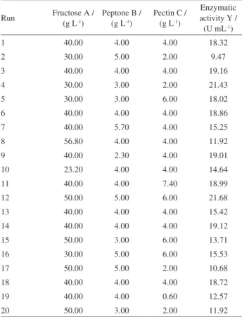
Table 1. Experimental range and levels of the independent process variables according to the central composite design
Variable Level of variable / (g L
-1)
−1.682 −1 0 −1 1.682
Fructose 23.20 30.00 40.00 50.00 56.80
Peptone 2.30 3.00 4.00 5.00 5.70
for exo-PG activity. The objective of this procedure was to purify the exo-PG present in the crude enzyme solution. The protein fraction with exo-PG activity was pooled, desalted overnight by dialysis at 4°C, freeze-dried and kept refrigerated until use.
Analytical electrophoresis
The relative molecular weight of the purified enzyme was determined by sodium dodecyl sulfate polyacrylamide gel electrophoresis (SDS-PAGE) in a Mini Protean II apparatus (10 × 8 cm) (Biorad). Electrophoresis was carried out in a vertical slab gel apparatus (Beijing Liuyi Instrument Factory, DYCZ-24DN) with a 5% (m/v) polyacrylamide stacking gel and 12% (m/v) resolving gel in Tris/glycine buffer (pH 8.3). Molecular mass of purified exo-PG was estimated using the Sigma molecular weight marker MP102 (14.4-94.0 kDa) in a parallel lane.19 The proteins were
visualized by silver staining.
Protein estimation
The protein concentration was determined in the concentration ranges of 1-10 and 10-100 µg mL−1 by the
Bradford microassay, using bovine serum albumin (BSA) as standard.20
Properties of purified enzyme
All enzyme catalytic properties were assayed with 1% citrus pectin [degree of esterification (D.E.) 67-70%] as substrate using the procedure for enzyme activity determination described above and carried out with three replicates. Exo-PG activity was assayed as a function of pH ranging from 3.0 to 8.0 in Na2HPO4-citric acid buffer
at 45°C, and temperature, in Na2HPO4-citric acid buffer
at the pH optimum, incubated at different temperatures between 35and 60 °C.
The thermal stability was investigated by remeasuring the activity of the purified enzyme solution after it had been kept for 2 h, in the absence of substrate, at different temperature in the range 30-60 °C. In these tests, the initial and final exo-PG activities were determined at optimum pH and temperature. The pH stability of the purified enzyme was evaluated by dispersing (1:1, v/v) enzyme solution in Na2HPO4-citric acid
buffer (pH 3.0-8.0) and maintaining these solutions at 45 °C for 4 hours. An aliquot was taken to determine the remaining activity at the optimum pH and temperature.
The Michaelis constant (Km) and maximum
velocity (Vmax) values of the enzyme were determined by
measuring the reaction velocity measured with 67-70% D.E. citrus pectin (Sigma) as substrate, at concentrations between 2.0 and 40.0 mg mL-1 at optimum pH and
temperature. According to the Michaelis-Menten enzyme kinetics, the reciprocal of the reaction velocity (1 / V) was plotted against the reciprocal of the substrate concentration (1 / [S]) to determine the Km and Vmax values by the
Lineweaver-Burke plot. The results were plotted with Excel.
Results and Discussion
Confirmation of enzyme type
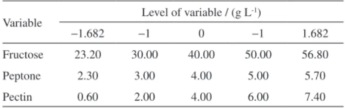
Cell-free supernatant of Aspergillus niger SW06 was found to be predominantly exo-PG activity with fewer amounts of endo-PG (Figure 1).
Production of exo-PG in flask culture
In general, enzyme production is influenced by the composition of the medium, in particular the carbon and nitrogen sources.21 Table 2 summarized the central
composite experimental plan along with the experimental responses for each individual experiment. By applying
Table 2. Central composite design of variables with exo-PG production as the response after 48 hours of incubation in flask culture
Run Fructose A / (g L-1)
Peptone B / (g L-1)
Pectin C / (g L-1)
Enzymatic activity Y / (U mL-1)
1 40.00 4.00 4.00 18.32
2 30.00 5.00 2.00 9.47
3 40.00 4.00 4.00 19.16
4 30.00 3.00 2.00 21.43
5 30.00 3.00 6.00 18.02
6 40.00 4.00 4.00 18.86
7 40.00 5.70 4.00 15.25
8 56.80 4.00 4.00 11.92
9 40.00 2.30 4.00 19.01
10 23.20 4.00 4.00 14.64
11 40.00 4.00 7.40 18.99
12 50.00 5.00 6.00 21.68
13 40.00 4.00 4.00 15.42
14 40.00 4.00 4.00 19.12
15 50.00 3.00 6.00 13.71
16 30.00 5.00 6.00 15.53
17 50.00 5.00 2.00 10.68
18 40.00 4.00 4.00 18.72
19 40.00 4.00 0.60 12.57
multiple regression analysis on the experimental data, the following second order polynomial equation was found to represent the exo-PG production adequately:
Y = 18.26 − 0.81A − 1.03B + 1.92C + 2.65AB +
1.27AC + 2.34BC − 1.75A2− 0.38B2− 0.86C2 (1)
where Y represents the response variable. A, B and C represent the coded values of fructose, peptone and pectin, respectively. The regression equation was optimized by the DESIGN EXPERT to get the optimum values. The optimal values of the test variables, in uncoded levels are as follows: fructose = 48.9, peptone = 5.0 and pectin = 6.0.
For testing the goodness of fit of the model, the multiple coefficient of correlation (R) and the determination coefficient (R2) were evaluated. The coefficient of
determination, R2, indicates that about 93.3% of the total
variability in the response could be explained by the model. The value of R is 0.9965, which indicates that the regression model explained the reaction well. The analysis of variance (ANOVA) of the quadratic regression model demonstrated that equation 1 is highly statistically significant model of exo-PG response, as was evident from the Fisher’s test with a very low probability value [(p model > F) = 0.0001]. The model F value of 25.68 implied that the model was significant. There was only a 0.01% chance that the “model F value” could occur because of noise.
In order to confirm the optimization results, the
suggested medium components were confirmed in triplicate. The 21.63 U mL-1 exo-PG was maximally
obtained under the optimum conditions just described, where the corresponding experimental response was 21.51 ± 0.11. This implied that the selected conditions were really the most suitable. Gattás et al.22 found that the
optimal pectin level was 2.0% (m/v) for exo-PG production by Aspergillus sp. CC1 in submerged culture, which suggests that the level of substance requirement for exo-PG production depends on the nature of the specific strain, though they belong to the same species (i.e., Aspergillus).
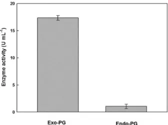
Purification of exo-PG
The exo-PG was purified through Sepharose CL-6B column and DEAE-Sepharose FFcolumn. As showed in Table 3, total protein content of the sample decreased from 91.2 mg in the crude sample to 1.96 mg in the final step. The specific activity had a marked increase in every step, i.e., from 140.35 U mg-1 in the crude sample to 382.65 U mg-1
in the final chromatographic step. Total enzyme activity in the crude sample was 12800 U. The yield of the enzyme was 5.9% with respect to the starting material. The enzyme solution separated on a Sepharose CL-6B column, afforded one single peak of exo-PG activity suggesting one fraction (Figure 2a). Exo-PG was collected for further purification to confirm its purity. When the exo-PG solution was concentrated and loaded on a DEAE-Sepharose FFcolumn, still only one PG peak was eluted (Figure 2b). The results were different with the reports of Kant et al.,9 who observed
two subunits of PG separated from A. niger MTCC 3323 by Sephacryl S-200 gel-filtration chromatography.
Characterization of exo-PG
The homogeneity of the purified exo-PG was demonstrated by the presence of one single protein band on polyacrylamide gel and its molar mass was estimated to be 66.2 kDa as single subunit (Figure 3). This observation was in the range reported for exo-PGs from several fungi, which have molecular weight ranging from 20 to 95 kDa.21,23
The molecular mass of the PG from A. niger NRRL3 was 32 kDa as estimated by gel filtration and sodium dodecyl
Figure 1. Comparative enzyme activity of exo-PG and endo-PG.
Table 3. Purification of exo-PG produced by A. niger SW06
Purification step Total activity / U Total protein / mg Specific activity /
(U mg-1) Purification fold Yield / %
Crude filtrate 12800 91.20 140.35 1.00 100.0
Sepharose CL-6B 1820 8.34 218.23 1.56 14.2
sulfate-polyacrylamide gel electrophoresis.24 In contrast,
a heterodimer of 34 and 69 kDa subunit was detected for PG from A. niger MTCC 3323,9 and two exo-PGs 1 and 2
from another A. niger had the molecular masses of 82 and 56 kDa, respectively.25
The effect of pH on the A. niger exo-PG activity toward polygalacturonic acid was examined at 45 °C. As shown in Figure 4a, the enzyme showed hydrolase activity from pH 3.0 to 8.0, and maximum activity (19.76 U mL-1) at
pH 5.0. The same pH optimum was reported for PGs from Aspergillus niger.26 The effect of pH on the stability
of A. niger exo-PG was investigated by incubating the enzyme at 45 °C at different pH’s for 4 h. The results showed that the enzyme was the most stable in the pH of 5.0, with 90-100% of the full activity in a broader pH range of 3.0-5.0 (Figure 4b). The results are very close to the results reported by Mallu et al.27 for A. niveus exo-PG.
They reported that exo-PG showed pH stability between 3.0 and 5.0. Sakamoto et al.25 reported that the optimum
activities occurred at pH 3.4-3.8 for exo-PG1 and 3.4-4.2 for exo-PG2 from another A. niger, respectively. In contrast,
Figure 2. Elution of exo-PG from Sepharose CL-6B gel filtration column previously equilibrated with 13 mmol L-1 Na
2HPO4-citric acid buffer
(pH 5.0) (a) and DEAE-Sepharose FF column based anion exchangers column equilibrated with 20 mmol L-1 Na
2HPO4-citric acid buffer
(pH 6.5) (b). Protein (); exo-PG activity ().
Figure 3. SDS-PAGE of standard proteins (a); crude exo-PG (b) and purified exo-PG (c) from DEAE-Sepharose FF chromatography.
the PG from A. kawachii had an optimum activity at low pH (2.0-3.0).28 The highest pH optimum value of pH 10.0
was observed for PG from Bacillus sp. MG-cp-2.29
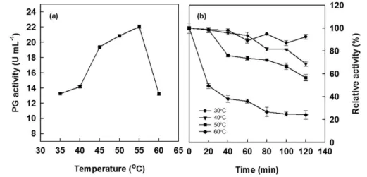
With respect to temperature, the purified exo-PG exhibited optimum activity of 55 °C as depicted in Figure 5a. Earlier similar results were obtained that the temperature optima for PGs from other A. niger PGs were around 37 and 45 °C.9,24,30 The effect of temperature on thermal stability of
A. niger exo-PG was investigated by incubation the enzyme for 2 h in 13 mmol L-1 Na
2HPO4-citric acid buffer, pH 5.0
at different temperatures ranging from 30 to 60 °C prior to substrate addition (Figure 5b). In the absence of substrate for 1 h, exo-PG showed 36-89% of the original activity at 30-60 °C. After 2 h, exo-PG showed 57-88% of the original activity at 30-50 °C, while at 60 °C, the enzyme lost 76% of its initial activity. Kant et al.9 observed that at 45 °C the
relative activity of A. niger PG after 30 min of incubation was to be 45.23%, i.e., it lost more than half of its activity.
The kinetic parameters of A. niger exo-PG affinity for citrus pectin in a range of 2.0 and 40.0 mg mL-1 at pH 5.0
and 45 °C were determined by a typical double reciprocal Lineweaver-Burk plot (Figure 6). According to the Figure 5, the Km and Vmax for the enzyme were calculated
as 0.58 mg mL-1 and 20.66 µmol (mL min)-1, respectively.
The Km values of A. niger exo-PG in this study were lower
than Km (2.5 mg mL-1) of PG from another A. niger.31
The reason for low Km may be due to the high affinity of
A. niger exo-PG using citrus pectin as substance.24 The V max
of A. niger exo-PG was in the range of Vmax, i.e., 13.0 to
2600 µmol (mL min)-1 from above three organisms.
Conclusion
In the present study, a statistical method, central composition design was applied to the optimization of medium composition for maximum exo-PG production
from A. niger SW06. This enzyme kept the stability in a pH range of 3.0-5.0 and at a temperature range of 30-60 °C. To our knowledge, this exo-PG from A. niger SW06 is more thermostable and acid-resisting, comparing the PGs from other several fungi. The thermostable and acidic nature for activity makes it possible to have wide range of industrial applications. Further works on scale-up fermentation optimization in bioreactor and industrial application are in progress in our laboratory.
Acknowledgments
This work was supported by the National Science Foundation of China (Grant No. B060806).
References
1. Ward, O. P.; Moo-Young, M.; Crit. Rev. Biotechnol.1989, 8, 237.
2. Niture, S. K.; Biologia2008, 63, 1.
Figure 5. Effect of temperature on exo-PG activity (a) and exo-PG stability (b).
3. Joslyn, N. A.; Mist, S.; Lambart, E.; Food Technol.1952, 6, 133.
4. Sakai, T.; Sakamoto, T.; Hallaert, J.; Vandamme, E. J.; Adv. Appl. Microbiol.1993, 39, 213.
5. Dey, T. B.; Adak, S.; Bhattacharya, P.; Banerjee, R.; LWT -- Food Sci. Technol.2014, 59, 591.
6. Mathew, A.; Eldo, A. N.; Molly, A. G.; J. Ind. Microbiol. Biotechnol.2008, 35, 1001.
7. Murad, H. A.; Azzaz, H. H.; Res. Microbiol.2011, 6, 246. 8. Hoondal, G. S.; Tiwari, R. P.; Tewari, R.; Dahiya, N.; Beg, Q.
K.; Appl. Microbiol. Biotechnol. 2002, 59, 409.
9. Kant, S.; Vohra, A.; Gupta, R.; Protein Expression Purif. 2013,
87, 11.
10. Borin, M. D. F.; Said, S.; Fonseca, M. J. V.; J. Agric. Food Chem. 1996, 44, 1616.
11. Niture, S. K.; Pant, A.; Microbiol. Res.2004, 159, 305. 12. Tuttobello, B. R.; Mill, P. J.; Biochem. J. 1961, 79, 51. 13. Miller, G. L.; Anal. Bioanal. Chem.1959, 31, 426.
14. Martins, E. S.; Silva, D.; Da Silva, R.; Gomes, E.; Process Biochem.2002, 37, 949.
15. Wang, H.; Zhang, X.; Dong, P.; Luo, Y.; Cheng, F.; Int. J. Pharmacol. 2013, 9, 288.
16. Hong, Z.; Lin, Z.; Liu, Y.; Tan, G.; Lou, Z.; Zhu, Z.; Chai, Y.; Fan, G.; Zhang, J.; Zhang, L.; J. Chromatogr. A2012, 1254, 14. 17. Zhao, Q.; Kennedy, J. F.; Wang, X.; Yuan, X.; Zhao, B.; Peng,
Y.; Huang, Y.; Int. J. Biol. Macromol.2011, 49, 181. 18. Mohamed, S. A.; Farid, N. M.; Hossiny, E. N.; Bassuiny, R. I.;
J. Biotechnol.2006, 127, 54.
19. Laemmli, U.; Nature1970, 227, 680.
20. Bradford, M. M.; Anal. Biochem.1976, 72, 248.
21. Gomes, E.; Leite, S. R. R.; Da Silva, R.; Silva, D.; Int. J. Food Microbiol.2009, 10, 1155.
22. Gattás, E. A. L.; Bueno, M. R.; Ribeiro, M. H. L.; Eur. Food Res. Technol. 2009, 229, 923.
23. Silva, D.; Martins, E. S.; Leite, R. S. R.; Da Silva, R.; Ferreira, V.; Gomes, E.; Process Biochem.2007, 42, 1237.
24. Fahmy, A. S.; El-beih, F. M.; Mohamed, S. A.; Abdel-Gany, S. S.; Abd-Elbaky, E. A.; Appl. Biochem. Biotechnol. 2008,
149, 205.
25. Sakamoto, T.; Bonnin, E.; Quemener, B.; Thibault, J. F.;
Biochim. Biophys. Acta, Gen. Subj.2002, 1572, 10.
26. Behere, A.; Satyanarayan, V.; Padwal-Desai, S. R.; Enzyme Microb. Technol.1993, 15, 158.
27. Mallu, A.; Damasio, A. R.; Da Silva, T. M.; Jorge, J. A.; Terenzi, H. F.; Mde, L.; Enzyme Res.2011, 10, 4061.
28. Contreas-Esquivel, J. C.; Voget, C. E.; J. Biotechnol, 2004, 110, 21.
29. Kapoor, M.; Beg, Q. K.; Bhushan, B.; Dadhich, K. S.; Hoondal, G. S.; Process Biochem.2000, 36, 467.
30. Gomes, J.; Zeni, J.; Cence, K.; Toniazzo, G.; Treichel, H.; Food Bioprod. Process.2011, 89, 281.
31. Parenicova, L.; Benen, J. A. E.; Kester, H. C. M.; Visser, J.; Eur. J. Biochem.1998, 251, 72.
Submitted: April 14, 2016