Article
0103 - 5053 $6.00+0.00*e-mail: [email protected]
A New Bianthron Glycoside as Inhibitor of
Trypanosoma cruzi
Glyceraldehyde
3-Phosphate Dehydrogenase Activity
Edângelo M. S. de Macedo,a Helton J. Wiggers,b Maria G. V. Silva,*,a,b Raimundo Braz-Filho,c
Adriano D. Andricopulod and Carlos A. Montanarib
aDepartamento de Química Analítica e Físico-Química, Departamento de Química Orgânica e Inorgânica,
Universidade Federal do Ceará, 60455-970 Fortaleza-CE, Brazil
bInstituto de Química de São Carlos and dInstituto de Física de São Carlos, Universidade de São Paulo,
13560-970 São Carlos-SP, Brazil
cSetor de Química de Produtos Naturais, CCT, Universidade Estadual do Norte Fluminense,
28013-602 Campos dos Goytacazes-RJ, Brazil
A investigação fitoquímica do extrato etanólico do caule de Senna martiana Benth. (Leguminoseae), espécie nativa do nordeste do Brasil, permitiu o isolamento e caracterização espectroscópica de uma biantrona inédita denominada martianina (1), 10,10’-H-bicrisofanol-10-ox-10,10’-biglucosil. Sua identificação estrutural inequívoca foi estabelecida por HRMS, IV e RMN de 1H e 13C uni e bi-dimensional. A atividade tripanocida de martianina foi avaliada por seu comportamento frente à enzima GAPDHTc através da obtenção de sua constante de inibição (Ki) determinada por titulação calorimétrica isotérmica (27,3 ± 2,47 µmol L-1). Estes resultados sugerem que esta classe de molécula tem potencial no desenvolvimento de novos fármacos a serem utilizados contra a doença de Chagas.
A phytochemical investigation of the ethanolic extract of stalks of Senna martiana Benth. (Leguminoseae), native specie of northeast Brazil, resulted in the isolation and spectroscopic characterization of a new bianthrone glycoside, martianine 1 (10,10’-il-chrysophanol-10-oxi-10,10’-bi-glucosyl). Its identification was established by HRMS, IR and 2D NMR experiments. The evaluation of martianine trypanocidal activity was carried out against gliceraldehyde 3-phosphate dehydrogenase enzyme from Trypanosoma cruzi. Its inhibitory constant (Ki) is in the low micromolar concentration and it was determined by isothermal titration calorimetry to be 27.3 ±2.47 µmol L-1. The non-competitive mechanism is asserted to be putative of the mode of action martianine displays against T. cruzi GAPDH. Results show that martianine has a great potential to become new lead molecule by inhibiting this key enzyme and for the development of new drugs against Chagas disease.
Keywords:Senna martiana, Leguminoseae, bianthrone, anthraquinone, Trypanosoma cruzi,
isothermal titration calorimetry
Introduction
Senna martiana (Benth) I&B (Leguminoseae), is a native specie of northeast Brazil and it is a shrub presenting abundant foliages and exuberant yellow flowers. This plant is locally known as “canafístula brava” and “caixão de canafístula”.1 Previous chemical studies do not exist
about S. martiana but others species of the genus Senna, have shown to be in great occurrence of anthraquinone
compounds, isolated from several parts of the plant with less incidence in the leaves.2,3 The anthraquinone
compounds are distributed widely in the nature, mainly as being responsible for the pigmentation and are found in fungi, superior plants and lichens.4 There are reports
of several biological activities attributed to the presence of anthraquinones such as antioxidant, laxative, antiviral, antimicrobial and trypanocidal.5-9
countries of Latin America and in the US 100,000-675,000 immigrants from Latin America are infected with T. cruzi. It is one of the most neglected diseases in the world and lack of financial support is one of the major characteristic avoiding the fulfillment of proper investigations. At present, the available drugs nifurtimox (Lampit™, Bayer) and benznidazole (Rochagan™, Radanil™, Roche) have strong side effects along with low efficacy in addition of being used only in the acute stage of the disease.10,11
New molecules with trypanocidal activity are scarce and most of them have low affinity for their biochemical targets. Among the available targets claimed to be of importance in the search for inhibitors is the gliceraldehyde-3-phosphate dehydrogenase (GAPDH), an enzyme of the T. cruzi glycolytic pathway. Although there is a growing interest in GAPDH as trypanocidal target against T. cruzi, a literature survey of known GAPDH inhibitors reveals none identified to date by Aronov et al.,12
Tomazela et al.,13 De Marchi et al.14 and Ladame et al.15
Thus, the need for discovering inhibitors to act against T. cruzi glicossomal GAPDH of interest in the Chagas disease has stimulated our group to begin a structure-based drug design study using isothermal titration calorimetry (ITC). In doing so, the discovery of new trypanocidal lead compounds can be dealt with.16
In the present work, we describe the isolation and structural elucidation of a new bianthron glycoside for us dubbed martianine (1) from stalks of Senna martiana Benth. Its spectroscopic 1H and 13C NMR data with 2D
NMR experiments (1H-1H COSY, HSQC, and HMBC)
confirmed the proposed structure. Its activity against T. cruzi was evaluated by isothermal titration calorimetry using the enzyme glyceraldehyde-3-phosphate dehydrogenase (GAPDH) as a potential target.
Experimental
General
IR spectra were recorded on Perkin Elmer, model FT-IR Spectrum 1000. NMR spectra were obtained on a Bruker, DPX-500 instrument operating at 500 MHz for
1H and 125 MHz for 13C. Chemical shifts (d) are reported
in ppm using solvent signal as the internal standard. 1H-, 13C-NMR, DEPT-135 (to determine carbon multiplicities)
spectra were recorded, as well as two-dimensional NMR spectra: COSY to investigate 1H-1H correlations, HSQC
for one-bond 13C-1H correlations, and HMBC for a
long-range 13C-1H correlations. Two-dimensional NMR
experiments were carried out using pulsed field gradients.
Mass spectra were recorded on an ultrOTOFQ ESI-TOF Mass Spectrometer, instrument of Bruker Daltonics, Billerica, MA, EUA, under the following conditions: infusion pump, flow 300 µL h-1, NA-TFA at 10 mg mL-1
was used for internal calibration and HCO2Na 10 mmol L-1
for external calibration.
Plant material
The stalks of S. martiana was collected at in Apodi plateau, Rio Grande do Norte State, Brazil in May 2003, and identified by Prof. Edson de Paula Nunes of the Biology Department, University Federal of Ceará (UFC). A voucher specimen (32440) has been deposited in the Herbarium of Biology Department-UFC, Fortaleza, CE, Brazil.
Extraction and isolation
Air-dried powdered stem of S. martiana (3020 g) was exhaustively extracted with EtOH by maceration. The extract was concentrated under vacuum to afford 70 g residue and was chromatographed on sand (60 mesh) previously treated. The measurements of the used column were of 8.0 cm of diameter for 60.0 cm of height, the amount of used fixed phase was of 500 g. Using C6H14, CH2Cl2, EtOAc, MeOH as movable phase, where was collected 11 fractions of 125 mL each. The CH2Cl2 fraction (10.7 g) was purificated using 130 g of silica gel (VETEC,
10,10’-il-chrysophanol-10-oxi-10,10’-bi-glucosyl (Martianine 1)
[α]D25: (c 1.0 mg mL-1, acetone); dark orange solid, acetone,
168.2-170.1 °C, 1H-NMR (500 MHz, acetone-d6, d), 13C NMR
(125 MHz, acetone-d6, d): see Table 1.
Isothermal titration calorimetry and enzyme kinetics
All titration experiments were carried out using an Isothermal Titration Calorimetry instrument (VP-ITC, MicroCal, Northampton, USA).17 Reaction cells
(1.43 mL) were filled with solutions and equilibrated at 25 oC. After this equilibration, an additional delay
period was allowed to generate the baseline used in the subsequent data analyses. All solutions were degassed by use of a vacuum degasser (ThermoVac, MicroCal) for 5 min prior to any experimental run. Stirring speed was maintained at 307 rpm and heat-flow (µcal s-1) was
recorded as a function of time.
Enzyme assays
Expression and purification of the glycossomal Trypanosoma cruzi gliceraldehyde-3-phosphate dehydrogenase GAPDH (EC 1.2.1.12) were carried out as described previously.18 The concentration of GAPDH
after the dialysis against TEA buffer was determined by Bradford Method,19 glycerol 10% v/v was added in
the solution at the final concentration of 2 µmol L-1 and
stocked at –80 ºC. Isothermal titration experiments were carried out with stock solutions of GAPDH in glycerol kept at −80 oC. The solution was diluted to final enzyme
concentration of 5.0 nmol L-1. The concentration of glycerol
was negligible after stock solution dilution, since only 5 µL of the stock solution was mixed with 2.0 mL enzyme reaction buffer and no significant change in the instrument thermal power was observed. The
D,L-glyceraldehyde-3-phosphate (G3P), TEA-HCl, β-mercaptoethanol,
NaHAsO4•7H
2O, NAD
+, DMSO (99.9%) and methanol
(99.9) were purchased from Sigma (USA). GAPDH normally forms 1,3-diphosphoglycerate when phosphate is present. Arsenate is the substitute for phosphate in this study reaction for minimizing the enzyme product inhibition. The enzyme GAPDH was dialyzed against a solution of TEA-HCl buffer pH 7.5 0.1 mol L-1, β-mercaptoethanol
1.0 mmol L-1, NaHAsO
4•7H2O, 30 mmol L
-1 and NAD+
1.5 mmol L-1. Due to stability, activity and reproducibility
of the assays not seen in other buffered solutions like Tris-HCl and phosphate the TEA-HCl was elected to be the choice for all experiments being carried out using this stock solution.
The multiple injection method was chosen due presence of product inhibition.20 There are two fundamentally
different calorimetric approaches: single injection and multiple injection assay. The advantage of the latter approach is that the amount of product formed is negligible. Hence, it is assured that product inhibition does not significantly influence the measurements (see references 3 and 4 for more details on these methods).
The apparent enthalpy (∆Happ) of conversion of G3P to 1-arseno-3-phosphoglycerate was determined by filling the sample cell with the D,L-glyceraldehyde-3-phosphate (G3P) 1.0 mmol L-1, TEA-HCl buffer pH 7.5 0.1 mol L-1
β-mercaptoethanol 1.0 mmol L-1, NaHAsO 4•7H2O
30.0 mmol L-1 and NAD+ 1.4 mmol L-1. The syringe was
loaded with the enzyme 1 µmol L-1 in the same cell solution
and titrated by an injection of 10 µL; the final enzyme concentration in the cell was 7 nmol L-1. The complete
G3P depletion was observed making the reaction come to an end and the instrumental thermal power returned to the initial baseline. The dilution of the enzyme was determined at same experimental conditions, in absence of G3P. The integration of the area under the curve for enzyme assay, after subtracted on the dilution heat, gives the value of ∆Happ obtained for all biochemical reactions.
In the kinetic assays the sample cell was loaded with GAPDH 5 nmol L-1, TEA-HCl buffer pH 7.5 0.1 mol L-1,
β-mercaptoethanol 1.0 mmol L-1, NaHAsO
4•7H2O,
30 mmol L-1, NAD+ 1.4 mmol L-1 and DMSO 5.0% v/v.
The experiments were carried out in absence of martianine and presence of 50 µmol L-1 for inhibition enzyme assay.
The syringe was loaded with G3P (5 mmol L-1) in the
same buffer solution as for the one in the sample cell and titrated by 25 injections of 4 µL each. The interval between each injection was 60 s. Under these conditions, it was not observed any significant product inhibition.
Data analysis
Analyses of the raw data were performed with Origin version 7.0 software.21 To carry out the calculations, the data
Results and Discussion
Martianine (1)
Compound 1 was isolated as a dark orange pigment and identified from the ethanolic extract stem S. martiana. Its molecular formula C42H42O16 was established by HRMS [m/z 859 (M + H2O + Na+)]. The IR spectrum showed
a broad band at 3401 cm-1 for free OH groups, aromatic
system at 1609, 1487 and 1452 cm-1 and a sharp (C=O
stretch) peak at 1638 cm-1. The structure was deduced from
detailed analysis of 1H and 13C NMR data aided by mean of
2D NMR experiments (1H-1H COSY, HSQC, and HMBC)
(Table 1). The character dimeric is observed in the spectrum of 1H NMR and 13C NMR, because all the signs appear in
a duplicated way. In the NMR 1H spectrum, the signals at
d 3.1-3.5 was suggested to be attributed to the sugar groups present in martianine structure. Ten signals in the region of
d 6.7-7.6 indicated the presence of system aromatic. The NMR 13C spectrum confirms the aromatic character of the
compound in question for the presence of the signals in the region of d 115.1-162.5, of these four they are of the oxygenated type, what is proven for your displacement, region of d 162.2-162.9, due to your character dimeric, we have then one system aromatic type anthraquinonic
Table 1. 1H and 13C NMR data of the 2D NMR experiments (1H-1H COSY,
HSQC, and HMBC) of martianine
Position dC dH HMBC 2J
CH
3J CH ( C )
1 162.5 - H-2
1’ 162.9 - H-2’
3 148.9 - Me-3; H-4
3’ 148.1 - Me-3’
4a 146.5 - H-4 H-1’’
4’a 146.5 - H-1’’’
8 162.6 - H-6
8’ 162.2 - H-6
8a 117.5 - H-5; H-7
8’a 117.3 - H-5’; H-7’
9/9’ 194.3
-9a 115.1 - H-2; H-4
9’a 115.3 - H-2’; H-4’
10 76.8 - H-1’’ H-4; H-5; H-2’’ 10’ 76.8 - H-1’’’ H-4’; H-5’;
H-2’’’
10a 149.0 - H-6; H-1’’
10’a 149.1 - H-5’ H-6’’; H-1’’’
2 117.7 6.74(s) Me-3; H-4
2’ 117.9 6.75 (s) Me-3’; H-4’ 4 118.9 7.36 (s) Me-3; H-2 4’ 120.1 7.27 (s) Me-3’; H-2’ 5 118.8 7.4 (dd, J 7.6 and 0.7 Hz) H-7 5’ 117.6 7.5 (dd, J 7.6 and 0.7 Hz) H-7’ 6 136.2 7.6 (t, J 7.6 Hz) H-5; H-7 6’ 136.9 7.6 (t, J 7.6 Hz) H-5’
7 118.0 6.9 (d, J 7.6 Hz) H-5 7’ 118.0 6.9 (d, J 7.6 Hz) H-5’ Me-3 22.5 2.4 (s) H-2; H-4 Me-3’ 22.5 2.4 (s) H-2’; H-4’ 1’’ 84.5 3.3 (d, J 9.4 Hz) H-2’’ H-3’’ 2’’ 72.9 3.1 (t, J 9.4 Hz) H-1’’; H-3’’ H-4’’ 3’’ 79.3 3.4-3.3 (m) H-2’’; H-4’’ H-1’’ 4’’ 71.8 3.0-2.9 (m) H-5’’ 2H-6’’ 5’’ 81.1 3.0-2.9 (m) H-4’’; 2H-6’’ H-1’’ 6’’ 63.3 3.6 (dd, J 11.3 and
2.6 Hz); 3.4 (m)
H-5’’ H-4’’
1’’’ 84.5 3.3 (d, J 9.5 Hz) H-2’’’ H-3’’’ 2’’’ 72.9 3.1 (t, J 9.5 Hz) H-1’’’, H-3’’’ H-4’’’ 3’’’ 79.4 3.4-3.3 (m) H-2’’’; H-4’’’ H-1’’’ 4’’’ 71.8 3.0-2.9 (m) H-5’’’ 2H-6’’’ 5’’’ 81.0 3.0-2.9 (m) H-4’’’; 2H-6’’’ H-1’’’ 6’’’ 63.3 3.5 (dd, J 11.3 and
2.6 Hz); 3.4 (m)
H-5’’’ H-4’’’
1.8-dihydroxyl. The other sign d 22.5 that shows the presence of a methyl probably linked to the aromatic ring. Summarizing, the 13C NMR data indicated that the
molecule presented two carbonyls (d 194.3), twenty-four aromatic carbons (d 115.1-162.9), twelve sugar carbons (d
63.3-84.5), and two methyl groups (d 22.5). The absence of a signal near d 180, that it would be corresponding to another one carbonyl is in accordance with the anthron and affords the presence of a characteristic group as belonging to the scaffold of the compound in question.
This martianine structure is the product of the condensation of two molecules of 10-hydroxy-10-glucosyl-chrysophanolanthron, its dimeric character was confirmed by the presence of the signals duplicates in the NMR 1H and 13C spectra and for its molecular mass of
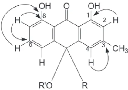
m/z 802, what it is in accordance with the total mass of two molecules of glucosyl-chrysophanolanthrons. The 2D NMR spectroscopic techniques had confirmed the assignments above cited and the presence of one unit of glucose to carbon 10, that it can be established by HMQC correlations of H-1’’ to C-10, H-2’’ to C-10, H-1’’ to C-5’’, H-2’’ to C-3’’, H1-6’’ to C-5’’, H2-6’’ to C-5’’ (Figure 1), which proves the connection of the chain of the sugar and C-10 anthron. The absence of the signal at about d 101.00 referring to anomeric carbon is not present, thus allowing concluding that the compound presents deoxy glucose in its structure. Long-range correlations in HMBC spectrum showed that at as correlations H-2 to C-1, H- 4 to C-3, H-6 to C-8, H-7 to C-6, H-7 to C-8 confirming the skeleton anthron for this compound. The long-range correlations H-4/H-5/H-2’’ to C-10 (Figure 2) and H-4’/H- 5’/H-2”’ to C-10’ confirm the position in C-10 of side chain of sugar.
The conversion rates of glyceraldehyde-3-phosphate to 1-arseno-3-phosphoglycerate catalyzed by T. cruzi GAPDH in solutions of TEA buffer were monitored via ITC. The experiments were carried out in presence and absence of martianine and results can be found in Figure 3. The exothermic sharp peaks that can be seen in Figure 3A
represent the heat of dilution for all injections. Among the peaks, the heat released from substrate depletion takes place at the steady state and the instrumental thermal power remains constant. There is a change in the steady state heat-flow as the reaction rate increases after each injection. This keeps going until the baseline reaches the final steady state Figure 2. Long-range correlations in HMBC spectrum.
Figure 3. (A) Typical raw data for thermal power change as a function of time in the multiple injections assay and (B) Nonlinear least-squares fit of experimental data (dotted line) to Michaelis-Menten equation to determine kinetic parameters. Experiment control () and presence of 50 µmol (), 25 injections of G3P 5.0 mmol L-1 were titrated into the sample cell filled
heat-flow level. The dependence of the reaction rate on glyceraldehyde-3-phosphate concentration follows typical Michaelis-Menten kinetics mechanism. The parameters kcat and KM were obtained by fitting the raw data points to the Michaelis-Menten equation, using non-linear regression, and results can be seen in Table 2.
The results depicted in Table 2 and Figure3 suggest that the GAPDH enzyme is inhibited by martianine in a non-competitive mechanism. The value of Michaelis-Menten constant (KM) does not change during the experiments and the catalytic constant (kcat) diminishes at no KM expenses, as compared to control experiments. The stocked enzyme shows a loss of activity in function of the storage time. Nevertheless, when a control experiment is carried out previously to each inhibition assay the determined inhibition constant (Ki) is reproducible. As can be seen in Table 1 even with the change in the kcat (experiments realized in different days) the Ki was still the same.
The enzyme glyceraldehyde-3-phosphate dehydrogenase (GAPDH) plays an important role in the life cycle of the Trypanosoma cruzi and it has become an important target in the drug discovery for Chagas disease.22 The high
martianine affinity for the GAPDH it agrees to the some anthraquinones have shown activity against the parasite.5,23
The potential isothermal titration calorimetry has in the search for new inhibitors are thus put forward. It provides a direct measure of small molecule binding to enzymes as molecular targets in the drug discovery and development. The relationship of kinetic parameters to the drug-receptor interactions allows a fast description of substrate binding. The contribution to GAPDH catalysis is easily accounted for all processes taking place during the conversion to product. The catalytic efficiency of GAPDH enzyme entails the shape complementarities between enzyme and transition state. The catalytic efficiency (kcat/KM 107mol L-1s-1)
for the first control experiment is 1.4 whereas it comes to 0.9 when the second control experiment is carried out. In the presence of martianine the first measurement results in the catalytic efficiency to be 1.2, and it comes down to 0.8 in the second one. Thus, the martianine small molecule
does not enhance the catalytic efficiency of GAPDH. On the contrary, it reduces the overall catalytic efficiency so indicating the diminution of the kcat not at the expenses of KM that remains thoroughly constant. Overall, despite the change in the catalytic efficiency due to GAPDH loosing its activity this is not accounted for when the inhibitory constant is to be determined via isothermal titration calorimetry.
Conclusions
Martianine 1 is aof a new bianthrone glycoside isolated from Senna martiana Benth and the high martianine affinity for the GAPDHTc enzyme suggests that this molecular class may be useful for the development of new drugs against Chagas disease.
Supplementary Information
Supplementary data of martianine structure as 1 and 2D 1H and 13C NMR spectra are available free of charge at
http://jbcs.sbq.org.br, as PDF file.
Acknowledgments
Authors acknowledge to Dr. N. P. Lopes (FCFRP, Universidade de São Paulo) for recording the mass spectra and also acknowledge to CNPq, FUNCAP and FAPESP for financial support.
References
1. Irwin, H. S.; Barneby, R. C.; Memoirs of the New York Botanical Garden; The American Cassiinae: A Synoptical Revision of
Leguminosae Tribe Cassieae, subtribe Cassiinae in the New
World, Vol. 35, Part 1, The New York Botanical Garden Press:
New York, 1982, p. 465.
2. Barbosa, F. G.; Oliveira, M. C. F.; Braz-Filho, R.; Silveira, E. R.; Biochem. Syst. Ecol.2004, 32, 363.
3. Alemayehu, G.; Abegaz, B.; Kraus, W. A.; Phytochemistry1998, 48, 699.
4. Koyama, J.; Morita, I.; Tagahara, K.; Aqil, M.; Phytochemistry
2001, 56, 849.
5. Mbwambo, Z. H.; Apers, S.; Moshi, M. J.; Kapingu, M. C.; Van Miert, S.; Claeys, M.; Brun, R.; Cos, P.; Pieters, L.; Vlietink, A.; Planta Med.2004, 70, 706.
6. Vila, J. L.; Mollinedo, P.; Sterner, O.; Rev. Bol. Quim. 2003, 20, 49.
7. Pivatto, M.; Crotti, A. E. M.; Lopes, N. P.; Castro-Gamboa, I.; Rezende, A.; Viegas Jr., C.; Young, M. C. M.; Furlan, M.; Bolzani, V. S.; J. Braz. Chem. Soc. 2005, 16, 1431.
Table 2. Kinetic parameters kcat, KM (observed values from curves in Figure 3) and Ki of the martianine
kcat / (s-1) K
M / (µmol L -1) K
i / (µmol L -1)
Control 1 177 (±2) 11.8 (±0.7)
-Martianine 1 156 (±3) 10.6 (±1.0) 28.4 (±0.5)
Control 2 117 (±1) 12.5 (±1.1)
-Martianine 2 105 (±1) 14.6 (±0.4) 26.2 (±0.9)
8. Silva, G. H.; Teles, H. L.; Trevisan, H. C.; Bolzani, V. S.; Young, M. C. M.; Pfenning, L. H.; Eberlin, M. N.; Haddad, R.; Costa-Neto, C. M.; Araújo, A. R.; J. Braz. Chem. Soc. 2005, 16, 1463.
9. Sepúlveda-Boza, S.; Cassels, B. K.; Planta Med.1996, 62, 98.
10. Coura, J. R.; Castro, S. L.; Mem. Inst. Oswaldo Cruz2002, 97, 3.
11. Campo, V. L.; Carvalho, I.; Allman, S.; Davis, B. J.; Field, R. A.; Org. Biomol. Chem. 2007, 5, 2645.
12. Aronov, A. M.; Suresh, S.; Buckner, F. S.; Van Voorhis, W. C.; Verlinde, C. L. M. J.; Opperdoes, F. R.; Hol, W. G. J.; Gelb, M. H.;Proc. Natl. Acad. Sci. U. S. A. 1999, 96, 4273.
13. Tomazela, D. M.; Pupo, M. T.; Passador, E. A. P.; Silva, M. F. G. F.; Vieira, P. C.; Fernandes, J. B.; Rodrigues, E. F.; Oliva, G.; Pirani, J. R.; Phytochemistry2000, 55, 643.
14. De Marchi, A. A.; Castilho, M. S.; Nascimento, P. G. B.; Archanjo, F. C.; Del Ponte, G.; Oliva, G.; Pupo, M. T.;Bioorg. Med. Chem. 2004, 12, 4823.
15. Ladame, S.; Fauré, R.; Denier, C.; Lakhdar-Ghazal, F.; Willson, M.; Org. Biomol. Chem.2005, 3, 2070.
16. Liñares, G. E. G.; Ravaschino, E. L.; Rodriguez, J. B.; Curr. Med. Chem.2006, 13,335.
17. Wiseman, T.; Williston, S.; Brandts, J. F.; Lin, L. N.; Anal. Biochem. 1989, 179, 131.
18. Hannaert, V.; Opperdoes, F. R.; Michels, P. A. M.; Protein Expression Purif. 1995, 6, 244.
19. Bradford, M. M.; Anal. Biochem. 1976, 72, 248.
20. Wiggers, H. J.; Cheleski, J.; Zottis, A.; Oliva, G.; Andricopulo, A. D.; Montanari, C. A.; Anal. Biochem.2007, 370, 107. 21. ITC Data Analysis in Origin®, Tutorial Guide, Version 7.0,
MicroCal, LLC: Northampton, MA, 2004.
22. Souza, D. H. F.; Garratt, R. C.; Araújo, A. P. U.; Guimarães, B. G.; Jesus, W. D. P.; Michels, P. A. M.; FEBS Lett.1998, 424, 131.
23. Cuellar, M. A.; Salas, C.; Corte´s, M. J.; Morello, A.; Maya, J. D.; Preite, M. D.; Bioorg. Med. Chem. 2003,11, 2489.
Received: March 3, 2008
Web Release Date: April 24, 2009
Supplementary Information
0103 - 5053 $6.00+0.00
*e-mail: [email protected]
A New Bianthron Glycoside as Inhibitor of
Trypanosoma cruzi
Glyceraldehyde
3-Phosphate Dehydrogenase Activity
Edângelo M. S. de Macedo,a Helton J. Wiggers,b Maria G. V. Silva,*,a,b Raimundo Braz-Filho,c
Adriano D. Andricopulod and Carlos A. Montanarib
aDepartamento de Química Analítica e Físico-Química, Departamento de Química Orgânica e Inorgânica,
Universidade Federal do Ceará, 60455-970 Fortaleza-CE, Brazil
bInstituto de Química de São Carlos and dInstituto de Física de São Carlos, Universidade de São Paulo,
13560-970 São Carlos-SP, Brazil
cSetor de Química de Produtos Naturais, CCT, Universidade Estadual do Norte Fluminense,
28013-602 Campos dos Goytacazes-RJ, Brazil