Article
Printed in Brazil - ©2016 Sociedade Brasileira de Química0103 - 5053 $6.00+0.00
*e-mail: [email protected]
Synthesis,
in vitro
Antiproliferative and Anti-
Mycobacterium tuberculosis
Activities
of Novel
β
-Carboline Derivatives
Flora M. F. Moreira,a Julio Croda,a Maria H. Sarragiotto,b Mary A. Foglio,c
Ana L. T. G. Ruiz,c João E. Carvalho c and Anelise S. N. Formagio*,d
aFaculdade de Ciências da Saúde and dFaculdade de Ciências Agrárias, Universidade Federal da
Grande Dourados, Rodovia Dourados - Itahum, Km 12, 79804-970 Dourados-MS, Brazil
bDepartamento de Química, Universidade Estadual de Maringá, Avenida Colombo, 5790,
Jardim Universitário, 87020-900 Maringá-PR, Brazil
cCentro Pluridisciplinar de Pesquisas Químicas, Biológicas e Agrícolas, Universidade Estadual de
Campinas, CP 6171, 13083-970 Campinas-SP, Brazil
A series of β-carboline derivatives with amino or guanidinium were synthesized and evaluated in vitro against anti-Mycobacterium tuberculosis and for antiproliferative activities against nine human cancer cell lines. The compounds 1-(4-hydroxyphenyl)-3-carboxamide(ethylamine) β-carboline (24.9 µg mL-1) and 1-(4-methoxyphenyl)-3-carboxamide(ethylamine) β-carboline (26.9 µg mL-1)
were the most active against M. Tuberculosis (MTB). Compounds 1-(4-hydroxyphenyl)-3-carboxamide(ethylamine) β-carboline and 1-(4-methoxyphenyl)-3-carboxamide(propylamine)
β-carboline, which had the same substituted groups, inhibited the growth of all human tumor cell lines with growth inhibitory activity (GI50) values from 1.37 to 9.20 mmol L-1. Also in this series,
compounds 1-(4-hydroxyphenyl)-3-carboxamide(propylamine) β-carboline and 1-(3-nitrophenyl)-3-carboxamide(propylamine) β-carboline demonstrated significant activity against NCI/ADR cells. Among compounds with a terminal guanidine group, compounds 1-(4-hydroxyphenyl)-3-carboxamide(ethyl)guanidine β-carboline (27.8 µg mL-1) and 1-(3-nitrophenyl)-3-carboxamide(ethyl)
guanidine β-carboline (37.4 µg mL-1) demonstrated the greatest activity against MTB. Additionally,
compounds 1-(4-methoxyphenyl)-3-carboxamide(ethyl)guanidine β-carboline (GI50 = 0.45 mmol L-1)
effectively inhibited growth and was highly selective against NCI/ADR. The in silico study revealed that 1-(4-hydroxyphenyl)-3-carboxamide(ethylamine) β-carboline, 1-(4-methoxyphenyl)-3-carboxamide(ethylamine) β-carboline, 1-(4-hydroxyphenyl)-3-carboxamide(propylamine)
β-carboline,1-(4-methoxyphenyl)-3-carboxamide(propylamine) β-carboline and 1-(3-nitrophenyl)-3-carboxamide(propylamine) β-carboline compounds follow the rules established by Lipinski, suggesting that this compound has no problems with oral bioavailability.
Keywords: synthesis, β-carboline, Mycobacterium tuberculosis, antiproliferative activity
Introduction
Tuberculosis (TB) exhibits high morbidity and mortality. The long-term treatment regimen can cause patients to be non-compliant in completing the treatment, thus leading to emergence of multidrug-resistant (MDR-TB) and extensively drug-resistant (XDR-(MDR-TB) TB strains. Infections caused by MDR-TB and XDR-TB do not respond to first-line drugs that are used to treat TB, and alternative treatment regimens include mostly injected drugs and prolonged treatments.1,2 Due to the appearance of
resistant strains and given high toxicity of anti-tuberculosis drugs, to the need to develop new drugs that are more effective and less toxic than current drugs, which would reduce time and complexity of treatment, is urgent.3,4 The
discovery of new drugs is also necessary for the treatment of cancer, because most chemotherapeutic agents exhibit severe toxicity and cause many undesirable side effects; additionally, current agents are very expensive, mutagenic, carcinogenic and teratogenic.5 Tuberculosis, caused by Mycobacterium tuberculosis (MTB),1 and cancer6 have
pathogens with cancer cells may be influenced by genetic alterations in the tumor cells.
Synthetic β-carboline derivatives exhibit a wide range of pharmacological activities, including antitumor and anti-tuberculosis activities.7-14 Our research group demonstrated
that β-carboline derivatives with various substituents at positions-1, 3 and 9 of the β-carboline skeleton presented significant in vitro antitumoral, antiviral, antitrypanosomal and antileishmanial activities.15-23 Other studies have shown
that β-carboline derivatives with a methyl-substituted group at position-1 and a guanidinium group-terminated side chain at C-3 exhibited anti-HIV-1 activity in MT4 cells by hindering the essential interaction of the regulatory protein Tat with trans-activation response region (TAR).24,25 Some results
cited above indicated that β-carbolinederivatives containing 4-hydroxyphenyl, 4-methoxyphenyl or 3-nitrophenyl group at C-1 showed potent anticancer activity for some of the human cancer cell lines tested. This led us to study novel
β-carboline analogs that might serve as antitumoral and anti-tuberculosis agents as part of our ongoing research program. Based on the idea that the addition of appropriate substituents at positions-1 and -3 might result in more potent compounds, we synthesized novel 1-substituted-phenyl-β-carboline with an amino or guanidinium group-terminated side chain at C-3 and evaluated the in vitro anti-tuberculosis, antiproliferative properties and in silico study.
Experimental
General procedure
1H and 13C nuclear magnetic ressonance (NMR)
spectra were recorded on a Varian Mercury Plus (Palo Alto, EUA) spectrometer operating at 300 and 75.5 MHz, respectively, using deuterated dimethyl sulfoxide (DMSO-d6), chloroform (CDCl3) and methanol (CD3OD) as solvent, and tetramethylsilane (TMS) as internal reference. Infrared (IR) spectra were recorded as potassium bromide pellets on a BOMEM spectrometer model MB-100 (Houston, USA). Melting points were determined in a Micro-Química apparatus MQAPF-301 model (Palhoça, Brazil) and are uncorrected. The reactions were monitored by thin layer chromatography (TLC) conducted on Merck TLC plates (Silica Gel 60 F254, Darmstadt, Germany). All reagents were purchased from commercial suppliers.
Preparation of 1-(substituted phenyl)-3-carboxamide-ethylguanidine-β-carboline(4a-c)
The 1-(substituted phenyl)-3-carbomethoxy-β-carboline were obtained as previously reported.21 The
1-(substituted-phenyl)-3-ethylamine-carboxamide β-carboline 2a-c
were obtained by reaction of the methyl esters 1a-c
(2.0 mmol L-1) with 1,2-ethylenediamine (6 mmol L-1)
at room temperature, stirred for 36 h. The formed solids were collected by filtration. The 1-(substituted-phenyl)-3-prophylamine-carboxamide β-carboline 3a-c were obtained
by reaction of the methyl esters 1a-c (1.7 mmol L-1) with
1,3-propanediamine (5 mmol L-1) in 25 mL MeOH/CHCl 3
was refluxed for 32 h. The formed solids were collected by filtration.26
To a solution of S-methylisothiourea sulfate (0.5 mmol L-1) in water (1.5 mL) and 2 mmol L-1 NaOH
(0.5 mL) was added a suspension of β -carboline-3-ethylamine-carboxamide 2a-c at 0 °C. The mixture was
kept under stirring, at room temperature by 1 h and later under reflux for 24 h, was again added a solution of S-methylisothiourea sulfate (0.5 mmol L-1) in water
(1.5 mL) and 2 mol L-1 NaOH (0.5 mL) and reflux for
24 h. The formed solids were collected by filtration and washed with cold water for obtained 1-(substituted phenyl)-3-carboxamide-ethylguanidine β-carboline (4a-c).26 The
characterization data of the compounds obtained are given bellow that corroborates with correlation spectroscopy (COSY), H1 detected multiquantum coherence (HMQC)
and heteronuclear multiple-bond correlation (HMBC) two-dimensional NMR spectra.
1-(4-Hydroxyphenyl)-3-carboxamide(ethylamine) β-carboline (2a)
Yield 68%; mp 171-174 °C; IR (KBr) νmax / cm-1
3090, 1684, 1480, 1460; 1H NMR (300 MHz, DMSO-d 6)
d 2.74 (t, 2H, J 6.0 Hz, CH2-ethyl), 3.07 (t, 2H, J 6.0 Hz, CH2-ethyl), 7.01 (d, 2H, J 8.7 Hz, Phenyl-H); 7.28 (t, 1H, J 7.0 Hz, H-6), 7.57 (td, 1H, J 7.7, 5.0 Hz, H-7), 7.67 (d, 1H, J 8.4 Hz, H-8), 7.99 (d, 2H, J 8.7 Hz, Phenyl-H), 8.37 (d, 1H, J 7.8 Hz, H-5); 8.73 (s, 1H, H-4); 13C NMR
(75.5 MHz, DMSO-d6) d41.2, 41.9, 112.1, 112.7, 115.7,
120.3, 121.5, 121.8, 128.5, 129.2, 129.7, 130.0, 134.8, 138.7, 141.4, 141.8, 157.9, 167.1.
1-(4-Methoxyphenyl)-3-carboxamide(ethylamine) β-carboline (2b)
Yield 74%; mp 180-182 °C; IR (KBr) νmax / cm-1
3354, 3106, 1686, 1590, 860; 1H NMR (300 MHz,
CDCl3/CD3OD) d2.84 (t, 2H, J 6.0 Hz, CH2-ethyl), 3.87
(t, 2H, J 6.0 Hz, CH2-ethyl); 3.95 (s, 3H, OCH3), 7.17 (d, 2H, J 8.1 Hz, Phenyl-H), 7.36 (ddd, 1H, J 7.8, 7.5, 2.1 Hz, H-6), 7.58 (d, 1H, J 7.5 Hz, H-8), 7.61 (ddd, 1H, J 7.8, 7.5, 2.1 Hz, H-7), 8.02 (d, 2H, J 8.1 Hz, Phenyl-H), 8.24 (d, 1H, J 7.8 Hz, H-5), 8.83 (s, 1H, H-4); 13C NMR (75.5 MHz,
121.0, 122.4, 122.8, 130.8, 131.6, 132.3, 132.9, 133.6, 139.5, 141.9, 142.0, 144.3, 162.5.
1-(3-Nitrophenyl)-3-carboxamide(ethylamine) β-carboline (2c)
Yield 66%; mp 172-175 °C; IR (KBr) νmax / cm-1 3190,
1636, 1555, 1496, 1420; 1H NMR (300 MHz, CDCl 3/
CD3OD) d2.98 (t, 2H, J 6.0 Hz, CH2-ethyl), 3.63 (t, 2H, J 6.0 Hz, CH2-ethyl), 7.37 (m, 1H, H-6), 7.60 (m, 2H,
H-8, Phenyl-H), 7.81 (t, 1H, J 7.8 Hz, H-7), 8.23 (dd, 1H, J 7.8 Hz, H-5), 8.37 (dd, 1H, J 7.0, 2.0 Hz, Phenyl-H), 8.40 (d, 1H, J 7.0 Hz, Phenyl-H), 8.86 (sl, 1H, Phenyl-H), 8,89 (s, 1H, H-4); 13C NMR (75.5 MHz, CDCl
3/CD3OD) d
39.9, 40.7, 112.4, 114.3, 120.8, 121.6, 121.7, 123.4, 123.5, 129.1, 129.9, 131.1, 134.7, 135.0, 138.6, 138.9, 139.4, 141.8, 148.5, 167.1.
1-(4-Hydroxyphenyl)-3-carboxamide(propylamine) β-carboline (3a)
Yield 72%; mp 180-182 °C; IR (KBr) νmax / cm-1 3226,
1640, 1550, 1496, 1440; 1H NMR (300 MHz, DMSO-d 6) d
1.65 (q, 2H, J 6.3 Hz, CH2-prophyl), 2.65 (t, 2H, J 6.3 Hz, CH2-prophyl), 3.45 (q, 2H, J 6.0 Hz, CH2-prophyl), 7.00 (d,
2H, J 8.1 Hz, Phenyl-H), 7.29 (t, 1H, J 7.2 Hz, H-6), 7.57 (t, 1H, J 7.2 Hz, H-7), 7.67 (d, 1H, J 8.1 Hz, H-8), 8.02 (d, 2H, J 8.1 Hz, Phenyl-H), 8.36 (d, 1H, J 8.1 Hz, H-5), 8.72 (s, 1H, H-4); 13C NMR (75.5 MHz, DMSO-d
6) d 32.8, 36.9,
39.5, 112.0, 112.6, 115.6, 120.1, 121.3, 121.9, 128.3, 128.4, 129.6, 130.1, 133.8, 139.7, 140.9, 141.5, 158.5, 164.9.
1-(4-Methoxyphenyl)-3-carboxamide(propylamine) β-carboline (3b)
Yield 60%; mp 188-193 °C; IR (KBr) νmax / cm-1
3200, 1678, 1550, 1520, 1448, 1010; 1H NMR (300 MHz,
DMSO-d6) d1.64 (qt, 2H, J 6.6 Hz, CH2-prophyl), 2.64
(t, 2H, J 6.6 Hz, CH2-prophyl), 3.44 (q, 2H, J 6.6 Hz, CH2-prophyl), 3.88 (s, 3H, OCH3), 7.18 (d, 2H, J 8.7 Hz,
Phenyl-H), 7.30 (t, 1H, J 7.2 Hz, H-6), 7.58 (t, 1H, J 8.1 Hz, H-7), 7.67 (d, 1H, J 8.1 Hz, H-8), 8.14 (d, 2H, J 8.7 Hz, Phenyl-H), 8.38 (d, 1H, J 7.2 Hz, H-5), 8.75 (s, 1H, H-4); 13C NMR (75.5 MHz, DMSO-d
6) d 33.0, 36.9, 37.0, 55.4,
112.3, 112.6, 114.2, 120.1, 121.3, 121.9, 128.4, 129.7, 130.0, 130.1, 133.9, 139.8, 140.4, 141.6, 159.9, 164.5.
1-(3-Nitrophenyl)-3-carboxamide(propylamine) β-carboline (3c)
Yield 55%; mp 178-180 °C; IR (KBr) νmax / cm-1 3160,
1686, 1534, 1474, 1440;1H NMR (300 MHz, CD 3OD) d
1.65 (qt, 2H, J 6.5 Hz, CH2-prophyl), 2.65 (t, 2H, J 6.0 Hz,
CH2-prophyl), 3.46 (q, 2H, J 6.0 Hz, CH2-prophyl), 7.33
(t, 1H, J 7.5 Hz, H-6), 7.63 (m, 2H, H-8, Phenyl-H), 7.72
(d, 1H, J 7.8 Hz, H-8), 7.91 (t, 1H, J 7.8 Hz, H-7), 8.33 (dd, 1H, J 8.1, 1.5 Hz, Phenyl-H), 8.45 (d, 1H, J 7.8 Hz, Phenyl-H), 8.64 (d, 1H, J 8.1 Hz, H-5), 8.86 (s, 1H, H-4); 13C NMR (75.5 MHz, CD
3OD) d 32.8, 36.8, 37.0, 112.6,
113.7, 120.4, 121.2, 122.2, 123.5, 123.6, 128.9, 130.2, 130.4, 134.7, 135.1, 138.2, 138.9, 139.6, 141.6, 166.5.
1-(4-Hydroxyphenyl)-3-carboxamide(ethyl)guanidine β-carboline (4a)
Yield 30%; mp 176-178 °C; IR (KBr) νmax / cm-1
3260, 1670, 1544, 1424, 1460, 1236;1H NMR (300 MHz,
DMSO-d6) d3.22 ( t,2H, J 6.5 Hz, CH2-ethyl), 3.49 (t, 2H, J 6.5 Hz, CH2-ethyl), 5.50 (s, 2H, NH2), 6.14 (s, 1H, NH),
6.63 (s, 1H, NH), 7.05 (d, 2H, J 8.4 Hz, Phenyl-H), 7.28 (t, 1H, J 7.5 Hz, H-6), 7.56 (t,1H, J 7.5 Hz, H-7), 7.67 (d, 1H, J 8.1 Hz, H-8), 8.05 (d, 2H, J 8.4 Hz, Phenyl-H), 8.37 (d, 1H, J 7.8 Hz, H-5), 8.72 (s, 1H, H-4), 8.81 (s, 1H, NH); 13C NMR (75.5 MHz, DMSO-d
6) d 37.3, 40.5, 112.4, 113.0,
116.0, 120.5, 121.5, 122.1, 128.6, 128.8, 129.9, 130.5, 134.1, 139.6, 141.3, 141.7, 158.6, 159.6, 163.1.
1-(4-Methoxyphenyl)-3-carboxamide(ethyl)guanidine β-carboline (4b)
Yield 45%; mp 190-194 °C; IR (KBr) νmax / cm-1 3320,
1670, 1550, 1520, 1448;1H NMR (300 MHz, DMSO-d 6)
d3.25 (q, 2H, J 6.0 Hz, CH2-ethyl), 3.45 (q, 2H, J 6.0 Hz, CH2-ethyl), 3.96 (s, 3H, OCH3), 5.54 (s, 2H, NH2), 6.18 (t,
1H, J 7.5 Hz, NH), 7.22 (d, 2H, J 8.7 Hz, Phenyl-H), 7.34 (t, 1H, J 7.0 Hz, H-6), 7.60 (t, 1H, J 7.5 Hz, H-7), 7.70 (d, 1H, J 8.0 Hz, H-8), 8.20 (d, 2H, J 8.7 Hz, Phenyl-H), 8.41 (d, 1H, J 8.1 Hz, H-5), 8.78 (s, 1H, H-4), 8.84 (t, 1H, J 7.5 Hz, NH), 11.81 (s, 1H, NH); 13C NMR (75.5 MHz,
DMSO-d6) d 38.6, 40.3, 55.4, 112.3, 112.6, 114.2, 120.8,
121.2, 121.9, 129.7, 129.9, 130.1, 133.8, 138.4, 139.6, 140.5, 141.5, 159.0, 159.9, 165.1.
1-(3-Nitrophenyl)-3-carboxamide(ethyl)guanidine β-carboline (4c)
Yield 38%; mp 192-194 °C; IR (KBr) νmax / cm-1
3204, 1668, 1530, 1520, 1448; 1H NMR (300 MHz,
DMSO-d6) d3.22 (m, 2H, CH2-ethyl), 3.42 (q, 2H, J 6.5 Hz,
CH2-ethyl), 5.49 (s, 2H, NH2), 6.15 (t, 1H, J 7.0 Hz, NH),
7.34 (t, 1H, J 7.0 Hz, H-6), 7.60 (d, 1H, J 7.8 Hz, H-8), 7.62 (t, 1H, J 7.8 Hz, Phenyl-H), 7.95 (t, 1H, J 8.0 Hz, H-7), 8.39 (d, 1H, J 7.7 Hz, H-5), 8.45 (d, 1H, J 8.1 Hz, Phenyl-H), 8.65 (d, 1H, J 7.5 Hz, Phenyl-H), 8.86 (s, 1H, Phenyl-H), 8.89 (s, 1H, H-4), 8.90 (s, 1H, NH), 12.02 (s, 1H, NH); 13C NMR (75.5 MHz, DMSO-d
6) d 38.7, 40.5,
Anti-Mycobacterium tuberculosis activity
M. tuberculosis H37Rv (ATCC27294) strains were
grown in Ogawa-Kudoh (OK)medium for 10 days at 37 °C. For testing, aliquots were removed and cultured in Middlebrook 7H9 broth (Difco, Sparks, USA) supplemented with oleic acid, bovine serum albumin, dextrose and catalase (OADC enrichment BBL/Becton-Dickinson); 0.5% glycerol was added as a carbon source and 0.5% Tween 80 was added to prevent the appearance of lumps. The culture was maintained for 15 days at 37 ºC. The bacterial suspensions were prepared and adjusted to the No. 1 of the McFarland scale.
Stock solutions of the test compounds were solubilized in dimethyl sulfoxide (DMSO) (Sigma-Aldrich, St. Louis, USA) and diluted in Middlebrook 7H9 broth (Difco, Sparks, USA) supplemented with OADC enrichment BBL/Becton Dickinson. Rifampicin and isoniazid were solubilized according to the manufacturer’s recommendations (Sigma-Aldrich, St. Louis, USA) and used as positive controls.
Antimycobacterial activity was determined using the resazurin microtiter assay (REMA).27 Briefly, 100 µL of
supplemented Middlebrook 7H9 broth (Difco, Sparks, USA) was dispensed into each well of a sterile flat-bottom 96-well plate; then, serial dilutions of the test compounds (0.98-250 µg mL-1) and reference drugs (0.004-1 µg mL-1)
were prepared. One hundred microliters of bacterial suspension (5 × 10⁵ UFC mL-1) was then added to each
well. Plates were incubated for 7 days at 37 °C, after was added 30 µL of resazurin (Sigma-Aldrich, St. Louis, USA) in sterile water (0.01%) in whole plate, and the samples were incubated for 24 h at 37 °C.
The change in absorbance, at 492 nm wavelength, was measured using a microplate reader TP-Reader (Thermo Plate®, Männedorf, Switzerland). Each compound was
analyzed in triplicate on alternate days. The minimum inhibitory concentration (MIC) was defined as the lowest concentration that resulted in 90% inhibition of the growth of M. tuberculosis.28 MIC values were used to classify a
compound’s activity as follows: inactive, > 150 µg mL-1;
moderate, between > 10 and < 100 µg mL-1; and active,
< 10 µg mL-1.
In vitro antiproliferative assay
The tested compounds were evaluated in vitro against a nine-cell line panel comprising melanoma UACC-62, breast MCF7, lung NCI-460, leukemia K-562, ovarian OVCAR, prostate PCO-3, colon HT29, renal 786-0 and adriamycin drug-resistant ovarian cancer NCI/ADR cells.
The tests were performed using the colorimetric method with sulforhodamine B according to the National Cancer Institute (NCI) standard protocol; doxorubicin was used as a positive control.29 Assays were performed in a 96-well
plate using four serial 10-fold dilutions (0.25-250 µg mL-1)
for each test compound. The anticancer activity was determined based on concentration-response curves, and three concentration response parameters, growth inhibitory activity (GI50), growth inhibition (TGI) and cytotoxic
activity (LC50) were calculated. The response parameter
GI50 refers to the drug concentration that produces 50%
reduction of cell growth when compared to untreated control cells. The TGI and LC50 parameters refer to the
drug concentrations required for total growth inhibition and for 50% cell mortality, respectively. Compounds with GI50
values < 100 µmol L-1 were considered active.
In silico study
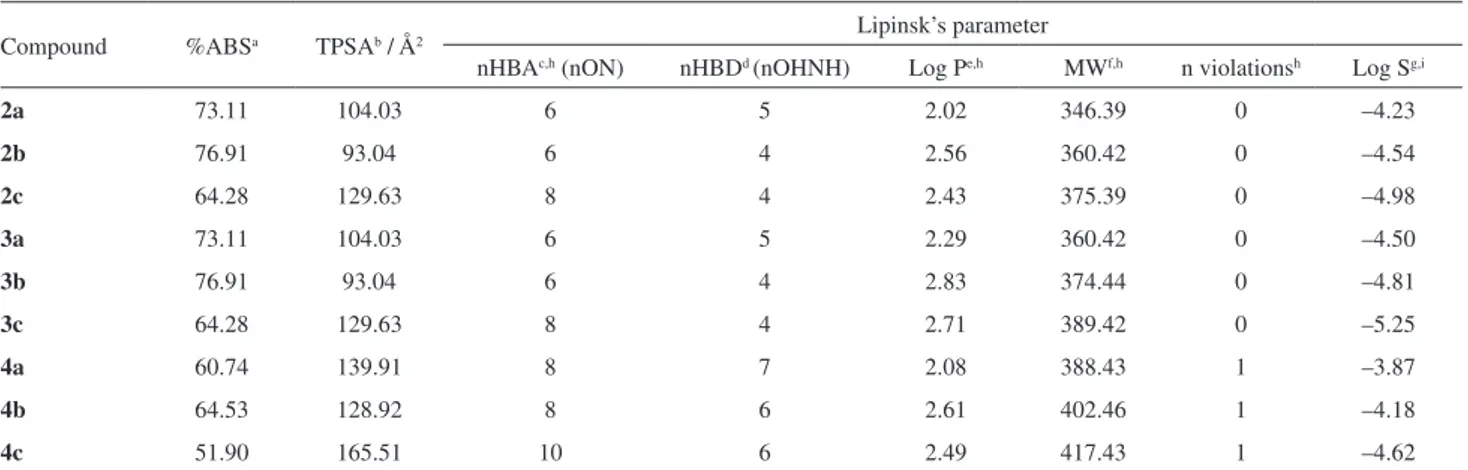
The in silico computational study of compounds were performed to determine Lipinski’s rules of five30(hydrogen
bond donors ≤ 5; hydrogen bond acceptors ≤ 10; molecular weight ≤ 500; the Log P is ≤ 5), topological polar surface area (TPSA) and percentage of absorption (%ABS). Calculations were performed using Molinspiration online property calculation toolkit software31 and
OSIRIS property explorer software.32 The percentage of
absorption was estimated using the following equation: %ABS = 10 – [0.345 × TPSA].
Results and Discussion
Chemistry
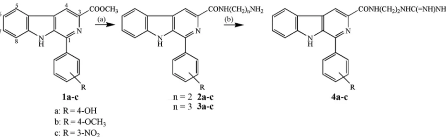
The synthetic pathway for the preparation of 1,3-disubstituted β-carbolines is presented in Scheme 1. The methyl esters in 1a-c were prepared by a Pictet-Spengler
condensation of L-tryptophan with the appropriate aromatic aldehydes in acidic media, subsequent esterification of the resulting carboxylic acids with methanol and sulfuric acid, and oxidation with sulfur in refluxing xylene.22
Compounds 2a-c and 3a-c were obtained by the reaction
of β-carboline methyl ester with 1,2-ethylenediamine and 1,3-propanediamine, respectively, and resulted in an amino group-terminated side chain at C-3. Finally, the coupling of β-carboline carboxamide derivatives 2a-c
with S-methylisothiourea yielded compounds 4a-c, which
include a terminal guanidinium group.
The novel compounds 2a-c, 3a-c and 4a-c were
characterized using 1H and 13C NMR spectroscopy, as
of carboxamides 2a-c and 3a-c showed additional signals at
dH 1.64-3.95 (reflecting the integration of two protons) and
at dH 7.01-8.94 (corresponding to aromatic hydrogens). The
presence of the ethylamine or propilamine carboxamide in position C-3 group was confirmed by 13C NMR, which
showed signals at dc 32.8-41.9 (CH2) and dc 162.5-167.1
(C=ON). Derivatives 4a-c was characterized by the
presence of an additional signal at dc 159.01-159.90,
corresponding to the guanidinium group.
Anti-Mycobacterium tuberculosis activity (MTB)
Derivatives 2a-c, 3a-c and 4a-c were evaluated
in vitro for their antimycobacterial activity against M. tuberculosis H37Rv (ATCC 27294) using the REMA method.27 The MIC values (µg mL-1 and µmol L-1) were
measured with respect to two standard antitubercular drugs, isoniazid (INH) and rifampicin (RFP), and the screening results are presented in Table 1. Among the nine compounds evaluated against MTB, seven presented moderate activity, with MIC values ranging from 58.3-24.9 µg mL-1; in particular, compounds 2a
(24.9 µg mL-1), 2b (26.9 µg mL-1), 4a (27.8 µg mL-1)
and 4c (37.4 µg mL-1) presented interesting activity.
Compounds 2a and 2b, which had p-hidroxyphenyl and p-methoxyphenyl substituents, respectively, at position-1 and ethylenediamine moieties at C-3 were the most active derivatives in this series. The length of the terminated side chains affected the activities of these compounds. Substituting the guanidinium group led to reduced activity (compare compounds 2a and 4a). The effect
of the guanidinium group was particularly significant when comparing compounds 2c (57.9 µg mL-1) and 4c
(37.4 µg mL-1). The substituents at positions-1 and 3
strongly affected anti-MTB activities of these compounds. Earlier studies reported the synthesis and investigations of the antimycobacterial activity, e.g., guanidinium-modified
compounds, which demonstrated potent antitubercular activity against M. tuberculosis, aminopyrimidine derivatives exhibit moderate to potent anti-MTB activity, with MIC values ranging from 12.5-3.12 µg mL-1.33
The introduction of an ethylguanidinium group at the upper rim resulted in high antimycobacterial activities for the unsubstituted, 5,5’-dimethyl-2,2’-bipyridyl and 4,4’-dimethyl-2,2’-bithiazolyl analogs, with MIC values of 1.51 and 2.69 µg mL-1, respectively, values that were
similar to those of current commercially available anti-tuberculosis agents.34
Antiproliferative activity
The antiproliferative activities of the synthesized 1,3-disubstituted β-carbolines derivatives (2a-c, 3a-c and 4a-c) were evaluated in vitro against nine human tumor cell lines. The results for compounds 2a-c and 3a-c,
Scheme 1. Synthetic route for the preparation of the β-carbolines derivatives. Reagents and conditions: (a) 1,2-ethylenediamine, at room temperature, 36 h or 1,3-propanediamine, CHCl3/MeOH, reflux, 32 h (55-72%); (b) S-methylisothiourea, 2N NaOH, 4 °C to room temperature, reflux, 48 h (30-45%).
Table 1. Anti-Mycobacterium tuberculosis H37RV activity of compounds
2a-c, 3a-c and 4a-c
Compound R n (µg mLMICa / -1) (µmol LMIC / -1)
2a 4-OH 2 24.9 75.4
2b 4-OCH3 2 26.9 74.6
2c 3-NO2 2 57.9 153.5
3a 4-OH 3 > 250 > 500
3b 4-OCH3 3 58.3 155.7
3c 3-NO2 3 > 250 > 500
4a 4-OH 2 27.8 74.4
4b 4-OCH3 2 57.5 142.6
4c 3-NO2 2 37.4 88.3
Isoniazid – – 0.05 0.3
Rifampicin – – 0.01 0.01
which were amino group-terminated at C-3, demonstrated that compounds 2a and 3b, with p-hydroxyphenyl and p-methoxyphenyl groups, respectively, at position-1, inhibited growth in all human tumor cell lines with GI50
values rangingfrom 1.37-9.20 µmol L-1. Also in this series,
compounds 3a (GI50 = 0.33 µmol L-1, TGI = 50.28 µmol L-1)
and 3c (GI50 = 0.71 µmol L-1, TGI = 11.08 µmol L-1,
LC50 = 26.62 µmol L-1) showed significant activity and
high selectivity against adriamycin drug-resistant ovarian cancer cells (NCI/ADR) (Tables 2 and 3).
The compounds with terminal guanidinium groups, including compound 4b (GI50 = 0.45 µmol L-1;
TGI = 72.09 µmol L-1) effectively inhibited growth and
was highly selective against adriamycin drug-resistant ovarian cancer cell lines (NCI/ADR) when compared
with compounds 2a-c, which are amino group-terminated.
However, the guanidinium-terminated compounds did not demonstrate any important interaction that could account for the cell growth inhibition. Substituting the phenyl group with electron-donating substituents at position-1 influenced each series differently.
A previous study evaluated the in vitro antitumor activities of several benzenesulfonamide derivatives with various substituted aminoguanidine groups. Compound 1-allyl-2-[4-chlorophenylcarbamoyl)-2-methylthiobenzenesulfonyl]-3-(5-nitrofurfurylideneamino) exhibited remarkable activity against 21 human tumor cell lines representing leukemia and melanoma and lung, colon, ovarian, renal, prostate and breast cancers (GI50 = 0.3-3.0 µmol L-1).35
Table 2.In vitro cell growth inhibition (GI50) of compounds 2a-c, 3a-c and 4a-c against neoplastic cells
Compound R n
GI50a / (µmol L-1)
UACC-62 melanoma
MCF7 breast
NCI-460 lung
K-562 leukemia
OVCAR ovarian
PCO-3 prostate
HT29 colon
786-0 renal
NCI/ADR ovarian-resistant
2a 4-OH 2 5.15 9.55 9.64 2.93 9.20 8.69 4.27 9.64 8.18
2b 4-OCH3 2 38.75 44.49 44.49 53.07 17.14 *c > 100 33.11 8.36
2c 3-NO2 2 10.34 11.72 88.26 > 100 9.36 * 25.82 21.64 2.42
3a 4-OH 3 25.14 16.19 13.59 13.01 22.75 23.57 23.57 18.71 0.33
3b 4-OCH3 3 9.66 6.98 7.35 5.20 7.09 2.39 9.02 4.40 1.37
3c 3-NO2 3 43.22 23.94 55.16 18.89 16.76 11.18 27.48 27.48 0.71
4a 4-OH 2 > 100 > 100 –b – > 100 – – 3.61 > 100
4b 4-OCH3 2 95.93 35.46 27.17 63.41 39.94 15.54 75.64 63.41 0.45
4c 3-NO2 2 51.85 99.17 > 100 31.03 87.51 91.82 25.02 31.03 1.71
aGI
50 = growth inhibitory activity; bnot determined; cnot tested.
Table 3. Total growth inhibition (TGI) and lethal concentration (LC50 - in parentheses) of compounds 2a-c, 3a-c and 4a-c (µmol L-1)
Compound R n UACC-62
melanoma
MCF7 breast
NCI-460 lung
K-562 leukemia
OVCAR ovarian
PCO-3 prostate
HT29 colon
786-0 renal
NCI/ADR ovarian-resistant
2a 4-OH 2 15.44
(37.5)
38.27 34.32 23.87 24.75
(51.7)
24.75 (74.42)
73.52 24.75 (64.37)
24.75
2b 4-OCH3 2 > 100 > 100 > 100 > 100 > 100 *b > 100 > 100 26.45
2c 3-NO2 2 20.35
(70.93)
22.41 (43.42)
> 100 44.27 19.33 (30.85)
* > 100 87.41 79.13
3a 4-OH 3 –a 42.55 – 36.01 – >100 – – 50.28
3b 4-OCH3 3 – 15.38
(32.8)
20.83 (51.25)
15.38 (37.3)
21.47 (46.84)
10.56 (35.2)
21.47 (49.15)
16.71 (46.84)
11.97 (28.94)
3c 3-NO2 3 > 100 > 100 – > 100 > 100 43.09 > 100 > 100 11.08
(26.62)
4a 4-OH 2 > 100 > 100 > 100 > 100 > 100 > 100 > 100 > 100 > 100
4b 4-OCH3 2 – > 100 > 100 > 100 > 100 > 100 > 100 > 100 72.09
4c 3-NO2 2 90.86 – – 55.07 – – 99.28 > 100 > 100
Lipinski’s rule of five
The drug-likeness concept helps optimize the pharmacokinetic properties of a compound, such as absorption, distribution, metabolism and excretion (ADME) in the human body.36 Lipinski’s rule of fiveis a refinement
of drug-likeness and is used to predict whether a chemical compound will have pharmacological or biological activity as an orally active drug in humans. This rule was formulated based on the observation that most medication drugs are relatively small and lipophilic molecules.33 The results
of the analysis are shown in Table 4 and indicate that the compounds are in agreement with the values determined by Lipinski, except 4a-c derivatives, which showed the number
of hydrogen bond donors (nHBD) > 5, in violation of the Lipinski rules. The calculated percent absorption (%ABS) of all compounds ranged from 51.90-76.91%, indicating good cell membrane permeability. Another important factor is obtained by the volume analysis and TPSA by the compounds showed lower than 140 Ų indicating that these derivatives have good absorption in the intestine, except the compound 4c (TPSA = 165.51). The compounds 2a-c, 3a-b
and 4a-c exhibited good solubility (Log S = –3.87 to –4.98),
except the compound 3c which showed a value of Log S
less than –5. Compounds with high solubility are easily metabolized and eliminated from the body, thus resulting in a lower probability of adverse effects and bioaccumulation.
Conclusions
Thus, our results showed for the first time the synthesis and antitumor and anti-MTBactivity of compounds with an amino or guanidinium group-terminated side chain at C-3 of a 1-substituted-phenyl-β-carboline nucleus. Compounds
2a, 2b and 4a were the most active against M. Tuberculosis H37Rv (ATCC27294). Compound 2a and 3b demonstrated
promising antiproliferative activity for all cancer cell lines. Eight compoundsinhibited thecell growth of adriamycin drug-resistant ovarian (NCI/ADR), showed activity and high selectivity for the 3a, 3c and 4b. Compound 2a
demonstrated promising antiproliferative and anti-MTB activity, in addition to follow as establishedLipinski’s rule of five, suggesting that this compound has no problems with oral bioavailability, and indicates good permeability a in the plasma membrane of the cell, which may represent a precursor to development of new molecules. Further studies are required to explore the mechanism of action of these compounds in detail.
Supplementary Information
Supplementary information is available free of charge at http://jbcs.sbq.org.br, as PDF file.
Acknowledgments
We are grateful to CNPq, FUNDECT and CAPES for providing financial support and UFGD.
References
1.World Health Organization (WHO); Global Tuberculosis Report, 20th ed.; Geneva, 2015, p. 1.
2. Gandhi, N. R.; Nunn, P.; Dheda, K.; Schaaf, H. S.; Zignol, M.; Van Soolingen, D.; Jensen, P.; Bayona, J.; TheLancet2010,
375, 1830.
3. Ma, Z.; Lienhardt, C.; Mcilleron, H.; Nunn, A. J.; Wang, X.; TheLancet2010, 375, 2100.
Table 4. Lipinski’s rule and percentage of absorption (%ABS), topological polar surface area (TPSA), for compounds 2a-c, 3a-c and 4a-c
Compound %ABSa TPSAb / Å2 Lipinsk’s parameter
nHBAc,h (nON) nHBDd (nOHNH) Log Pe,h MWf,h n violationsh Log Sg,i
2a 73.11 104.03 6 5 2.02 346.39 0 –4.23
2b 76.91 93.04 6 4 2.56 360.42 0 –4.54
2c 64.28 129.63 8 4 2.43 375.39 0 –4.98
3a 73.11 104.03 6 5 2.29 360.42 0 –4.50
3b 76.91 93.04 6 4 2.83 374.44 0 –4.81
3c 64.28 129.63 8 4 2.71 389.42 0 –5.25
4a 60.74 139.91 8 7 2.08 388.43 1 –3.87
4b 64.53 128.92 8 6 2.61 402.46 1 –4.18
4c 51.90 165.51 10 6 2.49 417.43 1 –4.62
4.Koul, A.; Arnoult, E.; Lounis, N.; Guillemont, J.; Andries, K.; Nature2011, 469, 483.
5.Jagetia, G. C.; Venkatesh, P.; Baliga, M. S.; Bio. Pharm. Bull.
2005, 28, 58.
6.World Health Organization (WHO); World Cancer Report, 1st ed.; Geneva, 2014, p. 1.
7.Ang, K. K.; Holmes, M. J.; Higa, T.; Hamann, M. T.; Kara, U. A.; Antimicrob. Agents Chemother.2000, 44, 1645. 8.Cao, R.; Chen, Q.; Hou, X.; Chen, H.; Guan, H.; Ma, Y.;
Peng, W.; Xu, A.; Bioorg. Med. Chem.2004, 12, 4613. 9.Cao, R.; Chen, H.; Peng, W.; Ma, Y.; Hou, X.; Guan, H.; Liu, X.;
Xu, A.; Eur. J. Med. Chem.2005, 40, 991.
10.Cao, R.; Peng, W.; Chen, H.; Hou, X.; Guan, H.; Chen, Q.; Ma, Y.; Xu, A.; Eur. J. Med. Chem.2005, 40, 249.
11.Wu, Q.; Cao, R.; Feng, M.; Guan, X.; Ma, C.; Liu, J.; Song, H.; Peng, W.; Eur. J. Med. Chem.2009, 44, 533.
12.Wu, J.; Li, C.; Zhao, M.; Wang, W.; Wang, Y.; Peng, S.; Bioorg. Med. Chem.2010, 18, 6220.
13.Begum, S.; Hassan, S. I.; Siddiqui, B. S.; Nat. Prod. Res.2004, 18, 341.
14.Begum, S.; Ali, S. N.; Siddiqui, B. S.; US 8.420.660, 2013. 15.Stefanello, T. F.; Panice, M. R.; Ueda-Nakamura, T.; Sarragiotto,
M. H.; Auzely-Velty, R.; Nakamura, C. V.; Antimicrob. Agents Chemother.2014, 58, 7112.
16.Savariz, F. C.; Foglio, M. A.; Ruiz, A. L.; Costa, W. F.; Silva, M.; Santos, J. C.; Figueiredo, I. M.; Meyer, E.; Carvalho, J. E.; Sarragiotto, M. H.; Bioorg. Med. Chem.2014, 22, 6867. 17.Savariz, F. C.; Foglio, M. A.; Carvalho, J. E.; Ruiz, A. L.; Duarte,
M. C.; Rosa, M. F.; Meyer, E.; Sarragiotto, M. H.; Molecules
2012, 17, 6100.
18.Barbosa, V. A.; Formagio, A. S.; Savariz, F. C.; Foglio, M. A.; Spindola, H. M.; Carvalho, J. E.; Meyer, E.; Sarragiotto, M. H.; Bioorg. Med. Chem.2011, 19, 6400.
19.Savariz, F. C.; Formagio, A. S. N.; Barbosa, V. A.; Foglio, M. A.; Carvalho, J. E.; Duarte, M. C. T.; Dias Filho, B. P.; Sarragiotto, M. H.; J. Braz. Chem. Soc.2010, 21, 288.
20.Tonin, L. T.; Panice, M. R.; Nakamura, C. V.; Rocha, K. J.; Santos, A. O.; Ueda-Nakamura, T.; Costa, W. F.; Sarragiotto, M. H.; Biomed. Pharmacother.2010, 64, 386.
21.Formagio, A. S. N.; Santos, P. R.; Zanoli, K.; Ueda-Nakamura, T.; Tonin, L. T. D.; Nakamura, C. V.; Sarragiotto, M. H.; Eur. J. Med. Chem.2009, 44, 4695.
22. Formagio, A. S. N.; Tonin, L. T. D.; Foglio, M. A.; Madjarof, C.; Carvalho, J. E.; Costa, W. F.; Cardoso, F. P.; Sarragiotto, M. H.; Bioorg. Med. Chem.2008, 16, 9660.
23. Pedroso, R. B.; Tonin, L. T. D.; Ueda-Nakamura, T.; Dias Filho, B. P.; Sarragiotto, M. H.; Nakamura, C. V.; Ann. Trop. Med. Parasitol.2011, 105, 549.
24. Yu, X.; Lin, W.; Li, J.; Yang, M.; Bioorg. Med. Chem. Lett.
2004, 14, 3127.
25. Yu, X.; Lin, W.; Pang, R.; Yang, M.; Eur. J. Med. Chem.2005, 40, 831.
26. Ishida, J.; Wang, H.; Bastow, K. F.; Hu, C.; Lee, K.; Bioorg. Med. Chem. Lett.1999, 9, 3319.
27. Palomino, J. C.; Martin, A.; Camacho, M.; Guerra, H.; Swings, J.; Portaels, F.; Antimicrob. Agents Chemother.2002,
46, 2720.
28. Pavan, F. R.; Poelhsitz, G. V.; Barbosa, M. I.; Leite, S. R.; Batista, A. A.; Ellena, J.; Sato, L. S.; Franzblau, S. G.; Moreno, V.; Gambino, D.; Leite, C. Q.; Eur. J. Med. Chem.
2011, 46, 5099.
29. Monks, A.; Scudiero, D.; Skehan, P.; Shoemaker, R.; Paull, K.; Vistica, D.; Hose, C.; Langley, J.; Cronise, P.; Vaigro-Wolff, A.; J. Natl. Cancer Inst.1991, 83, 757.
30. Lipinski, C. A.; Lombardo, F.; Dominy, B. W.; Feeney, P. J.; Adv. Drug Delivery Rev.2001, 46, 3.
31. http://www.molinspiration.com accessed in January 2016. 32. http://www.organic-chemistry.org/prog/peo accessed in January
2016.
33. Singh, N.; Pandey, S. K.; Anand, N.; Dwivedi, R.; Singh, S.; Sinha, S. K.; Chaturvedi, V.; Jaiswal, N.; Srivastava, A. K.; Shah, P.; Siddiqui, M. I.; Tripathi, R. P.; Bioorg. Med. Chem. Lett.2011, 21, 4404.
34. Mourer, M.; Dibama, M. H.; Constant, P.; Daffé, M.; Regnouf-de-Vains, J. B.; Bioorg. Med. Chem.2012, 20, 2035. 35. Brzozowski, Z.; Sączewski, F.; Sławiński, J.; Eur. J. Med. Chem.
2007, 42, 1218.
36. Vistoli, G.; Pedretti, A.; Testa, B.; Drug Discovery Today2008,
13, 285.
Submitted: November 4, 2015
Published online: February 26, 2016