CASE REPORT
Late onset Ito
’
s nevus
Cristina Resende, Catarina Araújo, Ana Paula Vieira, Celeste Brito
Department of Dermatology and Venereology, Hospital de Braga, Braga, Portugal
Correspondence to Dr Cristina Resende, [email protected]
To cite:Resende C, Araújo C, Vieira AP,et al. BMJ Case RepPublished online: [please includeDay Month Year] doi:10.1136/ bcr-2013-009746
SUMMARY
Dermal melanocytoses include a variety of congenital and acquired melanocytic lesions characterised by the presence of multiple spindle-shaped dendritic
melanocytes in the dermis. These lesions are commonly found in the skin of Asians, but they can also appear in Caucasians. The Mongolian spot, nevi of Ota and Ito are the most common morphological forms. We report a case of a 24-year-old Caucasian woman presented with a 10-months history of progressive darkening of the right side of her upper back. Cutaneous examination revealed a macular blue-grey hyperpigmentation of the right side of her upper back. Biopsy specimen from the macule showed multiple darkly pigmented, spindle-shaped dendritic melanocytes in the superficial dermis, interstitially arranged between collagen bundles. The diagnosis of nevus of Ito was established. Our patient is maintaining vigilance in dermatology consultation.
BACKGROUND
Dermal melanocytosis include a variety of congeni-tal and acquired melanocytic lesions characterised by the presence of multiple spindle-shaped den-dritic melanocytes in the dermis, migrating from the neural crest to the epidermis.1–3These lesions
are typically associated with Asians, but can also appear in Caucasians patients.4 5 Because of their similar histology, differentiating dermal melanocytic lesions is most often supported on clinical features and anatomic distribution.1 6
Dermal melanocytosis results in a brown or blue pigmentation, depending whether melanin is mainly in the upper or lower dermis, respectively.7
The Mongolian spot, nevi of Ota and Ito are the most common morphological forms of dermal mel-anocytosis.6 Nevus of Ito is often unilateral and
clinically similar to nevus of Ota,6except in ana-tomic location, which corresponds to the distribu-tion of the posterior supraclavicular and cutaneous brachial nerves.6 7
We report this case to salient the rarity of acquired Ito’s nevus, which appeared in a Caucasian adult and the importance of adequate follow-up.
CASE PRESENTATION
A 24-year-old Caucasian woman presented with a 10-month history of progressive darkening of the right side of her upper back. The patient denied any prior inflammation, significant sun exposure or trauma to the area. She also denied using any medica-tions or metals that could possibly alter skin pigmen-tation. Her medical history was unremarkable and there was no family history of cutaneous diseases.
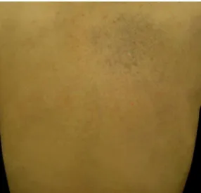
Cutaneous examination revealed a macular blue–
grey hyperpigmentation of the right side of her
upper back and shoulder (figure 1). There was no associated hypertrichosis. Examinations of the rest of the skin, mucous membranes, hair, nails and the remainder of the physical examination were normal.
Biopsy specimen from the macule showed mul-tiple darkly pigmented, spindle-shaped dendritic melanocytes in the superficial dermis, interstitially arranged between collagen bundles (figure 2). The diagnosis of nevus of Ito was therefore established.
OUTCOME AND FOLLOW-UP
Our patient refused to undergo any therapy. She is maintaining under vigilance in consultation of dermatology.
DISCUSSION
Nevus of Ito occurs most frequently in Asian popu-lations and the true incidence is unknown.4 The majority of cases are diagnosed in early infancy and in early adolescence.4 8 9 Few cases of acquired dermal melanocytosis after puberty have been reported.2 7A late-onset Ito’s nevus may be seen as in our patient.
There are several hypotheses to explain the pathogenesis of acquired dermal melanocytosis.1 7
One hypothesis is that dermal melanocytes migrat-ing from the neural crest durmigrat-ing the embryological development fail to reach their location in the basal layer of epidermis. In addition, dermal melanocytes may migrate from the basal layer of the epidermis or from hair bulb to dermis.1 7 10Other hypothesis
is that reactivation of pre-existing, latent dermal melanocytosis, may be activated by inflammation, trauma, pregnancy, hormone replacement therapy
Figure 1 Macular blue–grey hyperpigmentation of the right side of her upper back and shoulder.
Resende C,et al.BMJ Case Rep2013. doi:10.1136/bcr-2013-009746 1
or some unknown aging stimuli.1 10 11 Genetic factors are also important in the pathogenesis of dermal melanocytosis and a positive family history has been reported in literature.7
After the onset, nevus of Ito may progressively enlarge and darken in colour and its appearance usually remains stable once adulthood is reached.4
All dermal melanocytosis are generally considered benign, although rare cases of malignant transformation have been described in literature.6 Although nevus of Ito is much more prevalent in Asian populations, it is generally recognised that the risk of malignant transformation appears to be much more common in Caucasian individuals.3 6 Consequently, vigilance
may be necessary, particularly, when these lesions occur in Caucasian individuals.6 Skin biopsies after the diagnosis are
necessary only if clinical changes or malignant transformation are suspected within the involved skin, ocular or mucosal tissues.8
Pulsed Q-switched laser surgery is the current treatment for nevus of Ito and it works via selective photothermal and photo-mechanical destruction of dermal melanocytes and melano-phages.4 High success rates have been described with the Q-switched ruby, Q-switched alexandrite and Q-switched Nd: YAG lasers.4After 4–8 treatments, skin pigmentation is reduced or removed in almost 100% of cases, with a less than 1% risk of scarring.4Second-line therapies like dermoabrasion or cosmetic camouflage can also be used.7Other treatment modalities that
have been tried without any success are chemical peels, carbon
dioxide laser and topical bleaching.7 In the present case, the
patient is maintaining vigilance in dermatology consultation and she refused to undergo any therapy.
Learning points
▸ Nevus of Ito occurs most frequently in Asian populations,
but it can also occur in Caucasian patients.
▸ Nevus of Ito is usually diagnosed in early life, but it can be
diagnosed only in the adult life.
▸ There are several hypotheses to explain the pathogenesis of
acquired dermal melanocytosis.
Contributors CR contributed with the bibliographic research, the diagnosis and treatment of the patient and the elaboration of the article. CA contributed with the bibliographic research. APV helped in the diagnosis and the treatment of the patient. CB also contributed to the bibliographic research.
Competing interests None. Patient consent Obtained.
Provenance and peer reviewNot commissioned; externally peer reviewed.
REFERENCES
1 Harrison-Balestra C, Gugic D, Vincek V. Clinically distinct form of acquired dermal melanocytosis with review of published work.J Dermatol2007;34:178–82. 2 Stanford DG, Gerogouras KE. Dermal melanocytosis: a clinical spectrum.Australas J
Dermatol1996;37:19.
3 Zembowicz A, Mihm MC. Dermal dendritic melanocytic proliferations: an update. Histopathology2004;45:433–51.
4 Ogata H. Evaluation of the effect of Q-switched-ruby and Q-switched Nd-YAG laser irradiation on melanossomes in dermal melanocytosis.Keio J Med
1997;46:188–95.
5 Lee CS, Lim HW. Cutaneous diseases in Asians.Dermatol Clin2003;21:669–77. 6 Wise SR, Capra G, Martin P,et al. Malignant melanoma transformation within a
nevus of Ito.J Am Acad Dermatol2010;62:869–74.
7 Mataix J, López N, Haro R,et al. Late-onset Ito’s nevus: an uncommon acquired dermal melanocytosis.J Cutan Pathol2007;34:640–3.
8 Jimenez E, Valle P, Villegas C,et al. Unusual acquired dermal melanocytosis.J Am Acad Dermatol1994;30:277–8.
9 Ono T, Egawa K, Kayashima K,et al. Late onset dermal melanocytosis: an upper back variant.J Dermatol1991;18:97–103.
10 Buka R, Mauch J, Phelps R,et al. Acquired dermal melanocytosis in an African-American: a case report.J Am Acad Dermatol2000;43:934–6. 11 Mizushima J, Nogita T, Higaki Y,et al. Dormant melanocytes in the dermis:
do dermal melanocytes of acquired dermal melanocytosis exist from birth?Br J Dermatol1998;138:349.
Copyright 2013 BMJ Publishing Group. All rights reserved. For permission to reuse any of this content visit http://group.bmj.com/group/rights-licensing/permissions.
BMJ Case Report Fellows may re-use this article for personal use and teaching without any further permission.
Become a Fellow of BMJ Case Reports today and you can:
▸ Submit as many cases as you like
▸ Enjoy fast sympathetic peer review and rapid publication of accepted articles ▸ Access all the published articles
▸ Re-use any of the published material for personal use and teaching without further permission
For information on Institutional Fellowships contact [email protected]
Visit casereports.bmj.com for more articles like this and to become a Fellow Figure 2 Histopathological examination showing multiple darkly pigmented, spindle-shaped dendritic melanocytes in the superficial dermis, interstitially arranged between collagen bundles.
2 Resende C,et al.BMJ Case Rep2013. doi:10.1136/bcr-2013-009746