Received on 01/18/2011. Approved on 04/30/2011. Committee on Ethics of the Sociedade Evangélica Benefi cente de Curitiba (6824/08). Authors declare no confl icts of interest.
Service of Rheumatology of the Hospital Universitário Evangélico de Curitiba, state of Paraná, Brazil.
1. Ph.D. by the Instituto de Pesquisas Médicas, HUEC-PR; Full Professor of the Discipline of Rheumatology of the Faculdade Evangélica de Medicina do Paraná 2. M.Sc. Radiologist of the Imaging Service of the Hospital Universitário Evangélico de Curitiba
3. Specialist; Radiologist of the Clínica de Diagnóstico Avançado por Imagem 4. Medical student of the Faculdade Evangélica de Medicina do Paraná 5. Specialist in Rheumatology; M.Sc. by the Universidade Federal do Paraná
Correspondence to: Thelma L Skare. Rua João Alencar Guimarães, 796. Curitiba, PR. Brazil. CEP: 80310-420. E-mail: [email protected]
Pulmonary changes on high-resolution computed
tomography of patients with rheumatoid
arthritis and their association with clinical,
demographic, serological and therapeutic variables
Thelma Larocca Skare1, Irene Nakano2, Dante Luiz Escuissiato3,
Renato Batistetti4, Tales de Oliveira Rodrigues4, Marília Barreto Silva5
ABSTRACT
Background: Extra-articular manifestations are found in up to 50% of the patients with rheumatoid arthritis (RA). Objective: To assess the prevalence of pulmonary changes on high-resolution computed tomography (HRCT) in patients with RA and their association with demographic, clinical, serological and therapeutic variables. Method: Seventy-one patients with RA were assessed regarding their age at RA onset, duration of disease, gender, tobacco use, presence of rheumatoid nodules, secondary Sjögren’s syndrome, rheumatoid factor, presence of anti-CCP and antinuclear factor, respiratory complaints, use of medications, and pulmonary changes on HRCT. Results: HRCT changes were identifi ed in 55% of the patients, the most common being the presence of ground glass opacities, parenchymal bands, traction bronchiectasis, and honeycombing. None of the clinical variables studied associated with the HRCT fi ndings, except for duration of the disease, which was longer in patients with pulmonary nodules and reticular lesions (ground-glass opacity). Conclusions: There is a high prevalence of HRCT changes in patients with RA, which do not associate with clinical, serological, therapeutic and demographic variables, except for duration of disease.
Keywords: rheumatoid arthritis; pulmonary lesion; tomography.
[Rev Bras Reumatol 2011;51(4):325-37] ©Elsevier Editora Ltda
INTRODUCTION
Rheumatoid arthritis (RA) is a relatively common disease that affects approximately 1% of the population, mainly women.1
Characterized by the presence of an additive polyarthritis with a high deforming potential, RA is a systemic disease that, among other organs, affects the lungs.2
The spectrum of the pulmonary manifestations of RA is wide, ranging from pleuritis and nodules to interstitial lesions.2
The prevalence of pulmonary changes in RA patients varies
in the literature. Part of that variability can be explained by the genetic background of the population studied, due to the infl uence of genes, such as those of HLA DR4 and HLA DR1,2,3
on the phenotype of the disease. Genetic polymorphisms of the HLA-B40 and B54 are associated with the presence of pulmonary changes, especially fi brosis and bronchiolitis.4,5
Methotrexate6 and lefl unomide7 are considered possible
induc-ers of peripheral and visceral nodules. Tobacco, a potentially harmful agent to the lungs, is also an aggravating factor of RA, causing not only a more aggressive disease, but also the appearance of more extra-articular manifestations.3
The present study aimed at assessing the tomographic fi ndings of RA patients of a single rheumatology center in southern Brazil, and their possible correlation with the clinical and serological fi ndings of the disease.
MATERIAL AND METHODS
This study has been approved by the local Committee on Ethics in Research, and written informed consent has been provided by all participants. The study assessed 71 patients meeting at least four of the American College of Rheumatology classifi -cation criteria for RA.8 Their ages ranged from 31 to 84 years
(mean, 58.7 ± 10.4 years), ten were men and 61 women, and their mean duration of disease was 11.8 ± 6.9 years. Patients were chosen according to the scheduled visit order and decision to participate in the study. Patients with the fol-lowing characteristics were excluded from the study: pregnant patients; chronic pulmonary obstructive disease; previous his-tory of tuberculosis; hishis-tory of chest surgery and irradiation.
The data obtained through analysis of the medical re-cords were as follows: age at RA onset; presence of nodules; associated Sjögren’s syndrome;9 presence of autoantibodies,
such as rheumatoid factor (RF), anti-cyclic citrullinated peptide antibody (anti-CCP), and antinuclear antibody (ANA); use of medications; and tobacco exposure (current smokers and ex-smokers). Then, through structured interview, data about the current presence of respiratory complaints (cough, chest pain, and dyspnea) and functional index were obtained.10
All patients underwent high-resolution computed to-mography (HRCT), in the dorsal decubitus position, by using the Siemens Somatom SpiritT CT scanner, two channels, and the GE Healthcare LightSpeed Pro 16 CT scanner. The technique was as follows: axial slices were obtained during maximum inspiration, with 1- to 2-mm thickness, time interval of 500 ms-1.5 second, 10-mm increment; image reconstruction with high-resolution matrix (512 x 512); and mean level of the window ranging from -700 to -1.000 HU to assess pulmonary parenchyma, with win-dow width of 1,000 HU. The mediastinum was assessed with a window of approximately 30-50 HU, width of 400 HU, 120 Kvp, and automatically modulated amperage (120 to 250 mA). No intravenous iodinated contrast medium was administered in any phase of the exam. Some exams had multiplanar reformatting. The CT scans were read by two radiologists, one of whom (DLE)
exclusively dedicated to chest radiology, and they had no access to the patients’ other data. For the purpose of statistical analysis, the fi ndings were classifi ed into three major groups:
a) Pulmonary lesions with increased pulmonary density: posterior and peripheral typical ground-glass opacity; atypical glass opacity; focal typical ground-glass opacity; focal non-segmentary consolidation; multifocal non-segmentary consolidation; focal seg-mentary consolidation.
b) Pulmonary lesions with nodular pattern: perilymphatic nodules, centrilobular nodules, random nodules, tree-in-bud opacities, cavitary nodules, and masses. c) Pulmonary lesions with reticular pattern: peribronchial
thickening, septal thickening, intralobular interstitial thickening, parenchymal bands, architectural distortion, traction bronchiectasis, honeycombing.
The data obtained were collected in frequency and con-tingency tables. The Fisher and chi-square tests were used to assess the association of nominal variables, and the Mann-Whitney and non-paired Student t tests for the numerical variables with the aid of the Graph Pad Prism software. The signifi cance level of 5% was adopted.
RESULTS
Analysis of the population studied
In the sample studied, the age of RA onset ranged from 22 to 73 years (mean, 43.9 ± 11.0 years), 24/67 (35.8%) patients were smokers and ex-smokers, and 43/67 (64.1%) had no tobacco exposure. Rheumatoid nodules were observed in 11/68 (16.1%) patients, and secondary Sjögren’s syndrome in 9/61 (14.7%). The RF was identifi ed in 51/71 (71.8%) patients, the anti-CCP antibody was positive in 24/32 (75%) patients, and positivity for ANA was observed in 19/69 (27.5%) patients. Regarding the functional class, 27/66 (40.9%) patients were class 1, 29/66 (43.9%) were class 2, 3/66 (4.5%) were class 3, and 7/66 (10.6%) were class 4. The treatment included azathioprine for 4/71 (5.6%) patients, lefl unomide for 24/71 (33.8%), methotrexate for 45/71 (63.3%), anti-TNF-α for 7/71 (9.8%), sulfasalazine for 11/71 (15.4%), and antimalarials for 43/71 (60.5%) patients.
The respiratory complaints reported were as follows: dyspnea, 12/65 (18.4%) patients; cough, 6/66 (9.0%); and chest pain, 29/66 (43.9%). No respiratory complaints were reported by 30/67 (44.7%) patients.
Analysis of the association of the CT fi ndings of increased parenchymal density
The analysis of the patients with increased parenchymal density on CT (23/71 patients, 32.3%) is shown in Table 2. None of the variables studied showed an association with the CT changes in question, except for the presence of chest pain, whose as-sociation was negative.
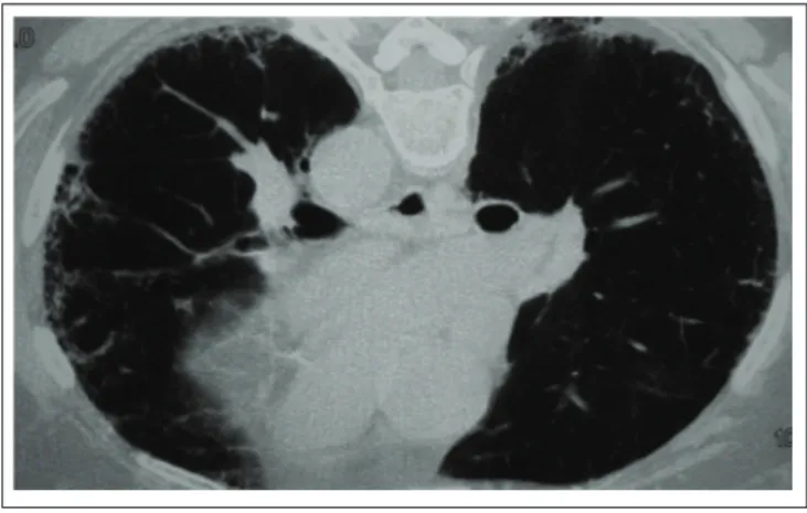
Figure 1A
Chest HRCT showing the reticular pattern of subpleural regions, pulmonary architectural distortion, and honeycombing areas.
Figure 1B
CT scan showing increased pulmonary attenuation with visu-alization of bronchi and vessels through the pulmonary area affected in the basal posterior segments of the lower lobes.
Figure 1C
CT scan showing thickening of the bronchial walls in the right lower and middle lobes, with centrilobular nodules accompa-nied by linear and nodular opacities.
Table 1 Chest HRCT fi ndings in 71 patients with rheumatoid arthritis
Finding N %
Normal 32/71 45.0
Peribronchial thickening 3/71 4.2
Septal thickening 5/71 7.0
Interstitial thickening 7/71 9.8
Parenchymal bands 14/71 19.7
Architectural distortion 11/71 15.4
Traction bronchiectasis 13/71 18.3
Honeycombing 9/71 12.6
Small perilymphatic nodules 1/71 1.4
Centrilobular nodules 1/71 1.4
Random nodules 5/71 7.0
Peripheral ground-glass opacity 17/71 23.9
Atypical ground-glass opacity 8/71 11.2
Focal ground-glass opacity 3/71 4.2
Note: No patient had consolidations, cavitary nodules, or tree-in-bud opacities.
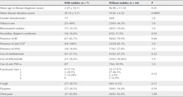
Analysis of the association of the nodular pattern pulmonary lesions
Tabela 2 Assessment of the demographic, clinical, serological and therapeutic variables in 71 patients with RA with and without increased pulmonary parenchymal density on chest HRCT
With increased pulmonary
parenchymal density (n = 23) Without increased pulmonary parenchymal density (n = 48) P
Mean age at disease diagnosis (years) 49.78 ± 10.23 44.9 ± 11.10 0.08
Mean disease duration (years) 12.57 ± 7.06 11.37 ± 6.96 0.50
Gender (female/male) 18/5 45/3 0.10
Tobacco use 6/21–28.5% 19/46–41.3% 0.41
Rheumatoid nodules 3/23–13.0% 8/45–17.7% 0.73
Secondary Sjögren’s syndrome 1/19–5.2% 8/41–19.5% 0.24
Presence of RF 18/22–81.8% 32/47–68.0% 0.26
Presence of anti-CCP 10/12–83.3% 17/20–85% 1.00
Presence of ANA 7/22–31.8% 11/45–24.4% 0.78
Use of methotrexate 11/22–50% 34/45 – 75.5% 0.052
Use of lefl unomide 8/22 – 36.3% 16/45–35.5% 1.00
Use of sulfasalazine 4/22–18.1% 7/45–16.6% 1.00
Use of anti-TNF-α 3/23–13.0% 4/48–8.3 % 0.67
Functional class 1 2 3 4
7–31.8% 10–45.4% 2–9.0% 3–13.3%
19–51.3% 13–35.13% 1–2.7% 4–10.8%
0.42
Cough 4/22–18.1% 2/43–4.65% 0.16
Dyspnea 5/22–22.7% 7/43–16.27% 0.52
Pain 5/22–22.7% 23/43–53.4% 0.03
anti-CCP: anti-cyclic citrullinated peptide antibody; ANA: antinuclear antibody; RF: rheumatoid factor; TNF: tumor necrosis factor.
Table 3 Assessment of the demographic, clinical, serological and therapeutic variables in 71 patients with RA with and without nodular lesions on chest HRCT
With nodules (n = 7) Without nodules (n = 64) P
Mean age at disease diagnosis (years) 3.29 ± 10.11 46.90 ± 11.10 0.41
Mean disease duration (years) 20.14 ± 5.11 10.82 ± 6.52 0.0005
Gender (female/male) 7/7 56/8 1.0
Tobacco use 2/5–40% 23/63–36.5% 1.0
Rheumatoid nodules 1/7–14.3% 10/51–19.6% 1.0
Secondary Sjögren’s syndrome 1/6–16.6% 6/52–11.5% 0.54
Presence of RF 6/7–85.7% 44/62–70.9% 0.66
Presence of anti-CCP 4/4–100% 23/28–82.1% 1.0
Presence of ANA 1/6–16.6% 17/61–27.8% 1.0
Use of methotrexate 4/7–57.1% 41/61–67.2% 0.23
Use of lefl unomide 2/7–26.6% 22/61–36.06% 1.0
Use of anti-TNF-α 0/7 7/64–10.9% 1.0
Functional class 1 2 3 4
4–57.1% 2–28.5% 1–14.28% 0
22-37.9 % 27-46.5% 2-3.4% 7-12.0%
0.32
Cough 2/7–28.5% 4/61–6.5% 0.11
Dyspnea 2/7–28.5% 10/61–16.4% 0.59
Chest pain 3/7–42.8% 26/61–42.6% 1.00
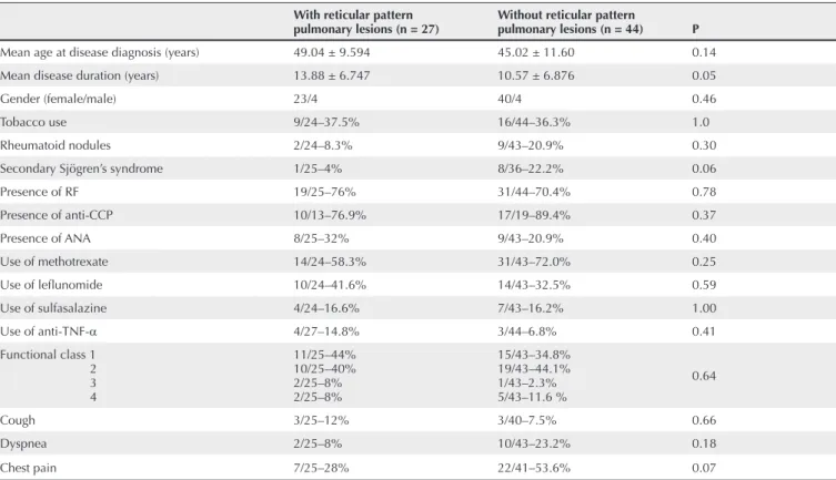
Table 4 Assessment of the demographic, clinical, serological and therapeutic variables in 71 patients with RA with and without reticular pattern pulmonary lesions on chest HRCT
With reticular pattern
pulmonary lesions (n = 27) Without reticular pattern pulmonary lesions (n = 44) P
Mean age at disease diagnosis (years) 49.04 ± 9.594 45.02 ± 11.60 0.14
Mean disease duration (years) 13.88 ± 6.747 10.57 ± 6.876 0.05
Gender (female/male) 23/4 40/4 0.46
Tobacco use 9/24–37.5% 16/44–36.3% 1.0
Rheumatoid nodules 2/24–8.3% 9/43–20.9% 0.30
Secondary Sjögren’s syndrome 1/25–4% 8/36–22.2% 0.06
Presence of RF 19/25–76% 31/44–70.4% 0.78
Presence of anti-CCP 10/13–76.9% 17/19–89.4% 0.37
Presence of ANA 8/25–32% 9/43–20.9% 0.40
Use of methotrexate 14/24–58.3% 31/43–72.0% 0.25
Use of lefl unomide 10/24–41.6% 14/43–32.5% 0.59
Use of sulfasalazine 4/24–16.6% 7/43–16.2% 1.00
Use of anti-TNF-α 4/27–14.8% 3/44–6.8% 0.41
Functional class 1 2 3 4
11/25–44% 10/25–40% 2/25–8% 2/25–8%
15/43–34.8% 19/43–44.1% 1/43–2.3% 5/43–11.6 %
0.64
Cough 3/25–12% 3/40–7.5% 0.66
Dyspnea 2/25–8% 10/43–23.2% 0.18
Chest pain 7/25–28% 22/41–53.6% 0.07
anti-CCP: anti-cyclic citrullinated peptide antibody; ANA: antinuclear antibody; RF: rheumatoid factor; TNF: tumor necrosis factor. Analysis of the association of the
reticular pattern pulmonary lesions
The analysis of the association of reticular pattern lesions (27/71 patients, 38%) with the variables studied is shown in Table 4. Once again, the only positive association was that of reticular lesions in patients with longer lasting disease.
DISCUSSION
Rheumatoid arthritis is traditionally considered a disease that involves the joints. Nevertheless, up to 50% of the patients have some type of extra-articular manifestations, such as serositis, pneumonitis, peripheral neuritis, nodules, and scleritis.2 This
study reviewed the prevalence of the pulmonary manifesta-tions seen on HRCT in a group of patients of southern Brazil, aiming at correlating such fi ndings with clinical, serological and treatment variables. The analysis of the data of the pres-ent study shows that most patipres-ents with RA (55%) have some type of CT changes. That high prevalence of fi ndings has been confi rmed by other authors, such as Zrour et al.,11 who have
reported changes in 49.3% of 75 patients from Tunisia, and Bilgicki et al.,3 who have reported them in 67.3% of 52 patients
in Turkey. In addition, Teraski et al.,12 studying patients with
RA and respiratory symptoms, have reported CT changes in 90% of them.
The most prevalent changes were those of the reticu-lar pattern, followed by increased parenchymal density. Positive associations with the demographic and clinical findings were scarce. Interstitial pulmonary disease has been reported as tending to be more common in male patients, those with high RF titers, and those with more severe and deforming joint disease.13,14 However, such
findings could not be confirmed in our sample, which showed an equal distribution in both genders, in the differ-ent functional classes, and in those with and without RF, anti-CCP, and ANA, although an analysis of the titers of those autoantibodies has not been performed. Similarly, the series studied by Bilgicki et al.3 has shown no association
The association between the pulmonary disease of RA and tobacco use is controversial. Tobacco use has been identifi ed as a predisposing factor to pulmonary disease in RA by several authors,15,16 but others have already shown a lack of association
between those two variables,3 such as observed in the present data.
It is worth noting that no association between the HRCT fi ndings and the respiratory complaints of pain, cough, and dyspnea could be found. Some possible explanations for those fi ndings are as follows: 1) patients with RA, suffering from several musculoskeletal changes, can have chest pain not related to pulmonary causes; 2) diffi culty in moving around, generated by the disability secondary to RA, does not allow the perception of dyspnea unless when more advanced.
Lastly, in the present series, none of the medications studied showed any association with the CT changes. Methotrexate is currently the disease-modifying anti-rheumatic drug most commonly used in the treatment of RA and has been associated with pulmonary toxicity. There is no data about the pulmonary involvement by methotrexate in our population. Cotin et al.17
have shown a deterioration of the pulmonary function in pa-tients on methotrexate, but such fi ndings were not clinically signifi cant. Other authors have found no changes in neither function nor CT fi ndings in patients with RA undergoing different types of treatment.3
The limitations of the present study were the size of the sample (n = 71) and the retrospective analysis of laboratory data (ANA, RF, and anti-CCP). The former can be responsible for type 2 statistical errors, hindering the appearance of some associations with the variables studied.
REFERENCES REFERÊNCIAS
1. Dedhia HV, DiBartolomeo A. Rheumatoid arthritis. Crit Care Clin 2002; 18:841-54
2. Nannini C, Ryu JH, Matteson EL. Lung Disease in Rheumatoid arthritis. Cur Opin Rheumatol 2008; 20:340-6.
3. Bilgici A, Ulusoy H, Kuru O, ÇelenkÇ-Unsal M, Danaci M. Pulmonary involvement in rheumatoid arthritis; Rheumatol Int 2005; 429-35.
4. Sugiyama Y, Ohno S, Kano S. Diffuse panbronchiolitis and rheumatoid arthritis: apossibler correlation with HLA B54. Inter Med 1994; 33:612-4
5. Charles PJ, Sweatman MC, Marckwick JR, Maini RN. HLA B40: a marker for susceptibility to lung disease in rheumatoid arthritis. Dis Markers 1991; 9:97-101.
6. Balbir-Gurman A, Guralnik L, Best LA, Vlodavsky E, Yigla M, Menahem-Nahir A et al. Accelerated pulmonary nodulosis and sterile pleural effusion in a patient with psoriatic arthropathy during methotrexate therapy: a case report. J Clin Rheumatol 2009; 15:29-30. 7. Rozin A, Yigla M, Guralnik L, Keidar Z, Vlodavsky E, Rozenbaum
M et al. Rheumatoid lung nodulosis and osteopathy associated with
lelunomide therapy. Clin Rheumatol 2006; 25:384-8.
8. Arnett FC, Edworthy SM, Bloch DA. McShane DJ, Fries JF, Cooper NS et al. The American Rheumatology Association 1987 revised
criteria for the classiication of rheumatoid arthritis. Arthritis Rheum
1988; 31:315-24.
9. Vitali C, Bombardieri S, Jonsson R. Moutsopoulos HM, Alexander EL, Carsons SE et al. Classification criteria for Sjögren’s syndrome: a revised version of the European criteria proposed by the American European consensus group. Ann Rheum Dis 2002; 61:554-8.
10. Klippel JH, Dieppe PA. Selected measures for outcome assessment of rheumatic diseases. In Klippel JH, Dieppe PA (Eds). Rheumatology 1998, 2nd Ed, Mosby, London, vol.2, S-A:1-12.
11. Zrour SH, Touzi M, Bejia I, Golli M, Rouatbi N, Sackly N et al. Correlations between high resolution computed tomography
of the chest and clinical function in patients with rheumatoid arthritis. Prospective study in 75 patients. Joint Bone Spine 2005; 72:41-7.
12. Teraski H, Fujimoto K, Hayabuchi N, Ogoh, Fukuda T. Respiratory symptons in rheumatoid arthritis: relation between
high resolution CT indings and functional impairment. Rad Med
13. Gabay E, Tarala R, Will R, Caroll G, Adler B, Cameron D et al. Interstitial lung disease in recent onset rheumatoid arthritis. Am J Respr Crit Care Med 1997; 156:528-5.
14. Mori S, Cho I, KogaY, Sugimoto M. Comparison of Pulmonary abnormalities on high resolution computed tomography in patients with early versus longstanding rheumatoid arthritis. J Rheumatol 2008; 35:1513-21.
15. Turesson C, Jacobsson LTH. Epidemiology of extra articular manifestations in rheumatoid arthritis.J Rheumatol 2004; 33:65-72.
16. Dawson JK, Fewins HE, Desmond J, Lynch MP, Graham DR. Fibrosing alveolitis in patients with rheumatoid arthritis as assessed by high resolution computed tomography, chest X Ray and pulmonary function tests. Thorax 2001; 56:622-7.