ABSTRACT
The objective of this systematic review was to characterize chest CT indings in patients with dysphagia and pulmonary aspiration, identifying the characteristics and the methods used. The studies were selected from among those indexed in the Brazilian Virtual Library of Health, LILACS, Indice Bibliográico Español de Ciencias de la Salud, Medline, Cochrane Library, SciELO, and PubMed databases. The search was carried out between June and July of 2016. Five articles were included and reviewed, all of them carried out in the last ive years, published in English, and coming from different countries. The sample size in the selected studies ranged from 43 to 56 patients, with a predominance of adult and elderly subjects. The tomographic indings in patients with dysphagia-related aspiration were varied, including bronchiectasis, bronchial wall thickening, pulmonary nodules, consolidations, pleural effusion, ground-glass attenuation, atelectasis, septal thickening, ibrosis, and air trapping. Evidence suggests that chest CT indings in patients with aspiration are diverse. In this review, it was not possible to establish a consensus that could characterize a pattern of pulmonary aspiration in patients with dysphagia, further studies of the topic being needed.
Keywords: Respiratory aspiration; Tomography, X-ray computed; Lung.
Chest CT indings in patients with
dysphagia and aspiration:
a systematic review
Betina Scheeren1, Erissandra Gomes2, Giordano Alves3, Edson Marchiori3,
Bruno Hochhegger1
Correspondence to:
Betina Scheeren. Rua Teixeira Mendes, 187, apto. 301, Chácara das Pedras, CEP 90050-170, Porto Alegre, RS, Brasil. Tel.: 55 51 9725-8226. E-mail: [email protected]
Financial support: None.
INTRODUCTION
The epidemiology of aspiration syndromes is not well described in the literature because of the lack of
speciicity and sensitivity markers; however, the literature
indicates that 5-15% of the cases of community-acquires pneumonia are due to aspiration.(1) Lung injury caused
by aspiration of saliva or food particles can often result
from dysphagia.(2,3) Dysphagia can be of neurogenic,
mechanical, or psychogenic origin and manifests itself through a series of signs and symptoms, such as cough, choking, and pharyngeal globus, being a major risk factor for malnutrition, dehydration, and aspiration
pneumonia.(4-6)
Evaluation of dysphagia involves clinical evaluation and speech pathology assessment, as well as ancillary tests, such as videoluoroscopic swallowing study (VFSS) and iberoptic endoscopic evaluation of swallowing (FEES), which serve to aid in the diagnosis of swallowing disorders,
such as aspiration.(7-9) In contrast, chest CT is used to
evaluate pulmonary lesions, being of great importance in the diagnosis of aspiration disorders, since pulmonary symptoms can be the irst manifestation of aspiration.(10)
Imaging indings of aspiration are numerous and usually nonspeciic, pulmonary infection being the most serious
complication of aspiration.(10,11) Therefore, knowledge about the different types of pulmonary aspiration is important for drawing correlations between the clinical
information and the main CT indings, including diffuse aspiration bronchiolitis, aspiration pneumonitis, aspiration
pneumonia, foreign body aspiration, and exogenous
lipoid pneumonia.(11) By speciically detailing the imaging
indings of aspiration pneumonia, segmental or lobar airspace consolidation can be observed, which may or
may not be associated with pleural effusion.(11)
Knowledge about the CT indings of aspiration is
essential for establishing the diagnosis of aspiration
disorders and for attempting to prevent lung injury. Here, we aimed to perform a systematic literature review of chest CT indings that characterize pulmonary aspiration in patients with dysphagia, identifying the characteristics
and the methods used.
METHODS
Research strategies
This systematic review followed the recommendations of the latest version of the Cochrane Handbook for Systematic Reviews of Interventions,(12) which involve
formulating the research question, inding and selecting scientiic articles, and critically assessing the selected
articles. The research question used was: “What chest
CT image indings are diagnostic markers of aspiration in patients with dysphagia?” The review was developed by three researchers, two of whom searched for articles
independently and blindly and one of whom was assigned
as a reviewer, being consulted in cases of uncertainty
so as to establish agreement. All of the researchers
involved—two speech-language pathologists and one
1. Programa de Pós-Graduação em Medicina, Universidade Federal de Ciências da Saúde de Porto Alegre, Porto Alegre (RS) Brasil.
2. Universidade Federal do Rio Grande do Sul, Porto Alegre (RS) Brasil. 3. Universidade Federal do Rio de Janeiro,
Rio de Janeiro (RJ) Brasil.
radiologist who works in thoracic radiology—have over 10years of clinical and practical experience in the study area. Studies were selected by using the following
search terms: “pneumonia aspirativa” and “aspiration
pneumonia”; “aspiração” and “aspiration”; “pulmonar”
and “pulmonary”; and “tomograia computadorizada” and “computed tomography”. These search terms
were obtained from DeCS and MeSH and were used to search the Brazilian Virtual Library of Health, LILACS,
Indice Bibliográico Español de Ciencias de la Salud, Medline, Cochrane Library, SciELO, and PubMed online
databases. The search was carried out between June
and July of 2016, on the basis of the intersection of
the chosen search terms.
Selection criterion
Studies in humans, written in English, Portuguese, or Spanish, regardless of the publication year, and whose title, abstract, or body of text contained any of the search terms chosen for this review were selected. Studies mentioning aspiration of food particles into the upper digestive tract and chest CT were included. Repeated studies were excluded, as were studies whose abstracts or full texts were not found in the databases consulted, review articles, dissertations, theses, case studies, and studies in which the underlying disease was tuberculosis. No search ilters were applied. The article selection process is described as a lowchart in Figure 1, as recommended in the PRISMA statement.(13)
Data analysis
After the abstracts of the studies found were selected, the full texts of the articles were retrieved. After full text reading, the following data were extracted: names of the authors; year of publication; country where the study was conducted; study design; study subjects;
sample size; diagnostic tests used; underlying disease; and chest CT indings, which were deined in accordance
with the glossary of terms for thoracic imaging from
the Fleischner Society.(14)
RESULTS
Five articles were selected for inclusion in the present systematic review, all of them carried out in the last 5 years, published in English, and coming from different countries (Italy, USA, Japan, China, and Brazil; Table 1). Sample sizes in the studies ranged from 43 to 56 patients, the predominant population being adults (19-59 years) and elderly subjects (≥ 60 years).
Most of the studies evaluated in this review had a retrospective, cross-sectional design.(15-18) The sample
characteristics varied. The patients studied had laryngeal cancer,(15) acute pneumonia associated with
dysphagia,(16) or chronic aspiration,(17,18) and a study of healthy subjects assessed the presence or absence of aspiration.(19) The diagnostic tests used in the studies
were VFSS,(15,16,18) FEES,(15,19) bronchoscopy,(17) HRCT,(15)
and conventional CT.(16-19)
The CT indings in patients with dysphagia-related aspiration were varied, including emphysema,(15)
bronchiectasis,(15,16,19) bronchial wall thickening,(15,16,18,19)
nodules,(15,16,18) tree-in-bud pattern,(15,19)
consolidation,(15-18) pleural effusion,(15-17)
ground-glass attenuation,(15,16,18) septal thickening,(15,16)
cavitary lesions,(15) lymph nodes,(15) atelectasis,(16-18)
bronchiolectasis,(18,19) ibrosis,(19) and air trapping.(18,19)
One of the studies demonstrated a higher frequency of indings in the right lung,(17) and two found changes
that were more prevalent in lower lung zones.(16,18) In
the study by Simonelli et al.,(15) it was not possible
to describe the proportion of indings, because they
Articles identified through database search
(N = 622)
Articles identified through other sources
(n = 0)
Articles excluded (n = 607)
Abstracts selected (n = 15)
Abstracts excluded (n = 10)
(Aspiration of gastric contents, absence of CT findings, and community-acquired pneumonia)
Full texts selected for inclusion in the study
(n = 5)
were reported by degree of aspiration. It is of note
that, in two studies, aspirators and non-aspirators
were compared.(18,19)
DISCUSSION
The selection, reading, and analysis of articles revealed that there have been few studies attempting to deine a pattern of chest CT indings related to dysphagia-related pulmonary aspiration. The ive articles selected in the present review were published in the last 5 years, which may explain the recent concern over early
identiication of patients with dysphagia who aspirate and over strategies that may intervene in the etiology. One study found a signiicant correlation between the degree of dysphagia and the relative risk of pneumonia,
demonstrating that patients with tracheal aspiration
are ten times more likely to develop pneumonia than individuals with normal swallowing.(20)
The most serious complication associated with aspiration in patients with dysphagia is pulmonary infection.(10) Studies indicate aspiration pneumonia as a cause of community-acquired pneumonia.(21,22) It is
important to note that, in addition to the respiratory
Table 1. Characteristics of the selected articles.
Study Year Country Population Study design
Sample size and characteristics
Diagnostic tests used
Chest CT findings
Simonelli et al.(15)
2010 Italy A/G RS 45 patients after partial laryngectomy (mean age = 67 years; 92.2% men) and 45 controls (patients with COPD and normal swallowing)
FEES, VFSS, and HRCT
Emphysema; bronchiectasis; bronchial wall thickening; pulmonary nodules or cysts; tree-in-bud pattern; consolidation; pleural effusion; septal thickening; cavitary lesions; and lymph nodes
Komiya et al.(16)
2013 Japan G RS 53 patients admitted to the hospital with pneumonia and dysphagia (mean age = 84 years; 66% of men)
VFSS and conventional CT
Centrilobular nodules (74%); ground-glass attenuation (74%); peribronchovascular thickening (42%); airspace consolidation (34%); atelectasis (17%); septal thickening (13%); pleural effusion (13%); and traction bronchiectasis (2%)
Lin et al.(17)
2014 China A/G RS 43 patients with aspiration (G = 17; A = 26; mean age = 56 years; 70% of men)
Bronchoscopy and
conventional CT
aConsolidation (93%/92%);
atelectasis (14%/23%); high-density airway lesion (29%/4%); pleural effusion (0%/8%); foreign body — food particles — (21%/35%): left lung (35%/31%) and right lung (65%/69%)
Butler et al.(19)
2014 USA G PS 50 healthy patients divided into 2 groups: aspirators (n = 25; mean age = 77 years; 15 women) and non-aspirators (n = 25; mean age = 76 years; 16 men)
FEES and conventional CT
bBronchiectasis (2%/8%);
bronchiolectasis (10%/6%); bronchial wall thickening (22%/12%); parenchymal band
(8%/4%); ibrosis (16%/16%);
air trapping (20%/26%); intraluminal airway debris (6%/8%); and tree-in-bud pattern (6%/4%) p > 0.05 for all
Scheeren et al.(18)
2016 Brazil A/G RS 56 patients divided into 2 groups: non-aspirators and aspirators (n = 28 in each group; mean age = 65 years; 29 men)
VFSS and conventional CT
bBronchial wall thickening
A B
complication, the swallowing disorder is a risk factor
for malnutrition and functional decline.(23) Aspiration pneumonia is the leading cause of death in patients with
dysphagia, a condition that affects 300,000-600,000 people per year in the USA.(1)
Simonelli et al.(15) addressed the relationship between
dysphagia and aspiration in laryngectomized patients, compared with a control group of patients with COPD, and found no signiicant differences in radiological indings between the groups. It is currently known that patients with COPD have dysphagia symptoms
related to airway protection because of changes in the breathing pattern and in the coordination of swallowing
and breathing, leading to a greater likelihood of developing pneumonia.(24) It should be noted here
that, of the 116 patients selected for the study,(15) only
45 had aspiration by VFSS and then underwent chest CT scans to assess the radiological manifestations of aspiration. In the two groups, the indings with the highest incidence rates were bronchial wall thickness, bronchiectasis, nodules, emphysema, consolidation, and septal thickening, with rates varying according to
the degree of aspiration in the study group.
In the study by Komiya et al.,(16) the pulmonary
CT indings were described in patients with an acute condition, that is, presenting with pneumonia at hospital admission, and dysphagia was conirmed by VFSS. The most frequent chest CT indings were airspace consolidation, ground-glass attenuation, centrilobular nodules, and peribronchovascular thickening. Pulmonary
opacities predominated in lower or diffuse areas of the lung and were distributed posteriorly. The authors
did not enroll a control group (without dysphagia/ aspiration). In the study,(16) there was a predominance
of elderly patients (geriatric population), among whom
the risk of aspiration of oropharyngeal secretions and food particles is increased.(25) There is evidence in the literature that the frequency of dysphagia is higher
in the elderly, and aspiration is an important etiologic
factor leading to pneumonia in this population.(26)
One of the studiesanalyzed in the present review did
not use ancillary tests to assess swallowing or detect
dysphagia; the presence of acute aspiration of large food particles was identiied solely by bronchoscopy.
(17) The most prevalent chest CT indings in the study
were consolidation, atelectasis, and high-density airway lesion, predominantly in the right lung and
lower lobe.(17)
In the study by Butler et al.(19) aspiration status was
prospectively evaluated by administering liquid boluses, and other bolus consistencies were not used, which
could result in an increased number of pulmonary
imaging indings. In addition, the authors did not use VFSS, which is considered the gold standard for detecting aspiration, choosing to use FEES. FEES is an exam that is performed with a nasal endoscope and allows direct observation and evaluation of laryngopharyngeal structures, as well as of swallowing; however, aspiration can only be observed after the swallow, through visualization of the presence of dyed
food particles in the trachea.(27) The authors did not
ind signiicant differences in the radiological pattern between the case and control groups, the indings being bronchiectasis, bronchiolectasis, bronchial wall thickening, air trapping, and ibrosis.(19)
The most recent study addressing pulmonary indings
in patients with chronic aspiration(18) included patients
with and without aspiration diagnosed by VFSS who underwent chest CT. A comparison of the two groups revealed that the patients with aspiration had a higher frequency of changes such as atelectasis, centrilobular nodules, bronchiolectasis, consolidation, and ground-glass
attenuation. Bronchial wall thickening and air trapping
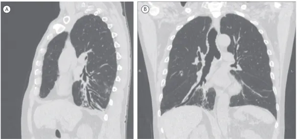
were the most prevalent indings in both groups; however, no signiicant differences were demonstrated. In addition, the authors reported that the indings were more prevalently distributed in lower lung zones. Figures 2 and 3 exemplify some of the CT indings described.
One of the limitations of the present systematic review was the lack of articles published on the subject,
Figure 2. In A, sagittal chest CT scan demonstrating bronchiolectasis, atelectasis, and areas of ground-glass opacity in
as well as the varied sample characteristics. In this review, it was not possible to establish a consensus that could characterize a pattern of pulmonary aspiration in patients with dysphagia, and further studies on the subject are needed. Evidence suggests
that chest CT indings in patients with aspiration are diverse; however, the articles mentioning the location of pulmonary indings detected that the indings were more prevalently distributed in the right lung and in lower lung zones.
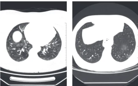
Figure 3. Axial chest CT scans showing areas of ground-glass attenuation in the left lower lobe.
REFERENCES
1. Marik PE. Aspiration pneumonitis and aspiration pneumonia. N Engl J Med. 2001;344(9):665-71. https://doi.org/10.1056/ NEJM200103013440908
2. Makharia GK, Seith A, Sharma SK, Sinha A, Goswami P, Aggarwal A,
et al. Structural and functional abnormalities in lungs in patients with achalasia. Neurogastroenterol Motil. 2009;21(6):603-8, e20.
3. Oue K, Mukaisho K, Higo T, Araki Y, Nishikawa M, Hattori T, et al. Histological examination of the relationship between respiratory disorders and repetitive microaspiration using a rat gastro-duodenal contents relux model. Exp Anim. 2011;60(2):141-50. https://doi. org/10.1538/expanim.60.141
4. Matsuo K, Palmer JB. Anatomy and physiology of feeding and swallowing: normal and abnormal. Phys Med Rehabil Clin N Am. 2008;19(4):691-707, vii. https://doi.org/10.1016/j.pmr.2008.06.001
5. Karkos PD, Papouliakos S, Karkos CD, Theochari EG. Current evaluation of the dysphagic patient. Hippokratia. 2009;13(3):141-6.
6. Carucci LR, Turner MA. Dysphagia revisited: common and unusual causes. Radiographics. 2015;35(1):105-22. https://doi.org/10.1148/ rg.351130150
7. Martin-Harris B, Jones B. The videoluorographic swallowing study.
Phys Med Rehabil Clin N Am. 2008;19(4):769-85, viii. https://doi. org/10.1016/j.pmr.2008.06.004
8. Kelly AM, Drinnan MJ, Leslie P. Assessing penetration and aspiration: how do videoluoroscopy and iberoptic endoscopic evaluation of swallowing compare? Laryngoscope. 2007;117(10):1723-7. https:// doi.org/10.1097/MLG.0b013e318123ee6a
9. Bours GJ, Speyer R, Lemmens J, Limburg M, de Wit R. Bedside screening tests vs. videoluoroscopy or ibreoptic endoscopic evaluation of swallowing to detect dysphagia in patients with neurological disorders: systematic review. J Adv Nurs. 2009;65(3):477-93. https://doi.org/10.1111/j.1365-2648.2008.04915.x
10. Franquet T, Giménez A, Rosón N, Torrubia S, Sabaté JM, Pérez C. Aspiration diseases: indings, pitfalls, and differential diagnosis. Radiographics. 2000;20(3):673-85. https://doi.org/10.1148/ radiographics.20.3.g00ma01673
11. Prather AD, Smith TR, Poletto DM, Tavora F, Chung JH, Nallamshetty
L, et al. Aspiration-related lung diseases. J Thorac Imaging. 2014;29(5):304-9. https://doi.org/10.1097/RTI.0000000000000092
12. Higgins JPT, Green S, editors. Cochrane Handbook for Systematic Reviews of Interventions Version 5.1.0 [updated 2011 Mar]. The Cochrane Collaboration; 2011. Available from:
http://www.cochrane-handbook.org
13. Moher D, Liberati A, Tetzlaff J, Altman DG; PRISMA Group. Preferred
reporting items for systematic reviews and meta-analyses: the PRISMA statement. Int J Surg. 2010;8(5):336-41. Erratum in: Int J Surg. 2010;8(8):658. https://doi.org/10.1016/j.ijsu.2010.02.007
14. Hansell DM, Bankier AA, MacMahon H, McLoud TC, Müller NL, Remy J. Fleischner Society: glossary of terms for thoracic imaging. Radiology. 2008;246(3):697-722. https://doi.org/10.1148/ radiol.2462070712
15. Simonelli M, Ruoppolo G, de Vincentiis M, Di Mario M, Calcagno P, Vitiello C, et al. Swallowing ability and chronic aspiration after supracricoid partial laryngectomy. Otolaryngol Head Neck Surg. 2010;142(6):873-8. https://doi.org/10.1016/j.otohns.2010.01.035
16. Komiya K, Ishii H, Umeki K, Kawamura T, Okada F, Okabe E, et al. Computed tomography indings of aspiration pneumonia in 53 patients. Geriatr Gerontol Int. 2013;13(3):580-5. https://doi. org/10.1111/j.1447-0594.2012.00940.x
17. Lin L, Lv L, Wang Y, Zha X, Tang F, Liu X. The clinical features of foreign body aspiration into the lower airway in geriatric patients. Clin Interv Aging. 2014;9:1613-8.
18. Scheeren B, Marchiori E, Pereira J, Meirelles G, Alves G, Hochhegger
B. Pulmonary computed tomography indings in patients with chronic aspiration detected by videoluoroscopic swallowing study. Br J Radiol. 2016;(1063):20160004. https://doi.org/10.1259/bjr.20160004
19. Butler SG, Clark H, Baginski SG, Todd JT, Lintzenich C, Leng X. Computed tomography pulmonary indings in healthy older adult aspirators versus nonaspirators. Laryngoscope. 2014;124(2):494-7. https://doi.org/10.1002/lary.24284
20. Pikus L, Levine MS, Yang YX, Rubesin SE, Katzka DA, Laufer I, et al.
Videoluoroscopic studies of swallowing dysfunction and the relative risk of pneumonia. AJR Am J Roentgenol. 2003;180(6):1613-6. https://doi.org/10.2214/ajr.180.6.1801613
21. Torres A, Serra-Batlles J, Ferrer A, Jiménez P, Celis R, Cobo E, et al. Severe community-acquired pneumonia. Epidemiology and prognostic factors. Am Rev Respir Dis. 1991;144(2):312-8. https:// doi.org/10.1164/ajrccm/144.2.312
22. Moine P, Vercken JB, Chevret S, Chastang C, Gajdos P. Severe
23. Serra-Prat M, Palomera M, Gomez C, Sar-Shalom D, Saiz A, Montoya JG, et al. Oropharyngeal dysphagia as a risk factor for malnutrition and lower respiratory tract infection in independently living older persons: a population-based prospective study. Age Ageing. 2012;41(3):376-81. https://doi.org/10.1093/ageing/afs006
24. Chaves Rde D, Carvalho CR, Cukier A, Stelmach R, Andrade CR. Symptoms of dysphagia in patients with COPD. J Bras Pneumol. 2011;37(2):176-83.
25. Augusto DK, Miranda LF, Cruz CE, Pedroso ER. Comparative
study of elderly inpatients clinically diagnosed with
community-acquired pneumonia, with or without radiological conirmation. J Bras Pneumol. 2007;33(3):270-4. https://doi.org/10.1590/S1806-37132007000300007
26. Marik PE, Kaplan D. Aspiration pneumonia and dysphagia in the elderly. Chest. 2003;124(1):328-36. https://doi.org/10.1378/ chest.124.1.328