Severe virus inluenza A H1N1 related pneumonia
and community-acquired pneumonia: diferences
in the evolution
INTRODUCTION
Traditionally, viral pneumonia is considered less severe than bacterial community-acquired pneumonia (CAP). However, with the inluenza A H1N1 (H1N1) outbreak in 2009, this assertion underwent a signiicant change because most of the infected individuals progressed to Acute Respiratory
Distress Syndrome (ARDS) and, in many cases, death.(1)
CAP is the leading cause of death from infectious diseases. he mortality rates are approximately 1% for outpatients and as high as 14% for hospitalized Paula Nardocci1, Caio Eduardo Gullo1, Suzana
Margareth Lobo1
1. Faculdade de Medicina de São José do Rio Preto - FAMERP - São José do Rio Preto (SP),
Brazil. Objective: To analyze the clinical,
laboratory and evolution data of patients with severe inluenza A H1N1 pneumonia and compare the data with that of patients with severe community-acquired bacterial pneumonia.
Methods: Cohort and retrospective study. All patients admitted to the intensive care unit between May 2009 and December 2010 with a diagnosis of severe pneumonia caused by the inluenza A H1N1 virus were included in the study. hirty patients with severe community-acquired pneumonia admitted within the same period were used as a control group. Severe community-acquired pneumonia was deined as the presence of at least one major severity criteria (ventilator or vasopressor use) or two minor criteria.
Results: he data of 45 patients were evaluated. Of these patients, 15 were infected with H1N1. When compared to the group with community-acquired pneumonia, patients from the H1N1
group had signiicantly lower leukocyte counts on admission (6,728±4,070 versus 16,038±7,863; p<0.05) and lower C-reactive protein levels (Day 2: 15.1±8.1 versus 22.1±10.9 mg/dL; p<0.05). he PaO2/FiO2 ratio values were lower in the irst week in patients with H1N1. Patients who did not survive the H1N1 severe pneumonia had signiicantly higher levels of C-reactive protein and higher serum creatinine levels compared with patients who survived. he mortality rate was signiicantly higher in the H1N1 group than in the control group (53% versus 20%; p=0.056, respectivelly).
Conclusion: Diferences in the leukocyte count, C-reactive protein concentrations and oxygenation proiles may contribute to the diagnosis and prognosis of patients with severe inluenza A H1N1 virus-related pneumonia and community-acquired pneumonia.
This study was conducted at the Division of Intensive Care, Hospital de Base de São José do Rio Preto, Faculdade de Medicina de São José do Rio Preto - FAMERP - São José do Rio Preto (SP), Brazil.
Conflicts of interest: None. Submitted on January 11, 2013 Accepted on June 30, 2013
Corresponding author:
Suzana Margareth Lobo
Serviço de Terapia intensiva do Hospital de Base Faculdade de Medicina de São José do Rio Preto Avenida Brigadeiro Faria Lima, 5.544
Zip code: 15090-000 - São José do Rio Preto (SP), Brazil
E-mail: [email protected]
ABSTRACT
Keywords: Pneumonia, viral; Pneumonia, bacterial; Inluenza A virus, H1N1 subtype
Pneumonia grave por vírus inluenza A H1N1 e pneumonia
comunitária grave: diferenças na evolução
patients; mortality rates are even higher for those who require hospitalization in intensive care units (ICU).(2,3)
he 2009 inluenza A H1N1 pandemic had signiicant morbidity,(4) and the irst reports suggested high mortality
even in young patients and in previously healthy middle-aged patients.(5) he hospitalization rates in countries in the
southern hemisphere ranged from 23.6 to 30.6% for individuals with inluenza A H1N1 2009; among these, 11.7 to 18.5% (3.6 to 4.4% of the total number of cases) were admitted to ICUs.(6) he mortality rates among those
admitted to the ICU were 16(7) to 41%(1), and most of those
patients required ventilatory support.(8)
Considering the severely ill patients, there was a high incidence of ARDS, the most common cause of death. Its lethality seems to be similar to that of seasonal inluenza,(1)
but it is greater than that reported for the respiratory
coronavirus registered during the 2003 outbreak.(9) A
study conducted in 11 ICUs from six cities in the state of Paraná (Brazil) included 63 suspected and 37 conirmed H1N1 cases and reported that most of the patients were young and that the mortality rate was 39.7%.(10)
he investigation of diagnostic and severity markers related to the nature of the infectious agent (bacterial versus viral) that caused the community-acquired pneumonia and those markers’ association with the clinical outcomes will allow that prediction of an unfavorable evolution, if present, and can guide earlier interventions and treatment. he present study aimed to determine the clinical, epidemiological and laboratory diferences between critically ill individuals with community-acquired pneumonia and those with severe pneumonia caused by the inluenza A H1N1 virus.
METHODS
Retrospectively and prospectively collected data from adult patients admitted to the Division of Critical Care (Mixed-24 bed ICU) of the Hospital de Base de São José do Rio Preto were analyzed. Data were collected between May 2009 and December 2010 from patients with a diagnosis of severe pneumonia caused by the inluenza A virus H1N1 (H1N1). his study was approved by the Institutional Research Ethics Committee and informed consent was waived due to its observational nature.
he infection with inluenza A H1N1 was diagnosed using the real time polymerase chain reaction (RT-PCR) of the nasopharyngeal secretion or respiratory tract specimens. Severe pneumonia caused by inluenza
A H1N1 was deined in the presence of fever >38°C,
compatible with pneumonia and signs of worsening of the disease, which included the following: tachypnea (respiratory rate >25), hypoxia (arterial oxygen saturation
- SaO2 ≤92% at room air and ≤94% in pregnant women),
cyanosis, oliguria, altered level of consciousness, worsening of chronic disease and hypotension (systolic blood pressure <90 mmHg) or the use of vasopressor drugs (dopamine >5 µg/kg/minute or any dose of norepinephrine).(11,12)
H1N1 patients were matched at a 2:1 ratio with cases of severe CAP admitted to the ICU. hus, the data used for comparison came from consecutive patients who were admitted to the ICU with severe CAP during the same period and for whom the diagnosis of inluenza A H1N1 had not been cogitated or the RT-PCR of nasopharyngeal or respiratory tract specimens were negative. Severe CAP requiring ICU admission was characterized by the presence of one major severity criteria (the need for mechanical ventilation or vasopressors) or two minor criteria (systolic blood pressure <90 mmHg or mean blood pressure <70 mmHg, arterial oxygen pressure/fraction of inspired oxygen [PO2/FiO2] <250, respiratory rate ≥30/minute, urea >19.6 mg/dL, altered mental status, multilobar
pneumonia, platelets <100,000 cells/mm3, leukopenia -
≤4 x 10⁹ or hypothermia [body temperature ≤36
°C]).(2)
he laboratory data included in the analysis were obtained from the routine morning collection, which occurred between 5 and 6 am. Serum PCR levels were measured using turbidimetric immunoassay.
Statistical analysis
he results were expressed as the mean and standard
deviation or median and 25th-75th percentiles. he
statistical analysis used Student’s t test to compare two groups of normally distributed continuous variables, and the Mann-Whitney test was used for non-normal distributions. Descriptive statistics were calculated for the quantitative variables, and they were analyzed using Fisher’s test. P values <0.05 were considered signiicant.
RESULTS
In patients with H1N1, the most frequent comorbidities were obesity, 53%; cardiovascular disease, 20%; and diabetes, 20%. In the CAP group, the most frequent comorbidities were cardiovascular diseases, 40%; tobacco smoking, 30%; and diabetes, 27% (Table 1). Obesity was signiicantly more prevalent in the H1N1 group (53%) than in the CAP group (6.6%), with p=0.02 (Table 1).
Compared with survivors, nonsurvivors of H1N1 were signiicantly older (40.2±9.2 versus 27.6±8.7 years; p=0.018), had higher serum lactate levels on admission (4.35±3.61 versus 1.53 ± 0.85 mEq/L; p=0.06) and had longer hospitalizations (not signiicant; Table 1).
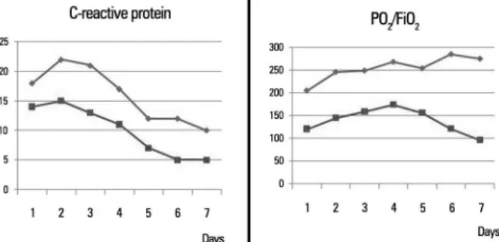
Compared with patients from the control group, patients from the H1N1 group had signiicantly lower leukocyte counts (Day 1: 6,728±4,070 versus 16,038±7,863 and Day 2: 7,957±5,981 versus 14,130±6,514; p<0.05 for both) and signiicantly lower serum PCR levels (Day 2: 15.1±8.1 versus 22.1±10.9 mg/dL, p<0.05; Table 2). H1N1 nonsurvivors had signiicantly higher CRP levels compared with survivors (Day 1: 21.9±2.9 versus 6.9±4.8 mg/dL; Day 2: 20.5±3.5 versus 10.6±8.2 mg/dL; Day 3: 20.3±4.1 versus 8.1±9.3 mg/dL; Day 4, 16.1±5.5 versus 5.6±6.8 mg/dL; p<0.05 for all; Table 2).
he PaO2/FiO2 ratio was signiicantly lower during
the irst week for patients with H1N1 (Table 3).
Nonsurvivors had lower PaO2/FiO2 values (Day 2: 54±43
versus 165±94) and higher plasma creatinine levels (Day 3: 1.72±0.59 versus 1.06±0.27 mg/dL; p<0.05) compared with survivors (Table 3). he platelet count values are described in table 3. Figure 1 shows the mean CRP (mg/
dL) and PaO2/FiO2 for the H1N1 and CAP groups.
DISCUSSION
In the present study, we observed that, compared with patients who were admitted with bacterial CAP, the H1N1 group had younger patients and a higher prevalence of obesity. Signiicant diferences were observed in the leukocyte count, CRP and oxygenation proiles, and higher mortality was observed in the H1N1 group.
Patients with diabetes mellitus, cancer, cardiovascular, respiratory and autoimmune diseases and pregnant women (especially those in the second and third trimesters of pregnancy) also showed increased susceptibility to more severe disease.(10) In a Brazilian study, 27% of the patients
with H1N1 were obese.(10) Obesity has been reported as a
risk factor for severity and mortality among patients with swine lu.(8) A probable reason for this fact is the decrease
in the functional residual capacity, which likely has an
important impact during the evolution of H1N1.(10)
Table 1 - Demographic data, clinical characteristics and outcomes from both groups
H1N1 CAP
Total S NS Total S NS
Number of patients 15 7 8 30 24 6
Male 9 (60.0) 6 (85.7) 3 (33.3) 21 (70.0) 17 (70.8) 4 (66.6)
Age (years) 34.3±10.9* 27.6±8.7 40.2±9.2# 55.9±16.4 58.2±18.9 55.3±16.1
Lactate at admission (mEq/L) 3.6±2.8 (N=7) 1.53±0.85 (N=3) 4.35±3.61 N= 4) 3.1±1.9 (N=28) 3.22±2.13 (N=22) 2.7±0.98 (N=6)
Comorbidities
Obesity 8 (53.3)* 3 (42.8) 5 (62.5) 2 (6.6) 2 (8.3) 0 (0.0)
CVD 3 (20.0) 1 (14.2) 2 (25.0) 12 (40.0) 10 (41.6) 2 (33.3)
Diabetes 3 (20.0) 2 (28.5) 1 (12.5) 8 (26.6) 5 (20.8) 3 (50.0)
Tobacco smoking 2 (13.3) 0 (0.0) 2 (25.0) 9 (30.0) 8 (33.3) 1 (16.6)
COPD 1 (6.6) 0 (0.0) 1 (12.5) 6 (20.0) 4 (16.6) 2 (33.3)
Alcoholism 0 (0) 0 (0.0) 0 (0.0) 4 (13.3) 3 (12.5) 1 (16.6)
CRF 0 (0) 0 (0.0) 0 (0.0) 1 (3.3) 1 (4.1) 0 (0.0)
Outcomes
Need for ventilator 13 (86.7) 7 (53.8) 6 (43.2) 3 (10.0) 2 (66.6) 1 (33.3)
Days of hospitalization 8.0 (5.0-15.0) 8.0 (6.0-11.0) 8.5 (2.7-24.0) 24 (15.7-28.2) 27.0 (19.2-9.5) 18.0 (9.0-3.7)
Table 2 - Inflammatory parameters during the first week of hospitalization for the two groups, H1N1 and community-acquired pneumonia survivors and nonsurvivors
H1N1 CAP
Total S NS Total S NS
Leukocytes D1 6,728±4,070 (N=14) 6,656±4,853 (N=7) 6,800±3,511 (N=7) 16,038±7,863 (N=29) 16,587±8,157 (N=23) 13,933±6,837 (N= 6)
Leukocytes D2 7,957±5,981 (N=14)* 5,886±3,458 (N=7) 10,029±7,453 (N=7) 14,130±6,514 (N=27) 15,376±6,841 (N=21) 9,767±2,026 (N=6) Leukocytes D3 9,867±6,874 (N=12) 8,050±4,529 (N=6) 11,683±8,691 (N=6) 13,363±6,257 (N=29) 13,410±6,270 (N=23) 13,182±6,799 (N=6)
Leukocytes D4 9,565±5,962 (N=12) 9,530±4,873 (N=6) 96,00±7,379 (N=6) 12,636±7,792 (N=28) 13,070±8,513 (N=23) 10,640±2,285 (N=5)
Leukocytes D5 10,691±5,657 (N=11) 9,933±6,868 (N=6) 11,600±4,375 (N=5) 13,179±8,180 (N=28) 13,665±8,826 (N=23) 10,940±3,955 (N=5) Leukocytes D6 11,210±5,821 (N=10) 8,983±5,894 (N=6) 14,550±4,355 (N=4) 12,708±7,820 (N=13) 13,291±8,375 (N=11) 9,500±2,828 (N=2)
Leukocytes D7 10,919±4,460 (N=7) 10,167±4,994 (N=3) 11,483±4,707 (N=4) 16,557±10,511 (N=7) 15,800±8,004 (N=18) 12,575±3,783 (N=4)
CRP D1 14.4±8.7 (N=12) 6.9±4.8 (N=6) 21.9±2.9 (N=6)# 18.0±10.9 (N=30) 18.2±11.7 (N=24) 17.2±8 (N=6) CRP D2 15.1±8.1* (N=13) 10.6±8.2 (N=7) 20.5±3.5 (N=6)# 22.1±10.9 (N=27) 22.1±11.7 (N=22) 22±6.8 (N=5)
CRP D3 13.0±9.5 (N=10) 8.1±9.3 (N=6) 20.3±4.1 (N=4)# 20.5±10.5 (N=30) 20.8±10.7 (N=24) 19.4±10.1 (N=6) CRP D4 11.3±8.1 (N=11) 5.6±6.8 (N=5) 16.1±5.5 (N=6)# 16.7±10.3 (N=29) 16.9±10.3 (N=24) 15.7±11.2 (N=5)
CRP D5 6.7±7.0 (N=11) 3.2±3.4 (N=6) 10.8±8.2 (N=5) 12.3±9.8 (N=28) 12.7±10.6 (N=23) 10.5±5.3 (N=5)
CRP D6 5.3±7.2 (N=9) 2.5±2.3 (N=5) 8.8±10.1 (N=4) 12.3±10.1 (N=26) 13.1±11 (N=21) 9.3±4.7 (N=5) CRP D7 5.2±6.2 (N=8) 2.4±2.5 (N=4) 8.0 ±7.8 (N=4) 10.6±8.6 (N=28) 10.7±9.1 (N=23) 10.3±6.8 (N=5) CAP - community-acquired pneumonia; S - survivor; NS - nonsurvivor; CRP - C-reactive protein. Results are expressed as number (%), mean±standard deviation (SD) (number of measures). *p<0.05 versus CAP; #p<0.05 versus survivors.
Table 3 - Oxygenation parameters, renal function and platelet count during the first week of hospitalization for both groups
H1N1 CAP
Total S NS Total S NS
P/F D1 121±92 (N=5) 165±94 (N=3) 54±43 (N=2) 205±94 (N=30) 208±93 (N=24) 192±106 (N=6)
P/F D2 145±113* (N=6) 283±4 (N=2) 76±46 (N=4)# 246±90 (N=25) 250±92 (N=19) 234±91 (N=6)
P/F D3 159±135 (N=6) 196±48 (N=2) 141±168 (N=4) 249±91 (N=27) 254±96 (N=23) 218±59 (N=4)
P/F D4 174±68* (N=5) 202 (N=1) 167±76 (N=4) 268±85 (N=29) 263±84 (N=24) 289±97 (N=5)
P/F D5 156±61 (N=5)* 217 (N=1) 140±59 (N=4) 254±68 (N=29) 265±68 (N=25) 201±37 (N=5)#
P/F D6 122±47 (N=6)* 138±81 (N=2) 113±34 (N=4) 285±87 (N=29) 283±83 (N=24) 294±113 (N=5)
P/F D7 97±31 (N=4)* 81±35 (N=2) 114±2 (N=2) 275±92 (N=29) 278±94 (N=24) 259±88 (N=5)
Creatinine (mg/dL) D1 1.23±0.3 (N=12) 1.2±0.37 (N=6) 1.2 ±0.33 (N=6) 1.39±0.6 (N=29) 1.34±0.64 (N=23) 1.62±0.43 (N=5)
Creatinine (mg/dL) D3 1.39±0.5 (N=10) 1.06±0.27 (N=5) 1.72±0.59 (N=5)# 1.22±0.7 (N=29) 1.32±0.84 (N=4) 1.15±0.39 (N=4)
Creatinine (mg/dL) D5 1.28±0.6 (N=11) 0.98±0.21 (N=6) 1.64±0.77 (N=5) 1.07±0.6 (N=28) 1.10±0.7 (N=23) 1.00±0.29 (N=4) Creatinine (mg/dL) D7 1.25±0.6 (N=6) 0.95±0.35 (N=2) 2.40±0.69 (N=4) 1.03±0.5 (N=28) 1.01±0.61 (N=24) 1.13±0.44 (N=4)
Platelets (x103) D1 181±57 (N=14) 158±58 (N=7) 203±50 (N=7) 238±143 (N=28) 282±174 (N=23) 205±114 (N=6)
Platelets (x103) D2 174±43 (N=8) 160±44 (N=3) 182±45 (N=5) 214±96 (N=28) 244±109 (N=21) 163±48 (N=6) Platelets (x103) D3 179±73 (N=7) 207±68 (N=5) 109±13 (N=2) 208±198 (N=28) 220±101 (N=23) 195±56 (N=6)
Platelets (x103) D4 188±63 (N=6) 194±59 (N=3) 182±78 (N=3) 198 ± 82 (N=28) 204±83 (N=23) 182±110 (N=5) Platelets (x103) D5 274±149 (N=5) 312±141 (N=4) 121 (N=1) 219±102 (N=28) 209±87 (N=22) 211±121 (N=5)
Platelets (x103) D6 285±135 (N=6) 294±190 (N=3) 276±97 (N=3) 226±110 (N=28) 216±100 (N=16) 137±30 (N=2)
Platelets (x103) D7 210±35 (N=4) 240±6 (N=2) 215±62 (N=3) 270±101 (N=28) 287±117 (N=16) 235±71 (N=4) CAP - community-acquired pneumonia; S - survivor; NS - nonsurvivor; P/F - PaO2/FiO2 ratio. Results are expressed as number (%), mean±standard deviation (SD). *p<0.05 versus CAP; #p<0.05
versus survivors.
CRP is an acute-phase protein released immediately after the start of the inlammatory or tissue injury process. CRP serum concentration is determined by its synthesis
monitoring of the response to CAP and nosocomial
pneumonia treatment.(13-15) A high CRP serum level on
admission to the emergency room was a predictor of
ICU admission and need for mechanical ventilation.(16)
However, the kinetics of serum CRP levels in patients with severe H1N1 pneumonia remain unknown to date.
Compared with patients from the CAP group, patients from the H1N1 group had signiicantly lower serum CRP levels and leukocyte counts, which suggest a lower inlammatory response compared with acute bacterial conditions and a possible viral infection. However, patients with H1N1 who did not survive had CRP levels that were higher than those of survivors and were similar to those found in the bacterial process. hese data suggest that the patients who died had a stronger inlammatory response or a secondary bacterial infection.
A secondary bacterial infection was implicated in the increased morbidity and mortality of patients with
inluenza A H1N1 2009.(17,18) he most commonly
identiied agents were Streptococcus pneumoniae,
Staphylococcus aureus, Haemophilus inluenzae and gram-negative bacilli. In fatal cases of H1N1 virus infection, the most evident histopathological indings were diferent degrees of alveolar injury with the presence of hyaline membrane and septal edema, necrotizing tracheitis and bronchiolitis, pulmonary vascular congestion, alveolar hemorrhage, pulmonary thromboembolism and bacterial coinfection in 26 to 38%.(19) A recent study reported that among patients with
H1N1 infection, CRP serum levels were signiicantly higher in those who developed pneumonia compared
with patients who did not develop pneumonia.(20)
PaO2/FiO2 values were signiicantly lower in patients with H1N1 than in patients with severe CAP who received mechanical ventilatory support. he main cause of death in patients with H1N1 is respiratory failure refractory to the usual mechanical ventilation.(21) Currently, the use of
extracorporeal membrane oxygenation in these patients seems to be one of the most efective measures for
reducing mortality.(21) he presence of severe hypoxemia
in patients who died suggests that the medical centers that receive these patients should be prepared to ofer this type of advanced life support to patients with severe hypoxemia.
he mortality rate in this series was 53% for patients with H1N1. Among ICU patients, the mortality rate is 16(7) to 41%(1) in diferent series, and most of the patients
required prolonged ventilatory support.(8)
he major limitations of this study are its observational nature, the small sample size and the pairing performed by consecutive sample. hus, other more adequate selection methods (such as the propensity score or by severity criteria) were not possible, which represent a potential selection bias). Other limitations were the inability to assess other markers of severity, such as procalcitonin and interleukin-6, and the fact that the study was conducted in a single center. However, the inclusion of a homogeneous population with severe forms of pneumonia supports the indings of the present study.
CONCLUSION
Usual laboratory tests may contribute to the diferential diagnosis of severe community-acquired pneumonia and severe inluenza A H1N1 pneumonia.
ACKNOWLEDGEMENTS
he authors thank the Scientiic Initiation Scholarship fund from the Faculdade de Medicina de São José do Rio Preto.
REFERENCES
1. Domínguez-Cherit G, Lapinsky SE, Macias AE, Pinto R, Espinosa-Perez L, de la Torre A, et al. Critically ill patients with 2009 influenza A(H1N1) in Mexico. JAMA. 2009;302(17):1880-7.
2. Mandell LA, Wunderink RG, Anzueto A, Bartlett JG, Campbell GD, Dean NC, Dowell SF, File TM Jr, Musher DM, Niederman MS, Torres A, Whitney CG; Infectious Diseases Society of America; American Thoracic Society. Infectious Diseases Society of America/American Thoracic Society consensus guidelines on the management of community-acquired pneumonia in adults. Clin Infect Dis. 2007;44 Suppl 2:S27-72.
3. Mongardon N, Max A, Bouglé A, Pène F, Lemiale V, Charpentier J, et al. Epidemiology and outcome of severe pneumococcal pneumonia admitted to intensive care unit: a multicenter study. Crit Care. 2012;16(4):R155. [Epub ahead of print]
4. AlMazroa MA, Memish ZA, AlWadey AM. Pandemic influenza A (H1N1) in Saudi Arabia: description of the first one hundred cases. Ann Saudi Med. 2010;30(1):11-4.
5. Louie JK, Acosta M, Winter K, Jean C, Gavali S, Schechter R, Vugia D, Harriman K, Matyas B, Glaser CA, Samuel MC, Rosenberg J, Talarico J, Hatch D; California Pandemic (H1N1) Working Group. Factors associated with death or hospitalization due to pandemic 2009 influenza A(H1N1) infection in California. JAMA. 2009;302(17):1896-902.
6. Maritz J, Maree L, Preiser W. Pandemic influenza A (H1N1) 2009: the experience of the first six months. Clin Chem Lab Med. 2010;48(1):11-21. 7. Bellomo R, Pettilä V, Webb SA, Bailey M, Howe B, Seppelt IM. Acute
kidney injury and 2009 H1N1 influenza-related critical illness. Contrib Nephrol. 2010;165:310-4.
8. Rello J, Rodríguez A, Ibañez P, Socias L, Cebrian J, Marques A, Guerrero J, Ruiz-Santana S, Marquez E, Del Nogal-Saez F, Alvarez-Lerma F, Martínez S, Ferrer M, Avellanas M, Granada R, Maraví-Poma E, Albert P, Sierra R, Vidaur L, Ortiz P, Prieto del Portillo I, Galván B, León-Gil C; H1N1 SEMICYUC Working Group. Intensive care adult patients with severe respiratory failure caused by Influenza A (H1N1)v in Spain. Crit Care. 2009;13(5):R148. Objetivo: Analisar dados clínicos, laboratoriais e de evolu-ção de pacientes com pneumonia grave por vírus inluenza A H1N1 em comparação à pneumonia bacteriana grave adquirida na comunidade.
Métodos: Estudo de coorte, retrospectivo. Todos os pa-cientes admitidos na unidade de terapia intensiva, entre maio de 2009 e dezembro de 2010, com diagnóstico de pneumonia grave por inluenza A H1N1 foram incluídos. Trinta pacientes com pneumonia adquirida na comunidade grave admitidos no mesmo período foram usados como grupo controle. Pneumonia adquirida na comunidade grave foi deinida como presença de ao menos um critério maior de gravidade (uso de ventilador ou vasopressor) ou de dois critérios menores.
Resultados: Foram avaliados os dados de 45 pacientes. Den-tre eles, 15 pacientes com H1N1. Em comparação ao grupo com pneumonia adquirida na comunidade, pacientes do grupo H1N1
tiveram contagens de leucócitos signiicativamente menores na admissão (6.728±4.070 versus 16.038±7.863; p<0,05) e níveis de proteína C-reativa mais baixos (dia 2: 15,1±8,1 vs. 22,1±10,9 mg/dL, p<0,05). Os valores da relação PaO2/FiO2 foram menores na primeira semana em pacientes com H1N1. Não sobreviventes de pneumonia grave por H1N1 tiveram níveis signiicativamente mais elevados de proteína C-reativa do que os sobreviventes, além de níveis séricos mais altos de creatinina. A taxa de mortalidade foi signiicativamente mais elevada no grupo H1N1 do que no grupo controle (53% versus 20%, p=0,056, respectivamente.
Conclusão: Diferenças nos peris de contagem de leucócitos, proteína C-reativa e de oxigenação podem auxiliar no diagnós-tico e na avaliação do prognósdiagnós-tico de pacientes com pneumonia grave por vírus inluenza A H1N1 e por pneumonia adquirida na comunidade.
RESUMO
Descritores: Pneumonia viral; Pneumonia bacteriana; Vírus da inluenza A subtipo H1N1
9. Centers for Disease Control and Prevention (CDC). Revised U.S. surveillance case definition for severe acute respiratory syndrome (SARS) and update on SARS cases--United States and worldwide, December 2003. MMWR Morb Mortal Wkly Rep. 2003;52(49):1202-6.
10. Duarte PA, Venazzi A, Youssef NC, Oliveira MC, Tannous LA, Duarte CB, et al. Pacientes com infecção por vírus A (H1N1) admitidos em unidades de terapia intensiva do Estado do Paraná, Brasil. Rev Bras Ter Intensiva. 2009;21(3):231-6.
11. Influenza A (H1N1). Situação epidemiológica da nova influenza A (H1N1) no Brasil, até semana epidemiológica 33 de 2009. Informe Epidemiológico. 2009;1(6):1-10. [Internet]. [citado2009 Abr 1]. Disponível em http://portal. saude.gov.br/portal/arquivos/pdf/informe_influenza_se_33_25_08_2009.pdf 12. Brasil. Ministério da Saúde. Secretaria de Atenção a Saúde. Departamento
de Atenção Básica. Emergência de Saúde Pública de Importância Internacional – ESPII. Diretrizes para o enfrentamento à pandemia de influenza A (H1N1): ações da atenção primária à saúde. Brasília: Ministério da Saúde; 2009. Disponível em http://portal.saude.gov.br/portal/arquivos/ pdf/protocolo_influenzaa_aps_atualizado.pdf
13. Bruns AH, Oosterheert JJ, Hak E, Hoepelman AI. Usefulness of consecutive C-reactive protein measurements in follow-up of severe community-acquired pneumonia. Eur Respir J. 2008;32(3):726-32.
14. Lisboa T, Seligman R, Diaz E, Rodriguez A, Teixeira PJ, Rello J. C-reactive protein correlates with bacterial load and appropriate antibiotic therapy in suspected ventilator-associated pneumonia. Crit Care Med. 2008;36(1):166-71.
15. Moreno MS, Nietmann H, Matias CM, Lobo SM. C-reactive protein: a tool in the follow-up of nosocomial pneumonia. J Infect. 2010;61(3):205-11. 16. Zimmerman O, Rogowski O, Aviram G, Mizrahi M, Zeltser D, Justo D, et
al. C-reactive protein serum levels as an early predictor of outcome in patients with pandemic H1N1 influenza A vírus infection. BMC Infect Dis. 2010;10:288.
18. Palacios G, Hornig M, Cisterna D, Savji N, Bussetti AV, Kapoor V, et al. Streptococcus pneumoniae coinfection is correlated with the severity of H1N1 pandemic influenza. PLoS One. 2009;4(12):e8540.
19. Writing Committee of the WHO Consultation on Clinical Aspects of Pandemic (H1N1) 2009 Influenza, Bautista E, Chotpitayasunondh T, Gao Z, Harper SA, Shaw M, Uyeki TM, et al. Clinical aspects of pandemic 2009 influenza A (H1N1) virus infection. N Engl J Med. 2010;362(18):1708-19. Erratum in N Engl J Med. 2010;362(21):2039.
20. Milosevic I, Korac M, Zerjav S, Urosevic A, Lavadinovic L, Milosevic B, et al. Non-specific inflammatory parameters in patients with pandemic H1N1 influenza. Biomed Pharmacother. 2013;67(3):218-20.