Extracorporeal membrane oxygenation in acute
respiratory distress syndrome due to inluenza
A (H1N1)pdm09 pneumonia. A single-center
experience during the 2013-2014 season
Nithya Menon1, Carlos M. Perez-Velez2, Jennifer A. Wheeler3, Michael F. Morris4, Orazio L. Amabile3, Mark R. Tasset3, Robert A. Raschke1
1. Division of Pulmonary and Critical Care Medicine, Department of Medicine, Banner - University Medical Center Phoenix - Arizona, United States.
2. Division of Infectious Diseases, Department of Medicine, Banner - University Medical Center Phoenix - Arizona, United States.
3. Division of Cardiothoracic Surgery, Department of Surgery, Banner - University Medical Center Phoenix - Arizona, United States.
4. Division of Thoracic Radiology, Department of Radiology, Banner - University Medical Center Phoenix - Arizona, United States.
Objective: his report aimed to describe the outcomes of the patients with severe H1N1 associated acute respiratory distress syndrome who were treated with extracorporeal membrane oxygenation therapy.
Methods: his retrospective review analyzed a single-center cohort of adult patients with H1N1-related acute respiratory distress syndrome who were managed with veno-venous extracorporeal membrane oxygenation during the winter of 2013/2014.
Results: A total of 10 patients received veno-venous extracorporeal membrane oxygenation for H1N1 inluenza between January 2013 and March 2014. Seven patients were transferred to our center for extracorporeal membrane oxygenation consideration (all within 72 hours of initiating mechanical ventilation). he median patient age was forty years, and 30% were female. he median arterial oxygen partial pressure to fraction of inspired oxygen ratio was 62.5, and the median RESP score was 6. hree
Conflicts of interest: None.
Submitted on November 20, 2016 Accepted on January 15, 2017
Corresponding author:
Nithya Menon
Virginia Mason Memorial Hospital 2811 Tieton Drive,
Yakima, WA 98902 United States E-mail: [email protected]
Responsible editor: Luciano César Pontes de Azevedo
Oxigenação por membrana extracorpórea na síndrome do
desconforto respiratório agudo devido à pneumonia por inluenza
A (H1N1)pdm09. Experiência em um único centro durante a
temporada de 2013-2014
ABSTRACT
Keywords: Extracorporeal
membrane oxygenation; Respiratory distress syndrome, acute; Inluenza A virus, H1N1 subtype
patients received inhaled nitric oxide, and four patients were proned as rescue therapy before extracorporeal membrane oxygenation was initiated. he median duration of mechanical ventilation was twenty-two days (range, 14 - 32). he median length of stay in the intensive care unit was twenty-seven days (range, 14 - 39). he median hospital length of stay was 29.1 days (range, 16.0 - 46.9). Minor bleeding complications occurred in 6 of 10 patients. Eight of the ten patients survived to hospital discharge.
Conclusion: he survivors were relatively young and discharged with good functional status (i.e., enhancing quality-adjusted life-years-saved). Our experience shows that even a relatively new extracorporeal membrane oxygenation program can play an important role in that capacity and provide excellent outcomes for the sickest patients.
INTRODUCTION
Extracorporeal membrane oxygenation (ECMO) has been a therapeutic option for severe acute respiratory distress (ARDS) for approximately forty years.(1) he
eicacy of ECMO for treating severe ARDS in adults has been supported by a single randomized controlled trial, which demonstrated a signiicant improvement in survival, with good neurological function in 180 patients randomized for referral to ECMO consideration versus conventional ventilator support (CESAR trial). his study was published in the fall of 2009, just as a global pandemic of a newly emergent H1N1 inluenza virus, or swine lu, peaked.
his pandemic originated in Mexico in March 2009,(2)
and it was found to have resulted from a quadruple re-assortment between 2 swine, 1 human, and 1 avian inluenza strains.(3,4) he epidemiological expression
of this pandemic was distinct compared to previous typical seasonal inluenza activity.(5) he inluenza-related
hospitalization rate for adults 18 - 49 years of age increased six-fold.(6-8) Unusually high numbers of young, previously
healthy adults sufered severe ARDS requiring mechanical ventilation and, in some centers, ECMO. he Centers for Disease Control and Prevention estimated that 12,500 deaths resulted in the United States,(6) including 85%
younger than 65 years of age. When the relative youth of the patients who succumbed was considered, it was estimated that 334,000 - 1,973,000 years of life were lost in the pandemic.(7) he predilection for severe disease in
young adults was attributed to the immune naïve status of younger patients in relation to H1N1 inluenza strains. Fourteen studies from Europe, Japan, the United States and Australia/New Zealand reported ECMO treatment results from a cumulative population of 487 patients with H1N1 inluenza during the 2009 pandemic, with reported survival rates ranging from 32 - 92%.(9-12)
he CESAR trial and 2009 lu pandemics were important motivators in the initiation of our ECMO program in May 2010.(13) From 2010-2012, however,
H3N2 emerged as the predominant clinical strain of inluenza A, and the apparent need for ECMO support for inluenza patients at our institution dropped sharply.(14) H1N1 then re-emerged in the fall of 2013.(15)
Seasonal inluenza A activity in the United States began to increase in mid-November 2013, and referrals of patients with severe ARDS due to inluenza A quickly increased
at our institution in January 2014. Other ECMO centers in Arizona simultaneously experienced peak ECMO demand. Although we coordinated our eforts, our combined capacity to provide ECMO was nearly completely engaged in February 2014.
his report aimed to describe the outcomes of the patients with severe H1N1 associated acute respiratory distress syndrome who were treated with extracorporeal membrane oxygenation therapy.
METHODS
he Banner Health Institutional Review Board approved this study, and informed consent and ethical approval requirements were waived (Project # 01-15-0112).
All adult patients who were treated with veno-venous ECMO (vvECMO) for severe microbiologically proven inluenza A during the winter of 2013-14 in our medical/surgical intensive care unit at Banner University Medical Center were included. Inluenza A virus was detected through polymerase chain reaction, direct luorescent antigen, rapid enzyme immunoassay test, and/ or viral cultures, in clinical specimens obtained through nasopharyngeal swabs, suction of endotracheal secretions or bronchoalveolar lavage. Patients were triaged to vvECMO at the discretion of intensivist and cardiothoracic surgeons. Our triage policy favors vvECMO in patients with an arterial oxygen partial pressure to fraction of inspired oxygen (PaO2/FiO2) ratio < 100, who do not have life-threatening co-morbidities and who have not received prolonged injurious mechanical ventilation (> 7 days) likely to have resulted in severe ventilator-associated lung injury. Venous access is typically achieved with 27 - 31 F Avalon catheters placed in the right internal jugular vein. he Seldinger technique was used to insert the catheter. Correct positioning was conirmed through luoroscopy and chest X-ray. Maquet Rotalow®
and Maquet Cardiohelp®
We collected the following data: the patient demographics, risk factors for severe inluenza H1N1 pneumonia and major co-morbidities, respiratory parameters before the ECMO initiation, technical characteristics of ECMO therapy, complications and outcomes. Illness severity was determined using the Sequential Organ Failure Score Assessment (SOFA) and the Acute Physiology and Chronic Health Evaluations score (APACHE IV). Secondary pneumonias were diagnosed through clinical and radiographic indings, in conjunction with quantitative bronchoalveolar lavage cultures.
RESULTS
A total of 10 patients received vvECMO for H1N1 inluenza between January 2013 and March 2014. Seven patients were transferred to our center for ECMO consideration, all within 72 hours of initiation of mechanical ventilation. he clinical features and management, complications and outcomes details are described in table 1. he median patient age was forty years, and 30% were female. Seven patients were obese, with a body mass index greater than 30kg/m2.
Four patients had signiicant comorbidities, including chronic obstructive pulmonary disease and coronary artery disease. Most patients, however, were otherwise previously healthy, with few comorbidities. One patient was pregnant and underwent an emergency Caesarean section before starting ECMO. None of our patients had received seasonal inluenza vaccinations.
Inluenza virus was diagnosed in all patients using at least one of the following viral diagnostic tests: positive reverse-transcriptase polymerase chain reaction (Quest Diagnostics, nine patients), positive viral culture (Quest Diagnostics, two patients), and positive direct luorescent antibody staining (Quest Diagnostics, three patients). Co-infection with other viruses was found in two patients, one with metapneumovirus and another with respiratory syncytial virus. Nine patients had a bronchoscopy with bronchial wash and lavage performed within 24 hours after hospitalization at our center. Respiratory cultures were sent for viral, bacterial and fungal analysis.
Most patients demonstrated typical symptoms of inluenza for at least a week before medical attention was sought but then rapidly deteriorated over 48 - 72 hours. Upon admission, nine patients presented with septic
shock requiring vasopressors. he median PaO2/Fi02 ratio was 62.5. Six patients had severe and bilateral air space disease involving three or four quadrants. he most common radiological patterns were dense consolidation in all ten patients, ground glass opacities in two patients and pleural efusions in one patient. Interestingly, one patient had a normal chest radiograph upon admission and then, forty-eight hours after admission, progressed to having iniltrates involving four quadrants.
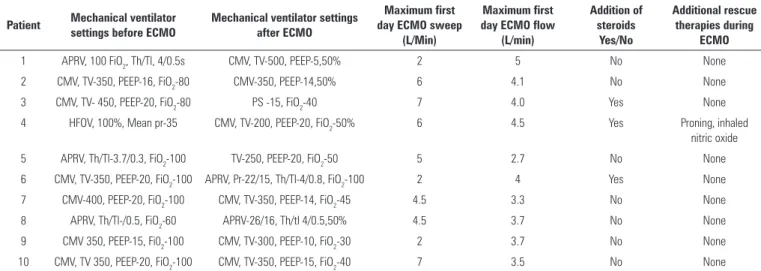
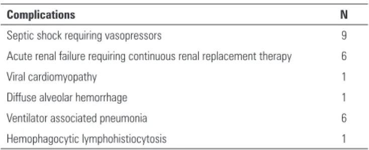
Half of the patients received volume-controlled ventilation, and half received airway pressure release ventilation prior to the initiation of ECMO (Table 2). Oseltamivir was started after a median 24 hours following symptom onset. hree patients received inhaled nitric oxide, and four patients were prone-positioned as rescue therapy before ECMO was initiated. Nine patients were placed on ECMO within 48 hours of admission, and one was placed on ECMO after six days of mechanical ventilation. he median SOFA score at the time of ECMO initiation was 12. Hemorrhagic complications occurred in a total of 6 patients. Four patients sufered bleeding from the ECMO catheter site, which required the temporary discontinuation of heparin. Two patients required transfusions of at least two units of packed red blood cells. Gastrointestinal bleeding was noted in two patients, and it resolved with blood transfusion and temporary withholding of heparin. Entrainment of air into ECMO circuit occurred in one case, but it did not result in systemic air embolization. Six patients sufered seven episodes of secondary bacterial ventilator-associated pneumonia: ive due to Pseudomonas aeruginosa, one due to methicillin-susceptible Staphylococcus aureus and one due to Enterococcus cloacae. One patient each sufered acute cardiomyopathy and difuse alveolar hemorrhage presumably related to inluenza. One patient who had underlying lymphoproliferative malignancy developed hemophagocytic lymphohistiocytosis shortly before his death. No patient sufered barotrauma.
Table 1 - Baseline characteristics
Patient Age Sex BMI Co morbidities Viral testing Rescue therapy p/f ratio SOFA at ECMO initiation
Days on ventilator prior to ECMO
1 27 M 26.5 None Rapid antigen Proning 62 13 < 24 hours
2 31 M 41.6 None Rapid antigen Prone, Inhaled nitric oxide 44 13 < 24 hours
3 42 M 40 Hypertension,
smoking Viral culture None 45 10 48 hours
4 44 F 52 Asthma, heart failure Rapid antigen
Neuromuscular blocking agents, HFOV, inhaled nitric
oxide, Prone
43 11 72 hours
5 30 F NA Pregnant DFA Neuromuscular blocking
agents 69 13 24 hours
6 58 M 32 CLL, COPD, smoking Viral PCR and culture Bilevel ventilation 66 9 5 days
7 66 M 37 Hypertension, WPW DFA Inhaled nitric oxide 56 12 48 hours
8 34 M 33.3 None PCR Bilevel ventilation 57 14 24 hours
9 41 F 25.9 Asthma, ulcerative
colitis PCR None 63 10 24 hours
10 40 M 50 None PCR None 49 16 24 hours
BMI - body mass index; SOFA - Simplified Organ Function Assessment; ECMO - extracorporeal membrane oxygenation; M - male; F - female; HFOV - high frequency oscillator ventilation; NA - no assessment; DFA - direct fluorescent antigen testing; CLL - chronic myeloid leukemia; COPD - chronic obstructive pulmonary disorder; WPW - Wolf-Parkinson White; PCR - polymerase chain reaction.
Table 2 - Additional ventilator settings
Patient Mechanical ventilator settings before ECMO
Mechanical ventilator settings after ECMO
Maximum first day ECMO sweep
(L/Min)
Maximum first day ECMO flow
(L/min)
Addition of steroids
Yes/No
Additional rescue therapies during
ECMO
1 APRV, 100 FiO2, Th/Tl, 4/0.5s CMV, TV-500, PEEP-5,50% 2 5 No None
2 CMV, TV-350, PEEP-16, FiO2-80 CMV-350, PEEP-14,50% 6 4.1 No None
3 CMV, TV- 450, PEEP-20, FiO2-80 PS -15, FiO2-40 7 4.0 Yes None
4 HFOV, 100%, Mean pr-35 CMV, TV-200, PEEP-20, FiO2-50% 6 4.5 Yes Proning, inhaled
nitric oxide
5 APRV, Th/Tl-3.7/0.3, FiO2-100 TV-250, PEEP-20, FiO2-50 5 2.7 No None
6 CMV, TV-350, PEEP-20, FiO2-100 APRV, Pr-22/15, Th/Tl-4/0.8, FiO2-100 2 4 Yes None
7 CMV-400, PEEP-20, FiO2-100 CMV, TV-350, PEEP-14, FiO2-45 4.5 3.3 No None
8 APRV, Th/Tl-/0.5, FiO2-60 APRV-26/16, Th/tl 4/0.5,50% 4.5 3.7 No None
9 CMV 350, PEEP-15, Fi02-100 CMV, TV-300, PEEP-10, FiO2-30 2 3.7 No None
10 CMV, TV 350, PEEP-20, FiO2-100 CMV, TV-350, PEEP-15, FiO2-40 7 3.5 No None
ECMO - extracorporeal membrane oxygenation; APRV - airway pressure release ventilation; FiO2 - fraction of inspired oxygen; Th/Tl- time high/time low; CMV - controlled mandatory ventilation;
TV - tidal volume; PEEP - positive end expiratory pressure, PS - pressure support; HFOV - high-frequency oscillator ventilation.
our eight survivors, all were ambulatory, and seven were on room air at the time of discharge to home.
DISCUSSION
Here, we describe the outcomes of a cohort of ten patients with severe H1N1 ARDS treated with vvECMO during the winter of 2013-2014 at our institution. he statewide need for ECMO support increased sharply this season compared to prior years, in conjunction with the
re-emergence of H1N1 inluenza. he observed increase in ECMO utilization was not relected in the overall mortality of inluenza during the 2012-2013 winter, which was not signiicantly increased compared to the 2010-2012 seasons.(16) It appears that a relatively small subset of
In many aspects, our patients were similar to those reported to have received ECMO for H1N1 inluenza in 2009. he median patient age of 40 in our series is similar to that previously reported.(18-20) Other characteristics,
such as obesity and comorbidities, were similar to those reported in previous studies.(9,18,21) he SOFA scores of our
patients were higher than those reported in a few recently published studies,(11,18) however, the PaO
2/FiO2 ratio and
lung injury score were similar to those in other reported studies.(10,17,18) ECMO was started within 48 hours of
mechanical ventilation in nine patients, similar to other studies.(11,17,18,21)
Life-threatening complications were common in our cohort (Table 3). he most frequent complication seen in patients treated with ECMO was bleeding.(22-27) Cannula
insertion sites, which were the most frequent bleeding site reported in the Extracorporeal Life Support Organization (ELSO) registry (occurring in 17% of patients), were also the most frequent bleeding site in our study.(26) Nine
patients sufered septic shock, and six required continuous renal replacement therapy. Our patients sufered a high rate of ventilator-associated pneumonia (VAP), despite active quality improvement eforts at our hospital to prevent VAP. he incidences of secondary bacterial VAP have ranged from 40 to 71% in previous studies of inluenza patients on ECMO,(9,12,23) and we are not the irst group to
report a preponderance of gram-negative pathogens.(9,28)
It is likely that post-inluenza bacterial pneumonias are fundamentally diferent than VAP in non-inluenza patients and more diicult to prevent. Less common complications, such as difuse alveolar hemorrhage, myocarditis and hemophagocytic lymphohistiocytosis, signiicantly impacted morbidity and mortality rates in our patients. Virus-associated hemophagocytic syndrome has been reported to be a major cause of death in 9 of 17 patients who received ECMO for H1N1 Inluenza related ARDS.(29) Renal dysfunction is also a common
complication, with six patients requiring continuous renal replacement therapy, and it reportedly occurred in 13% of patients in the ELSO registry.(26) In an analysis of 72
patients receiving ECMO for respiratory failure, only pre-ECMO serum creatinine levels correlated with survival.(30)
It is unclear if this inding is related to the overall severity of organ dysfunction and not speciically to ECMO.
Table 3 - Complications
Complications N
Septic shock requiring vasopressors 9
Acute renal failure requiring continuous renal replacement therapy 6
Viral cardiomyopathy 1
Diffuse alveolar hemorrhage 1
Ventilator associated pneumonia 6
Hemophagocytic lymphohistiocytosis 1
Despite the complications described above, our outcomes were encouraging and comparable to those reported during the 2009 H1N1 pandemic (Table 4). Our 80% survival rate is similar to that reported by Australia and New Zealand Extracorporeal Membrane Oxygenation (ANZ ECMO) Inluenza Investigators (79%), Zangrillo et al. (72%), Pham et al. (64%) and Holzgraefe et al. (92%).(9,11,18,21) A small study in a French
hospital (n = 12) reported a high survival rate, despite many complications, such as VAP, which was observed in 6/12 patients, and major hemorrhage, which was reported in 8/12 patients.(25) Our surviving patients were
all discharged with good functional status. Other small observational studies have reported lower survival rates, including a 35% survival rate reported in a Japanese series (n = 14), 39% survival in a series from Germany (n =18), 44.4% in some French centers (n = 9) and a 44.4% survival rate reported by Spanish hospitals (n = 9).(19,22,24,27) While these indings could be attributed
to hemorrhagic complications and longer durations to the initiation of ECMO, lack of experience and technical challenges could also have inluenced survival rates.(19,22,24)
Although ECMO is an expensive support modality, its cost efectiveness is enhanced in the treatment of relatively young patients likely to enjoy good functional status at discharge. Such patients are likely to enjoy many future years of high-quality life.
Table 4 - Outcomes
Outcomes N
Median duration of days on ECMO 12.5 (8 - 19)
Median duration of days on mechanical ventilation 22 (14 - 32)
Median duration of days in the intensive care unit 27.5 (14 - 39)
Survivors 8 (80%)
– H1N1 might re-emerge in the future and cause severe respiratory disease.
– In our experience, HIN1 primarily affected young adults who developed severe complications.
– Our mortality rate was encouraging despite the number of complications.
– Even a new ECMO program can play an important role in patient care and provide excellent outcomes.
– ECMO capacity should be regionally planned, and triage should be regionally coordinated.
Regional ECMO triage was an important factor related to our patient series. It was our shared statewide experience that inluenza A caused many more cases of severe ARDS requiring vvECMO in 2013-2014 compared to any previous recent year, with the possible exception of 2009. To the best of our knowledge, this result was a legitimate relection of the virulence of the strain and not due to triage bias. By January 2014, our ECMO utilization was the highest that we had experienced in the short history of our program. At several points over the winter, all four of our available ECMO units were in use, necessitating the loan of a backup circuit. A statewide organization of ECMO providers was formed to discuss management and provide a state-wide triage system in which patients could be transferred from one ECMO center to another (if needed), depending on the availability of ECMO circuits/ pumps. his system was efective in placing all patients for whom ECMO was indicated in a center (located somewhere in the state) that could provide the required care. We believe that this cooperation was instrumental in the high survival rate reported here.
CONCLUSION
H1N1 might re-emerge in the future and cause severe respiratory disease, despite the increasing immunity of our population. Other respiratory viruses, such as H5N1 avian inluenza and Middle Eastern respiratory virus MERS, may also emerge to burden regional extracorporeal membrane oxygenation capacities with unpredictable timing. We believe that extracorporeal membrane oxygenation capacity should be regionally planned and that triage should be regionally coordinated. Our experience shows that even a relatively new extracorporeal membrane oxygenation program can play an important role in that capacity and provide excellent outcomes for the sickest patients.
ACKNOWLEDGMENTS
We thank Rebecca Sunenshine, MD (CDR, US Public Health Service, CDC Career Epidemiology Field Oicer and Medical Director, Disease Control Division,
Maricopa County Department of Public Health, Phoenix, Arizona, USA) and Vjollca Berisha, MD, MPH (Senior Epidemiologist, Maricopa County Department of Public Health, Phoenix, Arizona, USA) for their technical assistance in providing us with the adult inluenza mortality rates in Maricopa County by age group from the 2009-2010 to 2013-2014 seasons.
We also thank the following physicians who participated in the care of patients who were included in this case series: homas Bajo, MD, Gregory Chu, MD, Leonor Echevarria, MD, Kenith Fang, MD, Roxanne Garcia-Orr, MD, and, Edwin Yu, MD.
Authors’ contributions
N Menon was involved in the study conception and data collection, as well as designing, writing and revising the manuscript, and its inal approval. C Perez-Velez contributed equally to the manuscript and should receive the same credit as the irst author, as he was involved in designing and preparing the manuscript, including designing, drafting and revising the manuscript, as well as its inal approval. J Wheeler was involved in data collection, as well as drafting and revising the manuscript and providing inal approval for publication of this version. M Morris was involved in analyzing the radiographic data, helped to draft the manuscript and provided inal approval for the manuscript. R Raschke was involved in the study conception and data acquisition and analysis, as well as designing, drafting and critically revising the manuscript and its inal approval. All authors have read and approved the manuscript.
Objetivo: Descrever os desfechos de pacientes com sín-drome do desconforto respiratório agudo associada à inluenza subtipo H1N1 grave tratados com oxigenação por membrana extracorpórea.
Métodos: Trata-se de revisão retrospectiva de uma coorte de pacientes oriunda de um único centro, constituída por adultos com síndrome do desconforto respiratório agudo relacionada com inluenza subtipo H1N1 e tratados com oxigenação veno-venosa por membrana extracorpórea durante a temporada de inverno no hemisfério norte de 2013/2014.
Resultados: Dez pacientes receberam oxigenação venoveno-sa por membrana extracorpórea para tratamento de inluenza subtipo H1N1 entre janeiro de 2013 e março de 2014. Sete de-les foram transferidos para nosso centro visando à utilização de oxigenação por membrana extracorpórea dentro de um período de 72 horas após o início da ventilação mecânica. A idade me-diana foi de 40 anos, sendo 30% dos pacientes do sexo femini-no. O valor mediano da proporção entre pressão parcial de oxi-gênio e fração inspirada de oxioxi-gênio foi de 62,5, sendo o escore
RESP mediano de 6. Três pacientes receberam inalação de óxido nítrico e quatro utilizaram posição prona como tratamento de resgate antes de ser iniciada a oxigenação por membrana extra-corpórea. A duração mediana da ventilação mecânica foi de 22 dias (variação de 14 - 32). O tempo mediano de permanência na unidade de terapia intensiva foi de 27 dias (variação de 14 - 39). O tempo mediano de permanência no hospital foi de 29,1 dias (variação de 16,0 - 46,9). Ocorreram complicações não impor-tantes de sangramento em seis dos dez pacientes. Oito dos dez pacientes sobreviveram até a alta hospitalar.
Conclusão: Os sobreviventes eram relativamente jovens e tiveram alta com boas condições funcionais, o que salienta os anos de vida ajustados pela qualidade que foram salvos. Nossa experiência demonstra que mesmo um programa ainda relativa-mente novo de oxigenação por membrana extracorpórea pode desempenhar um papel importante, e proporcionar resultados excelentes para os pacientes mais graves.
RESUMO
Descritores: Oxigenação por membrana extracorpórea; Sín-drome do desconforto respiratório do adulto; Vírus da Inluenza A subtipo H1N1
REFERENCES
1. Hill JD, O’Brien TG, Murray JJ, Dontigny L, Bramson ML, Osborn JJ, et al. Prolonged extracorporeal oxygenation for acute post-traumatic respiratory failure (shock-lung syndrome). Use of the Bramson membrane lung. N Engl J Med. 1972;286(12):629-34.
2. Centers for Disease Control and Prevention (CDC). Outbreak of swine-origin influenza A (H1N1) virus infection - Mexico, March-April 2009. MMWR Morb Mortal Wkly Rep. 2009;58(17):467-70.
3. Novel Swine-Origin Influenza A (H1N1) Virus Investigation Team; Dawood FS, Jain S, Finelli L, Shaw MW, Lindstrom S, Garten RJ, et al. Emergence of a novel swine-origin influenza A (H1N1) virus in humans. N Engl J Med. 2009;360(25):2605-15. Erratum in: N Engl J Med. 2009;361(1):102. 4. Garten RJ, Davis CT, Russell CA, Shu B, Lindstrom S, Balish A, et al.
Antigenic and genetic characteristics of swine-origin 2009 A(H1N1) influenza viruses circulating in humans. Science. 2009;325(5937):197-201.
5. Jhung MA, Swerdlow D, Olsen SJ, Jernigan D, Biggerstaff M, Kamimoto L, et al. Epidemiology of 2009 pandemic influenza A (H1N1) in the United States. Clin Infect Dis. 2011;52 Suppl 1:S13-26.
6. Shrestha SS, Swerdlow DL, Borse RH, Prabhu VS, Finelli L, Atkins CY, et al. Estimating the burden of 2009 pandemic influenza A (H1N1) in the United States (April 2009-April 2010). Clin Infect Dis. 2011;52 Suppl 1:S75-82. 7. Viboud C, Miller M, Olson D, Osterholm M, Simonsen L. Preliminary
estimates of mortality and years of life lost associated with the 2009 A/ H1N1 pandemic in the US and comparison with past influenza seasons. PLoS Curr. 2010;2:RRN1153.
8. Cox CM, D’Mello T, Perez A, Reingold A, Gershman K, Yousey-Hindes K, Arnold KE, Farley MM, Ryan P, Lynfield R, Morin C, Baumbach J, Hancock EB, Zansky S, Bennett NM, Thomas A, Schaffner W, Finelli L; Emerging Infections Programs Network. Increase in rates of hospitalization due to laboratory-confirmed influenza among children and adults during the 2009-10 influenza pandemic. J Infect Dis. 2012;206(9):1350-8.
9. Australia and New Zealand Extracorporeal Membrane Oxygenation (ANZ ECMO) Influenza Investigators, Davies A, Jones D, Bailey M, Beca J, Bellomo R, Blackwell N, et al. Extracorporeal membrane oxygenation for 2009 influenza A(H1N1) acute respiratory distress syndrome. JAMA. 2009;302(17):1888-95.
10. Töpfer L, Menk M, Weber-Carstens S, Spies C, Wernecke KD, Uhrig A, et al. Influenza A (H1N1) vs non-H1N1 ARDS: analysis of clinical course. J Crit Care. 2014;29(3):340-6.
11. Zangrillo A, Biondi-Zoccai G, Landoni G, Frati G, Patroniti N, Pesenti A, et al. Extracorporeal membrane oxygenation (ECMO) in patients with H1N1 influenza infection: a systematic review and meta-analysis including 8 studies and 266 patients receiving ECMO. Crit Care. 2013;17(1):R30. 12. Patroniti N, Zangrillo A, Pappalardo F, Peris A, Cianchi G, Braschi A, et al.
The Italian ECMO network experience during the 2009 influenza A (H1N1) pandemic: preparation for severe respiratory emergency outbreaks. Intensive Care Med. 2011;37(9):1447-57.
13. Peek GJ, Mugford M, Tiruvoipati R, Wilson A, Allen E, Thalanany MM, Hibbert CL, Truesdale A, Clemens F, Cooper N, Firmin RK, Elbourne D; CESAR trial collaboration. Efficacy and economic assessment of conventional ventilatory support versus extracorporeal membrane oxygenation for severe adult respiratory failure (CESAR): a multicentre randomised controlled trial. CESAR trialcollaboration. Lancet. 2009;374(9698):1351-63. Erratum in Lancet. 2009;374(9698):1330.
14. Lindstrom S, Garten R, Balish A, Shu B, Emery S, Berman L, et al. Human infections with novel reassortant influenza A(H3N2)v viruses, United States, 2011. Emerg Infect Dis. 2012;18(5):834-7.
15. Centers for Disease Control and Prevention (CDC). Update: influenza activity - United States, September 29-December 7, 2013. MMWR Morb Mortal Wkly Rep. 2013;62(50):1032-6.
17. Noah MA, Peek GJ, Finney SJ, Griffiths MJ, Harrison DA, Grieve R, et al. Referral to an extracorporeal membrane oxygenation center and mortality among patients with severe 2009 influenza A(H1N1). JAMA. 2011;306(15):1659-68.
18. Pham T, Combes A, Rozé H, Chevret S, Mercat A, Roch A, Mourvillier B, Ara-Somohano C, Bastien O, Zogheib E, Clavel M, Constan A, Marie Richard JC, Brun-Buisson C, Brochard L; REVA Research Network. Extracorporeal membrane oxygenation for pandemic influenza A(H1N1)-induced acute respiratory distress syndrome: a cohort study and propensity-matched analysis. Am J Respir Crit Care Med. 2013;187(3):276-85.
19. Papadopoulos N, Ahmad Ael-S, Marinos S, Moritz A, Zierer A. Extracorporeal membrane oxygenation for influenza-associated acute respiratory distress syndrome. Thorac Cardiovasc Surg. 2013;61(6):516-21.
20. Cianchi G, Bonizzoli M, Pasquini A, Bonacchi M, Zagli G, Ciapetti M, et al. Ventilatory and ECMO treatment of H1N1-induced severe respiratory failure: results of an Italian referral ECMO center. BMC Pulm Med. 2011;11:2.
21. Holzgraefe B, Broomé M, Kalzén H, Konrad D, Palmér K, Frenckner B. Extracorporeal membrane oxygenation for pandemic H1N1 2009 respiratory failure. Minerva Anestesiol. 2010;76(12):1043-51.
22. Takeda S, Kotani T, Nakagawa S, Ichiba S, Aokage T, Ochiai R, Taenaka N, Kawamae K, Nishimura M, Ujike Y, Tajimi K; Committee of Crisis Control, the Japanese Society of Respiratory Care Medicine and Committee of Pandemic H1N1 Surveillance, the Japanese Society of Intensive Care Medicine. Extracorporeal membrane oxygenation for 2009 influenza A(H1N1) severe respiratory failure in Japan. J Anesth. 2012;26(5):650-7.
23. Roncon-Albuquerque R Jr, Basílio C, Figueiredo P, Silva S, Mergulhão P, Alves C, et al. Portable miniaturized extracorporeal membrane oxygenation systems for H1N1-related severe acute respiratory distress syndrome: a case series. J Crit Care. 2012;27(5):454-63.
24. Bonastre J, Suberviola B, Pozo JC, Guerrero JE, Torres A, Rodríguez A, Martín-Loeches I; SEMICYUC-CIBERES-REIPI working group. [Extracorporeal lung support in patients with severe respiratory failure secondary to the 2010-2011 winter seasonal outbreak of influenza A (H1N1) in Spain]. Med Intensiva. 2012;36(3):193-9. Spanish.
25. Beurtheret S, Mastroianni C, Pozzi M, D’Alessandro C, Luyt CE, Combes A, et al. Extracorporeal membrane oxygenation for 2009 influenza A (H1N1) acute respiratory distress syndrome: single-centre experience with 1-year follow-up. Eur J Cardiothorac Surg. 2012;41(3):691-5.
26. Extracorporeal Life Support Organization. Registry report: international summary. Ann Arbor: ELSO; July 2012.
27. Roch A, Lepaul-Ercole R, Grisoli D, Besserau J, Brissy O, Castanier M, et al. Extracorporeal membrane oxygenation for severe influenza A (H1N1) acute respiratory distress syndrome: a prospective observational comparative study. Intensive Care Med. 2010;36(11):1899-905.
28. De Rosa FG, Corcione S, Pagani N, Stella ML, Urbino R, Di Perri G, et al. High rate of respiratory MDR gram-negative bacteria in H1N1-ARDS treated with ECMO. Intensive Care Med. 2013;39(10):1880-1.
29. Beutel G, Wiesner O, Eder M, Hafer C, Schneider AS, Kielstein JT, et al. Virus-associated hemophagocytic syndrome as a major contributor to death in patients with 2009 influenza A (H1N1) infection. Crit Care. 2011;15(2):R80. 30. Wagner K, Risnes I, Abdelnoor M, Karlsen HM, Svennevig JL. Is it possible