Study conducted at the Department of Neurology, Hangzhou Third People’s Hospital, Zhejiang, China
Submitted on: 11/09/2011 Approved on: 01/28/2012
Correspondence to: Lizhen Liang Department of Neurology,
Hangzhou Third People’s Hospital, 38# Xihu Avenue, Hangzhou Zhejiang, China – 310009 Phone: +86-571-87823126 [email protected]
Conflict of interest: None.
SUMMARY
Objective: To study the eicacy, safety, and feasibility of stent-assistant angioplasty (SAA) in the treatment of symptomatic vertebrobasilar artery stenosis in the elderly.
Methods: SAA was performed in 26 elderly patients with symptomatic vertebrobasilar artery stenosis. he success rate, perioperative complications, and long-term efective-ness were evaluated. Results: Atotal of 29 balloon expandable stents were implanted in these patients. he success ratio was 100%. he degree of stenosis decreased from 81.3 ± 8.8% to 3.7 ± 3.6% (p < 0.01). Complications were absent during the periopera-tive period. Follow-up was performed for seven to 36 months (median: 21.9 months). Two patients developed the recurrent symptoms of vertebrobasilar artery stenosis, and no cerebral ischemic events were noted in the remaining patients, suggesting a favorable outcome. Conclusion: SAA is a safe and efective strategy for the treatment of symptom-atic vertebrobasilar artery stenosis in the elderly.
Keywords: Stent-assistant angioplasty; vertebrobasilar artery stenosis; complication; therapy.
©2012 Elsevier Editora Ltda. All rights reserved.
Treatment of symptomatic vertebrobasilar artery stenosis with
stent-assistant angioplasty in the elderly
YONGXING YAN1, LIZHEN LIANG1, TAO CHEN1, CHANGYANG ZHONG1, PENG LI1
INTRODUCTION
Symptomatic vertebrobasilar artery stenosis (SVAS) is an important cause of posterior circulation ischemia. Tradi-tional treatments for SVAS are usually unlikely to achieve long-lasting efectiveness and the prognosis of SVAS is of-ten poor1,2. he selection of a proper therapeutic strategy
for SVAS has been challenging clinicians. In recent years, stent-assistant angioplasty (SAA) has been introduced for the treatment of cerebrovascular diseases. SAA can re-canalize the blood vessels, increase the cerebral blood low (CBF), and reduce the incidence of stroke3,4. he present
study retrospectively reviewed the clinical records of 26 el-derly patients with SVAS who did not responded to phar-macotherapy and received SAA in this hospital.
PATIENTSAND METHODS
GENERALINFORMATION
A total of 26 elderly SVAS inpatients aged 72.1 ± 3.9 years were recruited from the Department of Neurology from May 2008 to October 2010. Before admission, the risk factors of stroke were controlled in these patients with anti-platelet aggregation therapy and plaque stabiliza-tion therapy (statins). he group comprised 11 males and 15 females with a mean age of 72.1 ± 3.9 years (range: 65-78 years). 12 patients were characterized by transient ischemic attack (TIA) of vertebrobasilar system and 14 by posterior circulation infarct (POCI). Digital subtraction angiography (DSA) revealed stenosis in the irst part of the vertebral artery (n = 16), V4 segment of the vertebral artery (n = 3), and basilar artery (n = 7). his study was approved by the ethics committee of Hangzhou hird People’s Hospital. All patients met the indications for SAA and an informed consent was ob-tained before surgery.
he indications for SAA included: 1) patients were aged ≥ 60 years; 2) patients had symptomatic SVAS (TIA of vertebrobasilar system or non-disabling isch-emic stroke), the degree of stenosis in DSA was > 50%, and patients had concomitant contralateral occlusion; 3) the degree of stenosis in the dominant side was > 50%; 4) the vertebral artery stenosis was also found in the non-dominant side and this vertebral artery was connected to the posterior inferior cerebellar artery – the symptoms were related to the insuicient blood supply of the ipsi-lateral posterior inferior cerebellar artery; (5) the degree of stenosis of the basilar artery was > 50%; 6) patients and/or relatives accepted the SAA and patients were compliant to the treatment. Contra-indications included: 1) patients with residual severe neurological dysfunction ater stroke; 2) patients with concomitant severe liver, heart, kidney or lung diseases/failure; 3) patients with complete vascular occlusion; 4) patients who developed intracranial hemorrhage or internal bleeding within 3
months prior to surgery, or had hemorrhagic tendency; 5) patients with intracranial aneurysm or arteriovenous malformation which could not be managed before or during the SAA; 6) patients with intracranial tumors.
TREATMENT
hree days before surgery, oral aspirin (100 mg/d), clopi-dogrel (75 mg/d), and atorvastatin (20 mg/d) were ad-ministered, and patients fasted for 6 h before surgery. Extra-cranial angioplasty was performed under local anesthesia and intra-cranial angioplasty under general anesthesia. Patients were routinely monitored with an electrocardiogram monitor. Right femoral artery punc-ture was performed with modiied Seldinger technique; subsequently, a 6F arterial sheath was inserted. Systemic heparinization was performed with 5000 U of intrave-nous heparin. A 6F guiding tube was inserted along the arterial sheath. Under the direction of a guiding wire, the top of a guiding tube was inserted into the 1~2 cm proximal to the afected vessels. When the lesions were located at the beginning part of the vertebral artery, the top of guiding tube was inserted into the subclavian ar-tery; when the lesions were located at the intracranial segment of the vertebral artery and basilar artery, the guiding tube was inserted into the afected vertebral ar-tery. hen, the balloon expandable stent was guided into the lesioned artery, and released once the location was conirmed. Subsequently, angiography was carried out to detect the location of the stent, its relation with the blood vessel wall, forward blood low, and the improve-ment of stenosis. When the expansion of the stent was not acceptable, an intra-stent expansion was performed. he arterial sheath was remained and the heparin was naturally neutralized. Four hours later, the arterial sheath was removed. Post-operatively, the vital signs were moni-tored and the symptoms and signs of the nervous system were observed. Oral aspirin (100 mg/d) and clopidogrel (75 mg/d) were administered for three consecutive months. hereater, the aspirin was discontinued and clop-idogrel (75 mg/d) was given for maintenance. All patients were treated with atorvastatin 20 mg/d continuously.
EVALUATIONOFTHERAPEUTIC EFFICACY
MEASUREMENTOFSTENOSISDEGREE
he evaluation of stenosis was performed according to the North American Sypmptomatic Carotid Endarterectomy Trial (NASCET) criteria5. he luminal diameter at the point
of greatest stenosis (Dsten) and at the normal part of the ar-tery (Ddist) was measured, and the degree of stenosis was
MEASUREMENTOFBLOODFLOWVELOCITYATTHESTENOTIC
LESIONS
Transcranial Doppler (TCD) was employed to measure the blood low velocity. he media velocity (Vm) at the point of greatest stenosis was measured before surgery and one week as well as three, six, 12, and 24 months ater surgery.
EVALUATIONOFCLINICALEFFICACY
he success rate and the incidence of perioperative compli-cations were determined. For the 12 TIA patients, TIA was monitored post-operatively. For 14 patients with cerebral infarction, the National Institutes of Health Stroke Scale (NIHSS)6 was employed to assess the neurological
func-tion at admission, discharge, and one day ater surgery.
FOLLOW-UP
For the 26 patients receiving SAA, the score system of Malek et al.7 was used to evaluate the clinical eicacy.
Malek’s assessment is divided into ive scores - 1 (the best): no neurological deicit and no vertebrobasilar ischemic symptoms during the follow-up period; 2 (good): no neu-rological deicit, and vertebrobasilar TIA does not exceed one in three months; 3 (better): a slight neurological dei-cit, vertebrobasilar TIA does not exceed one per month; 4 (poor): no improvement in neurological deicit or ver-tebrobasilar ischemic symptoms are not relieved; 5: any cause of death. During the follow-up period, TIA, stroke, and death were recorded. Evaluation was performed at three, six, 12, and 24 months ater surgery. Follow-up was performed through hospital visit with the same clinician.
STATISTICALANALYSIS
he Statistical Package for Social Sciences (SPSS) 10.0 was used for statistical analysis. Data were expressed as mean ± standard deviation. Variance analysis (one-way ANOVA) was used in group while the Q test (post-hoc multiple comparisons between groups were made with LSD) was used between groups. A p-value < 0.05 was considered sta-tistically signiicant.
RESULTS
CLINICALEFFICACYANDPERI-OPERATIVECOMPLICATIONS
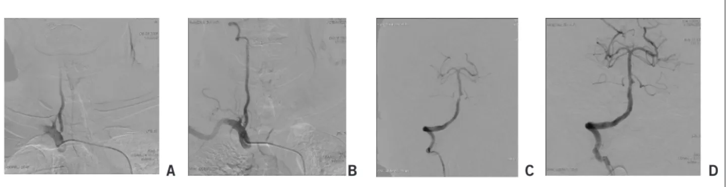
In the present study, a total of 29 stents were inserted: ten Genesis stents, two Cypher stents, seven Intec stents, and ten Apollo stents. As shown in Figures 1A, 1B, 1C, and 1D, the success rate was 100%. Before surgery, the mean degree of stenosis was 81.3 ± 8.8% (range: 65%~95%). Ater sur-gery, the mean degree of stenosis decreased to 3.7 ± 3.6% (range: 0%~10%). he degree of stenosis was signiicantly improved (p < 0.01). For 14 patients with cerebral infarc-tion, the neurological function was markedly improved, and the NIHSS score6 decreased remarkably (p < 0.01). On
discharge, the neurological function of these patients was further improved but similar to that of one day ater sur-gery. Before discharge, ischemia-related symptoms were not found, and the deterioration of original symptoms was also absent. TIA was not noted among these patients ater surgery. During the perioperative period, no severe com-plications were found.
BLOOD FLOWVELOCITYATTHEPOINTOFGREATESTSTENOSIS
he blood low signals in TCD were satisfactory in all patients. he blood velocity (Vm) before surgery and one week, and three, six, 12, and 24 months ater sur-gery is presented in Table 1. Results showed the Vm of stenotic arteries was extremely high before surgery, but dramatically reduced ater surgery and remained in nor-mal range. Signiicant diference was found in the Vm be-tween before and ater surgery (p < 0.01). However, the Vm was similar at one week, and three, six, 12, and 24 months ater surgery (p > 0.05).
FOLLOW-UP
he median follow-up period was 21.9 months (range: 7~36 months). he results showed that there were 23 patients with Malek score7 of 1, two with Malek score
of 2, and one with Malek score of 3. For one patient with vertebral artery stenosis and TIA, the degree of residual
Figure 1 – The stenosis of the vertebral artery and the clinical efficacy. Before surgery, ostial stenosis degree of the vertebral artery was 85% (A); after surgery, it was 0% (B). Before surgery, V4 stenosis degree of the vertebral artery was 90% (C); after surgery, it was 10% (D).
stenosis was 10%, TIA occurred once at seven months af-ter surgery, and the symptoms of TIA were similar to those before surgery. Re-examination with DSA showed that the degree of stenosis was still 10%, and anti-platelet/coagula-tion therapy was then performed without occurrence of TIA aterwards. For one patient with cerebral infarction, the pre-operative stenosis degree was 92% and the NIHSS score was 9. Immediately ater surgery, the degree of steno-sis was 10% and the NIHSS score was 4. On discharge, the NIHSS was 1. However, 16 months ater surgery, the initial symptoms deteriorated and the NIHSS score was 6 on re-examination. DSA showed that the degree of stenosis was similar to that immediately ater surgery. Intensive therapy was carried out and the patient achieved remission. Of the remaining patients, new ischemia-related symptoms and deterioration of initial symptoms were absent, and TIA and death were not observed.
Of note, ive patients received re-examination with DSA, of whom two were found to have developed stenosis (28.6%) at the irst part of arteries. he remaining 19 pa-tients were examined with CTA. Stent migration and frac-ture, and re-stenosis were not found. he forward blood low was acceptable.
DISCUSSION
he traditional treatments for SVAS include pharmaco-therapy and surgical intervention. However, the risk for stroke is still relatively high in vertebrobasilar artery ste-nosis patients receiving routine pharmacotherapy, and the prognosis of these patients is still poor. he warfarin-aspirin symptomatic intracranial disease (WASID) study demonstrated that the incidence of stroke events in the region supplied by the stenotic arteries was still high even though the patients were treated with warfarin or aspirin8.
he annual incidence of stoke due to stenosis of basilar ar-tery, vertebral arar-tery, posterior cerebral arar-tery, and poste-rior infeposte-rior cerebellar artery was 10%, 7%, 7.8%, and 6%, respectively. Rasmussen et al.9 reported that approximately
25% to 30% of patients with vertebrobasilar TIA had in-creased incidence of fatal events and elevated mortality and disability. In the present study, 26 patients received pharmacotherapy with statins and/or antiplatelet drugs to control the risk factors of stroke before admission, but the response was poor. hese results demonstrate that phar-macotherapy cannot improve the stenosis fundamentally.
In addition, the surgical intervention for stenosis has complicated procedures and potential complications, which signiicantly limits the wide application of surgi-cal intervention. In recent years, with the development of neurological intervention techniques and interventional materials, SAA has been an efective strategy for the treat-ment of vertebrobasilar artery stenosis due to the minimal invasion and high efectiveness10,11. In the present study, a
total of 29 lesions in 26 patients were treated with SAA, achieving a success rate of 100%. he clinical symptoms and signs were signiicantly improved, showing favorable short-term therapeutic efectiveness. Moreover, these pa-tients were followed-up for seven to 36 months (median: 21.9 months). Results showed that there were 23 patients with Malek score of 1, two with Malek score of 2, and one with Malek score of 3, which indicates that the inter-mediate and long-term efectiveness are also high.
he SAA for the stenotic arteries can re-canalize the arteries, improve the blood supply, elevate the perfusion of the brain tissues, and alleviate the symptoms. In addi-tion, the stent can prevent the atherosclerotic plaque from breaking and reduce the risk for stroke due to falling of the atherosclerotic plaque. In the present study, the de-gree of stenosis in these patients was 81.3 ± 8.8% before surgery and 3.7 ± 3.6% ater surgery. Hemodynamics test-ing showed a marked improvement followtest-ing SAA, and normal hemodynamics were maintained for at least 24 months, which was consistent with previously reported re-sults12. he 12 patients with TIA were followed-up for
sev-en to 36 months post-operatively, and the results showed signiicant improvement. Except for one patient who de-veloped TIA seven months ater surgery, similar symp-toms and cerebrovascular events were not observed in the remaining patients. In the 14 patients with cerebral infarc-tion, neurological function was dramatically improved post-operatively. Only one patient developed deteriora-tion of original symptoms at 16 months ater surgery, and re-examination with DSA showed the degree of stenosis was similar to that immediately ater surgery. his may be related to the falling of emboli in other sites. Improvement was achieved following intensive therapy.
SAA is an efective method for the treatment of SVAS, but still presents a risk for rupture, artery dissection, per-forating artery occlusion, thrombosis, cerebral hyperpefu-sion, and re-stenosis13-15, which may be associated with the
Table 1 – The blood velocity (Vm) of basilar and vertebral arteries before surgery and one week and three, six, 12, and 24 months after surgery (cm/sec)
Before surgery 1 week 3 months 6 months 12 months 24 months
Basilar 111.58 ± 8.48* 67.57 ± 7.00 68.57 ± 4.54 62.43 ± 7.09 66.17 ± 9.20 59.00 ± 7.81
Vertebral 100.42 ± 9.50* 51.42 ± 5.44 55.68 ± 5.88 47.79 ± 5.46 50.40 ± 4.81 54.75 ± 5.24
sample selection, therapeutic regimen, and experience of clinicians. In the present study, peri-operative complica-tions did not occur, which may be attributed to the simple lesions in these patients, good preparation before surgery, and small sample size.
It has been reported that the incidence of re-stenosis was as high as 32.4% within six months ater surgery13,
and some patients with re-stenosis were even asymptom-atic. In the present study, two patients were found to have developed stenosis among seven patients receiving DSA, but both were asymptomatic, which may be related to the mild degree of stenosis (20% and 35%, respectively). In one patient, the stenosis may be attributed to the large di-ameter of the stent, which results in the over-expansion of the afected artery and subsequent hyperplasia of intima. Although SAA has some complications, the incidence of these complications is relatively low, and some complica-tions can be avoided with the accumulation of experience.
Currently, large randomized controlled studies on en-dovascular stenting for symptomatic basilar artery steno-sis are still lacking16,17. he Carotid and Vertebral Artery
Transluminal Angioplasty Study (CAVATAS), the only randomized study to date to compare outcomes ater en-dovascular and medical treatment for patients with verte-bral artery stenosis, included only 16 such patients17,18, and
there was no diference in outcomes among those treated by stenting or drug therapy. Presently, there are mostly retrospective studies on the vertebrobasilar artery stenosis treated with SAA. Although the evidence of these indings cannot be compared to that of evidence-based random-ized controlled studies, SAA for vertebrobasilar artery stenosis has some advantages such as a high success rate, fewer complications, prevention of recurrent stroke, and better short-term clinical improvement16,19. Overall, SAA
is safe and efective. Following SAA, the degree of stenosis is signiicantly improved and the hemodynamics returns to normal. Follow-up also conirms the favorable long-term efectiveness.
REFERENCES
1. Levy EI, Turk AS, Albuquerque FC, Niemann DB, Aagaard-Kienitz B, Pride L, et al. Wingspan in-stent restenosis and thrombosis: incidence, clinical presen-tation, and management. Neurosurgery. 2007;61(3):644-51.
2. Chimowitz MI, Lynn MJ, Howlett-Smith H, Stern BJ, Hertzberg VS, Frankel MR, et al. Warfarin-Aspirin Symptomatic Intracranial Disease Trial Investigators. Comparison of warfarin and aspirin for symptomatic intracranial arterial ste-nosis. N Engl Med. 2005;352(13):1305-16.
3. Kung DK, Liu W, Smoker WR, Hasan DM. Duplication of the internal ca-rotid artery presenting with severe atherosclerotic stenosis. J Clin Neurosci. 2011;18(7):982-3.
4. Li W, Liu JM. Impact of intracranial stenting on the perforating artery. Chin J Neurosurg. 2007;23(8):636-8.
5. North American Symptomatic Carotid Endarterectomy Trial Collaborators. Beneicial efect of carotid endarterectomy in symptomatic patients with high-grade carotid stenosis. N Engl J Med. 1991;325(7):445-53.
6. Spilker J, Kongable G, Barch C, Braimah J, Brattina P, Daley S, et al. Using the NIH Stroke Scale to assess stroke patients. he NINDS rt-PA Stroke Study Group. J Neurosci Nurs. 1997;29(6):384-92.
7. Malek AM, Higashida RT, Phatouros CC, Lempert TE, Meyers PM, Gress DR, et al. Treatment of posterior circulation ischemia with extracranial percutane-ous balloon angioplasty and stent placement. Stroke. 1999;30(10):2073-85.
8. he Warfarin-Aspirin Symptomatie Intracranial Disease (WASID) Study Group. Prognosis of patients with symptomatic vertebral or basilar artery ste-nosis. Stroke. 1998;29(7):1389-92.
9. Rasmussen PA, Perl J, Barr JD, Markarian GZ, Katzan I, Sila C, et al. Stent-assisted angioplasty of intracranial vertebrobasilar atherosclerosis:an initial experience. J Neurosurg. 2000;92(5):771-8.
10. Liu JM, Hong B, Xu Y. Endovascular stenting for vertebrobasilar artery steno-sis. Chin Radiol. 2002;36(5):1063-7.
11. Chang BG, Xue DY, Li W. Intravascular treatment of symptomatic vertebro-basilar artery stenosis: report of 95 cases. Chin J Neurosurg. 2009; 25(2):106-9. 12. Fiorella D, Chow MM, Anderson M, Woo H, Rasmussen PA, Masaryk TJ.
A 7-year experience with balloon-mounted coronary stents for the treatment of symptomatic vertebrobasilar intracranial atheromatous disease. Neurosur-gery. 2007;61(2):236-43.
13. SSYLVIA Study Investigators. Stenting of symptomatic atherosclemtic le-sions in the vertebral or intracranial arteries (SSYLVIA): study results. Stroke. 2004;35(6):1388-92.
14. Rezende MT, Spelle L, Mounayer C, Piotin M, Abud DG, Moret J. Hyper-perfusion syndrome ater stenting for intracranial vertebral stenosis. Stroke. 2006;37(1):E12-4.
15. Yu W, Smith WS, Singh V, Ko NU, Cullen SP, Dowd CF, et al. Long-term out-come of endovascular stenting for symptomatic basilar artery stenosis. Neurol-ogy. 2005;64(6):1055-7.
16. Eberhardt O, Naegele T, Raygrotzki S, Weller M, Ernemann U. Stenting of ver-tebrobasilar arteries in symptomatic atherosclerotic disease and acute occlu-sion: case series and review of the literature. J Vasc Surg. 2006;43(6):1145-54. 17. Coward LJ, McCabe DJ, Ederle J, Featherstone RL, Cliton A, Brown MM, et al.
Long-term outcome ater angioplasty and stenting for symptomatic vertebral artery stenosis compared with medical treatment in the Carotid And Verte-bral Artery Transluminal Angioplasty Study (CAVATAS): a randomized trial. Stroke. 2007;38(5):1526-30.