Uso tópico do inteferon alfa-2b para neoplasia
escamosa da superficie ocular em um caso de
paciente com xeroderma pigmentoso
Nayara Queiroz Cardoso Pinto
1, Jhonatan de Paula Araujo Ferreira
1, Gustavo Jose Arruda Mendes Carneiro1, Jailton
Vieira Silva
1, Luanna Biana Costa Bezerra
2, Natália Ponte Nogueira
31 Hospital Universitário Walter Canídeo, Fortaleza, CE, Brazil. 2 Hospital Geral de Fortaleza, Ceará, Fortaleza, CE, Brazil.
3 Irmandade Santa Casa de Misericórdia de São Paulo, São Paulo, SP, Brazil.
Study carried out at the Ophthalmology Service, Hospital Universitário Walter Cantídio, Fortaleza, CE, Brazil.
A
BSTRACTReport of a case of xeroderma pigmentosum with squamous cell carcinoma of bilateral conjunctiva that showed a significant regression in tumor size with the use of interferon alfa-2b topic. Case report: Female patient with Xeroderma pigmentosum in an advanced stage, with no healthy skin, having been subjected to about 60 excisions of malignant skin lesions. The patient appeared with conjunctival tu-mors in both eyes, corresponding to squamous cell carcinoma of the conjunctiva. Due to surgical difficulties, high relapse rate and high probability of symblepharon formation, therapy was started with interferon alpha 2beta 1,000,000 topic units, obtaining good results with a significant decrease in lesion size and resolution of symptoms. Conclusion: Topical use of alpha-interferon in 2beta squamous neoplasia of the conjunctiva proved to be a good therapeutic option for high surgical risk and situations of postoperative complications.
Keywords: Xeroderma pigmentosum; Nevus, halo; Skin abnormalities; Skin neoplasms; Skin pigmentation; Eye neoplasms; Eye manifestations; Case reports
R
ESUMORelato de um caso clínico de Xeroderma Pigmentoso com carcinoma espinocelular de conjuntiva bilateral que apresentou regressão importante das dimensões tumorais com o uso de Interferon alfa-2b tópico. Relato de caso: Paciente feminina com Xeroderma Pig-mentoso em estágio avançado, com ausência de pele sadia, tendo sido submetida a cerca de 60 exéreses de lesões de pele malignas. A paciente compareceu com tumoração conjuntival em ambos os olhos, correspondendo a carcinoma espinocelular de conjuntiva e neoplasia intraepitelial de conjuntiva em olho esquerdo. Devido as diiculdades cirúrgicas, alta taxa de recidiva e elevada probabilidade de formação de simbléfaro foi-se iniciado terapêutica com Interferon alfa-2beta 1.000.000 unidades tópico, obtendo-se bons resulta-dos com importante regressão do tamanho da lesão e resolução resulta-dos sintomas. Conclusão: O uso tópico de interferon alfa-2beta em neoplasia escamosa de conjuntiva, mostrou-se uma boa opção terapêutica em situações de elevado risco cirúrgico e de complicações pós operatórias.
Descritores: Xeroderma pigmentoso; Nevus com halo; Anormalidades da pele; Neoplasias da pele; Pigmentação da pele; Neo-plasias oculares; Manifestações oculares; Relato de casos
Received for publication 14/10/2016 - Accepted for publication 07/12/2016.
Topical Interferon alfa-2b for ocular surface
squamous neoplasia in one case of patient
with xeroderma pigmentosum
I
NTRODUCTIONX
eroderma Pigmentosum (XP) is an recessive autosomal di-sorder of DNA repair, of genetically heterogeneous etio-logy and variable expressivity, resulting in sharp sensitivity to ultraviolet radiation (UV). The genetic heterogeneity relects molecular changes at gene level found in a set of genes encoding proteins involved in the repair pathway by nucleotide excision in cells in the replicative or quiescent state. There are eight altered genes described, with variable expressivity of the physical and clinical manifestations being the result of the interaction between genetic risk, genetic heterogeneity and environmental exposure.(1)It can occur in all ethnic groups with prevalence of 1:1,000,000 in Europe and the US, and 1:22,000 in Japan. There is also a predominance in countries of North Africa and the Middle East where consanguinity becomes more common. In Brazil, so far, there are no statistics showing the frequency of this syndrome, although some case reports have been described.(1)
The clinical manifestations of XP include extreme sun sen-sitivity (blistering burns with only a few minutes of exposure) and pigmentation changes (hyperpigmentation and depigmentation), ocular changes, presenting a risk greater than 10,000 times of developing neoplasms in areas exposed to the sun like the skin, mucous membranes and eyes. In addition, approximately 20% to 30% of patients may develop progressive neurological disease, being clinically manifested as progressive ataxia and spasticity and cognitive deterioration. The clinical manifestations of XP depend on both the molecular abnormality and environmental damage suffered, and they can evolve differently even within the same family group. (2,3)
Ocular manifestations are an important component of XP. They were irst described in the 19th century, and included xerosis and corneal opaciication. Ocular involvement related to XP involves the areas that are exposed to UV radiation such as eyelid and ocular surface. Its main complaints are photophobia, conjunctivitis, ectropion, exposure keratitis, which may lead to opaciication of the cornea or vascularization, pterygium, and neoplasia. (3)
The changes begin in early childhood. The skin of the eyelid may atrophy, and ectropion or entropion may occur. The bulbar conjunctiva can become thin, pigmented, and atrophic, with in-lammatory lesions and, which can eventually become malignant. The cornea may present a time of early tear ilm breakdown, edema, exposure keratitis, vascularization, pterygium (with accelerated growth), epithelial neoplasms and corneal opacity. Opaciication may result from changes in the epithelium/stroma, or may be secondary to endothelial decompensation as a result of damage due to chronic sun exposure. (4,5)
The ophthalmologic follow-up of these patients should be done since childhood, because the alterations derived from the disease begin after 1 to 2 years of onset of sun exposure. Early diagnosis and treatment is the most appropriate way of conducting cases, especially in the case of neoplasms where timely treatment may be essential. (6)
The objective of this report is to demonstrate favorable outcome of the use of Interferon Alpha-2b topic as a therapeutic option in case of a patient with xeroderma pigmentosum asso-ciated to bilateral conjunctival neoplasia of the squamous cell.
The report was based on regular appointments during the follow-up of the patient in the service of Ophthalmology and other specialties at Hospital Universitário Walter Cantídio. On
each visit, we recorded: visual acuity with Snellen chart, slit-lamp biomicroscopy with photography and neoplastic lesion measu-rement in millimeters, applanation tonometry and funduscopy.
C
ASER
EPORTFemale patient, 25 years diagnosed with XP for 15 years, attends the ophthalmology service complaining of low visual acuity (LVA) in both eyes complaining of worsening during the last 2 years associated to tearing and eye pain. Refresh Advan-ced® was used 5 times a day in both eyes, and there was excision of conjunctival lesion in the left eye 7 years ago. She mentioned previous history of excision of about 60 skin lesions in the der-matology service.
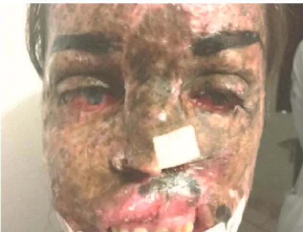
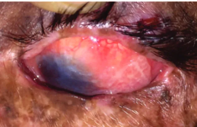
The physical examination showed visual acuity (VA) in the right eye (RE) of 20/200 and in left eye (LE) of hand movement. In the biomicroscopy RE, we can observe the presence of inferior symblepharo, conjunctival hyperemia, peripheral vascularization of the cornea, and vascularized lesion in conjunctiva, elevated and of papillomatous aspect in lower nasal area. The LE presented conjunctival hyperemia, corneal edema and peripheral vascula-rization, presence of vascularized lesion in the conjunctiva, irre-gular, lobed, invading the corneal surface in the upper temporal region (20x15 mm) (Figure 1). The patient’s appearance was of advanced dermal disease with severe involvement of the upper lips, nose and eyelids in ectoscopy (Figure 2).
Figure 1. Primary lesion of the left eye
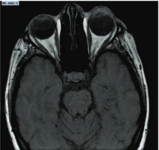
The hypothesis for the case was bilateral and extensive conjunctival involvement by XP associated to possible ocular tumor lesions (squamous conjunctiva neoplasia), and with proba-ble recurrence of the lesion in the LE, according to the American Joint Committee on Cancer Classiication, with ocular lesion RE Tis and LE T2. The hypothesis was having an incisional biopsy of the lesions to identify the histological subtype (this type of biopsy does not allow us to deine whether or not there is invasion of the lamina propria) and the performance of a Magnetic Nuclear Resonance (NMR) of the orbit to evaluate the extent of the le-sion (Figures 3 and 4). The results of the biopsies were bilateral
After diagnostic conirmation, topical therapy was initiated, using Interferon alfa-2 beta 1,000,000u/ml every 6h in both eyes. After one month of therapy, we noticed a signiicant improve-ment in the lesion (Figure 7). The treatimprove-ment was continued and reassessed 2 months afterwards (Figure 8). During this period, the patient reported visual and symptom improvement, with VA RE 20/100 and LE 20/400.
D
ISCUSSIONInterferon alfa-2B is an immunomodulator that has several clinical indications. Its main reported actions are as antiviral, immunomodulator and antiproliferative complexes. Due to its various actions the medication has been used in several areas of medicine. Many articles discuss the use of this medication and its squamous cell carcinoma “In Situ” (Figure 5) and High Grade
Intraepithelial Neoplasia (Grade 3) in the left eye (Figure 6). NMR showed signiicant ocular lesion in the left eye, but without invasion of palpebral and orbital tissue.
Figure 3. NMR showing hyperdense area in the anterior area of the left eye mode T1
Figure 4. NMR showing hyperdense area in the anterior area of the left eye mode T2
Figure 5. Histopathological aspect of right eye lesion demonstrating the appearance of squamous cell carcinoma “in situ”
effect on diseases such as dermal melanoma, polycythemia vera, hepatocellular carcinoma, and ocular diseases of the external and internal segments.(7-9)
Ocular treatments with Interferon alfa-2b have good thera-peutic responses demonstrated in some studies. Nanji A.A. et al. reported a comparative study between the use of Interferon and surgical removal of lesions diagnosed as ocular surface squamous neoplasia (OSSN) where no major recurrence was evidenced with use of eyedrops. The 49 patients obtained total cure of the lesions in an average time of 2.3 months with a total average of 4 months of treatment. Kusumesh R. reported the effectiveness and safety of interferon as a single therapy in OSSN lesions, reaching total remission in 91.6% of patients treated and presenting low side effects, with a case of spontaneous tumor bleeding three weeks after the use of Interferon every 6h. (10-11)
Other reports also include good results in cases of primary melanoma acquired with atypia, conjunctival intraepithelial ne-oplasia, kaposi sarcoma, ocular melanoma (intravitreal use).(12-13) In the current literature, there are still divergences regar-ding the best approach for neoplastic lesions of the ocular con-junctiva in these patients, but the most accepted treatment and best prognosis is the removal of all lesions with appearance and/ or diagnosis of malignancy with safety margin and therapy with cryotherapy on the margins of resection and/or intraoperative mitomycin C. Their withdrawal cannot be conservative because of the high risk of local recurrences. In these reports they further describe the preferential use of Interferon alfa-2b in patients with extensive ocular disease in an attempt to preserve the eye in cases
Figure 7. Left eye lesion after 30 days of use of Interferon eyedrops
Figure 8. Left eye lesion after 2 months of use of Interferon eyedrops
of non-invasion of the orbital content, in an attempt to avoid a more aggressive surgical procedure.(14)
In a study, 23 of the patients who had undergone some previous treatment for tumor lesion (excisional biopsy 9 [39%], cryotherapy 2 [9%], topical mitomycin C 1 [4%], and topical In-terferon alfa-2b 3 [13%]) presented recurrence and underwent a new therapy with Interferon alfa-2b. The classiication of tumors according to the AJCC classiication (table 1) included in the studies the following categories: Tis (3 [13%]), T3 (20, [87%]), N0 (23 [100%]), e M0 (23 [100%]). The primary indication for the use of Interferon alfa-2b was extensive, unresectable ocular lesion in 15 tumors (65%), with no surgical indication in 6 tumors (26%), and low visual acuity in the contralateral eye in 2 tumors (9%). The results of the study were total tumor resolution in 19 cases (83%), and in 4 (17%) there was a partial resolution of the lesion; one patient with XP in the study presented regression of 49% of the lesion size in 4 months, and the lesion remained stable for a period of 8 months.(15)
The surgical removal of the ocular lesions has a high chance of recurrence in this proile of patients, besides subjecting them to
Clinic Category Definition
Primary Tumor
TX Tumor cannot be assessed
T0 Tumor absent
Tis umor present as carcinoma in situ / Conjunctiva intraepithelial neoplasia T1 umor present with greatest basal
diameter ≤5mm
T2 Tumor present with greater basal
diameter ≥5mm, without invasion
of adjacent structures*
T3 Tumor invading adjacent structures T4 Tumor invading the orbit with or without
additional extension
T4a Tumor invades soft parts of the orbit without,
bone invasion
T4b Tumor invading the bone
T4c Tumor invading underlying paranasal sinuses
T4d Tumor invading the brain
Regional lymph node
Nx Nodules in regional lymph nodes
cannot be assessed
N0 Nodules in regional lymph nodes without metastasis
N1 Nodules in regional lymph nodes with metastasis Distant metastasis
M0 Distant metastasis absent
M1 Distant metastasis present
Table 1
. Classification of the “American Joint Committee on
Cancer” for ocular surface squamous neoplasia (15)
surgical stress by repeated interventions and having a great possi-bility of ocular sequelae such as symblepharon and other ocular surface alterations. Thus, the treatment with topical medication has shown to be a better option for the patient in question. Other studies corroborate the use of interferon as a good option for surgery, comparing the cost of the surgical treatment to the topical medication, showing similar results in both, with an additional visit to the ofice in the group of patients using Interferon alfa-2B.(16-18)
R
EFERENCES1. Ryan SJ. Retina: basic science and inherited retinal disease. 4th ed. Philadelphia: Mosby; 2006. Vol. 1
2. Vilela MA, Colossi CG. Retinosquises. Rev Bras Oftalmol. 2011; 70(2):125-32.
3. Caye LM, Barbosa PH, et al. Uso da dorzolamida tópica em paciente portador de retinosquise juvenil ligada ao X. Rev Bras Oftalmol. 2010; 69(2):129-131.
4. Forsius H, Vainio-Mattila B, Eriksson A. X-linked hereditary reti-noschisis. Br J Ophthalmol. 1962;46(11):678-81.
5. Sauer CG, Gehrig A, Warneke-Wittstock R, Marquardt A, Ewing CC, et al. Positional cloning of the gene associated withX-linked juvenile retinoschisis. Nat Genet. 1997;17(2):164-70.
6. Bastos AL, Freitas BP, Villas Boas O, Ramiro AC. Uso da dorzolamida tópica em pacientes com retinosquise juvenil ligada ao X. Arq Bras Oftalmol. 2008; 71(2): 286-90.
7. Kanski JJ, Bowling B. Clinical ophthalmology: a systematic approach. 7th ed. Edinburgh: Elsevier Saunders; 2011.
8. Nelson textbook of pediatrics. 19th ed. Philadelphia, PA; Elsevier/ Saunders; c2011. Disorders of the eye. Part 29.
9. Holing-Lima AL, Moeller CT, Freitas D, Martins EN. Manual de condutas em Oftalmologia. São Paulo: Atheneu; 2008.
10. Audo I, ,lder GE, Moore A T. Inherited stationary disorders of the retina. In: Puech B, De Laey J-J, Holder GE, editors. Inherited chorioretinal dystrophies. A textbook and atlas. 2014.p. 77-98
Corresponding Author:
Nayara Queiroz Cardoso Pinto
Rua Silva Paulet, 205 – Meireles, Ed. Studio 1 Fortaleza , CE, Brazil.
ZIP Code: 60.060-120 Phone No.: +55(84)99651-5533
11. Apushikin MA, Fishman GA. Use of dorzolamide for patients with X-linked retinoschisis. Retina. Retina. 2006; 26(7):741-5. Erratum in: Retina. 2007;27(1):128.
12. Ober MD, Freund KB, Shah M, Ahmed S, Mahmoud TH, Aaberg TM Jr, Zacks DN, GaoH, Mukkamala K, Desai U, Packo KH, Yannuzzi LA. Stellate nonhereditary idiopathic foveomacular retinoschisis. Ophthalmology. 2014;121(7):1406-13.
13. Garip A, Schaumberger MM, Wolf A, Herold TR, Miller CV, Klin-genstein A, et al. Evaluation of a short-term topical interferon -2b treatment for histologically proven melanoma and primary acquired melanosis with atypia. Orbit. 2016;35(1):29-34.
14. El Hamichi S, Messaoudi R, Belmalih M, Iferkhass S, Zerrouk R, Reda K, Oubaaz A. Bilateral enucleation avoided by excision with mitomycin C for bilateral iniltrating conjunctival squamous cell carcinoma in a girl with xeroderma pigmentosum. J Fr Ophtalmol. 2015;38(7):e135-7. 15. Shah SU, Kaliki S, Kim HJ, Lally SE, Shields JA, Shields CL. Topical interferon alfa-2b for management of ocular surface squamous ne-oplasia in 23 cases: outcomes based on American Joint Committee on Cancer classiication. Arch Ophthalmol. 2012 130(2):159-64. 16. Moon CS, Nanji AA, Galor A, McCollister KE, Karp CL. Surgical
versus medical treatment of ocular surface squamous neoplasia a cost comparison. Ophthalmology. 2016; 123(3):497-504.