498
IMAGEM EM MEDICINA
Rev Assoc Med Bras 2011; 57(5):498
Syphilitic meningoencephalitis associated with vasculitis
LUCAS ALVERNE FREITAS DE ALBUQUERQUE1, RENATA BRANT DE SOUZA1, PAULO PEREIRA CHRISTO2
1Interns, Neurosurgery, Santa Casa de Belo Horizonte, Belo Horizonte, MG, Brazil 2 Professor, Postgraduate Course, Santa Casa de Belo Horizonte, Belo Horizonte, MG, Brazil
Study conducted at Santa Casa de Belo Horizonte, MG, Brazil
Correspondence to: Lucas Alverne Freitas de Albuquerque – Rua Aimorés, 1006 - apto. 202 – Funcionários – Belo Horizonte – MG – CEP: 30140-071 Phone: 55 (31) 8327-5630 – [email protected]
©2011 Elsevier Editora Ltda. Todos os direitos reservados.
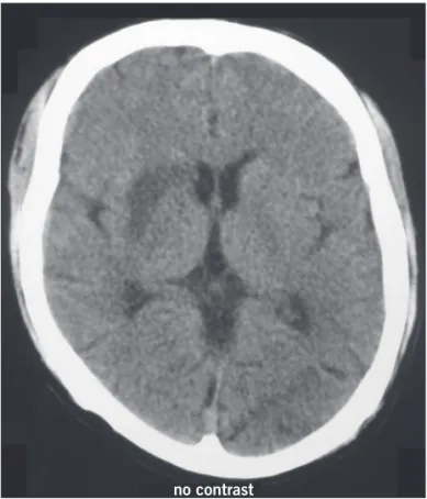
A 28 year-old man, with no previous comorbidity, pre-sented let side weakness and verbal luency reduction. A computed tomography (CT) (Figure 1) performed in an-other hospital revealed hipodensity in the basal ganglia. Ater 20 days the patient presented a generalized tonic-clonic seizure and became stuporous (Glasgow Coma Scale 11/15), with aphasia, anisocoria (right > let pupil) with mild light reaction (Argyll Robertson pupil), and tetraparesis (Medical Research Council Scale of Muscle Strength 2/5). Ten days later the patient was admitted at our service and a MRI revealed multiple areas of isch-emia (Figure 2) suggestive of central nervous system vasculitis. Laboratorial evaluation revealed positive
se-rology to HIV-1, HIV-1 RNA/mL 201.777 copies, CD4 count 97/mm3, serum VDRL 1/2048 and positive
FTA-Abs. Cerebrospinal luid analyses revealed 6 cells/mm3
(97% mononuclear), glucose 43 mg/dL, total protein 147 mg/dL and VDRL 1/16. he diagnosis of vascular meningoencephalitis syphilitic was made. he MRI was compatible with a case of Nissl-Alzheimer endarteri-tis syphilitica, a subtype that involves the small brain vessels1-3. Despite initiation of speciic treatment, the
pa-tient deteriorated to no visual or verbal contact, increase in muscle weakness (Muscle Strength 1/5) in the four limbs. Ater one month of medical support the patient died of a multi-resistent bacterial pneumonia.
REFERENCES
1. Lucato LT, Barbosa Júnior A, Andrade CS, Amato Filho AC. Doen-ças infecciosas e inlamatórias do sistema nervoso central. In: Cerri GG, editor. Neurorradiologia: diagnóstico por imagem das altera-ções encefálicas. Rio de Janeiro: Guanabara Koogan; 2008. p.213-87. 2. Hajjaj I, Kissani N. Status epilepticus revealing syphilitic
meningo-encephalitis. Acta Neurol Belg 2010;110:263-7.
3. Gaa J, Weidauer S, Sitzer M, Lanfermann H, Zanella FE. Cerebral vasculitis due to Treponema pallidum infection: MRI and MRA indings. Eur Radiol 2004;14:746-7.
Figure 1 – Computed tomography (CT) shows hipodensity in the basal ganglia.
Figure 2 – Axial FLAIR weighted MRI shows multiple areas of ischemia, affecting the territory of small vessels in the cortical, subcortical and basal ganglia area.