3 artigo 531
ORIgINAL ARtICLE
1 – Assistant Professor and Head of the Shoulder and Elbow Surgery Group, Department of Orthopedics and Traumatology, School of Medical Sciences, Santa Casa de São Paulo, São Paulo, Brazil.
2 – Assistant Professor and Attending Physician in the Shoulder and Elbow Surgery Group, Department of Orthopedics and Traumatology, School of Medical Sciences, Santa Casa de São Paulo, São Paulo, Brazil.
3 – Attending Physician in the Shoulder and Elbow Surgery Group, Department of Orthopedics and Traumatology, School of Medical Sciences, Santa Casa de São Paulo, São Paulo, Brazil.
4 – Trainee in the Shoulder and Elbow Surgery Group, Department of Orthopedics and Traumatology, School of Medical Sciences, Santa Casa de São Paulo, São Paulo, Brazil. 5 – Adjunct Professor, Academic Consultant and Member of the Shoulder and Elbow Surgery Group, Department of Orthopedics and Traumatology, School of Medical
Sci-ences, Santa Casa de São Paulo, São Paulo, Brazil.
6 – Assistant Professor in the Mechanical Engineering Course, Mackenzie Presbyterian University, São Paulo, Brazil. 7 – Assistant Professor in the Mechanical Engineering Course, Mackenzie Presbyterian University, São Paulo, Brazil.
Work performed in the Department of Orthopedics and Traumatology, School of Medical Sciences, Santa Casa de São Paulo (DOT-FCMSCSP), “Fernandinho Simonsen” Wing (Director: Prof. Dr. Osmar Avanzi), and in the Department of Mechanical Engineering, Mackenzie Presbyterian University (Coordinator: Prof. Dr. Antônio Gonçalves Mello), São Paulo (SP), Brazil.
Correspondence: Rua Dr. Cesário Mota Jr. 112, Vila Buarque, 01221-020 São Paulo, SP. E-mail:[email protected] / [email protected]
Work received for publication: June 2, 2011; accepted for publication: August 19, 2011.
ANALYSIS ON THE VARIATION OF mEDIAL ROTATION
VALuES ACCORDINg TO THE POSITION
OF THE HumERAL DIAPHYSIS
Alberto Naoki Miyazaki1, Marcelo Fregoneze2, Pedro Doneux Santos3, Luciana Andrade da Silva³, Guilherme do Val Sella3, Carina Cohen4, Taís Stedile Busin Giora4, Sergio Luiz Checchia5, Fabio Raia6, Hélio Pekelman6, Raquel Cymrot7
ABstRACt
Objective: To analyze the validity of measurements of medial rotation (MR) of the shoulder, using vertebral levels, according to the variation in the position of the humeral diaphysis, and to test the bi-goniometer as a new measuring instrument. Methods: 140 shoulders (70 patients) were prospectively evaluated in cases presenting unilateral shoulder MR limitation. The vertebral level was evaluated by means of a visual scale and was correlated with the angle obtained according to the position of the humeral diaphysis, using the bi-goniometer developed with the Department of Mechanical Engineering of Mackenzie University. Results: The maximum vertebral level reached through MR on the unaffected side ranged from T3 to T12, and on the affected side, from T6 to the trochanter. Repositioning of the affected limb in MR according to the angular values on the normal side showed that 57.13% of the patients reached
INtRODUCtION
Range of motion (ROM) is an important parameter in functional evaluations of the shoulder and precision
is needed in measuring it(1). These measurements can
be made as specified in the manual of the American
Academy of Orthopaedic Surgeons (AAOS), using
lower levels, between the sacrum, gluteus and trochanter. From analysis on the maximum vertebral level attained and the variation between the affected angle x (frontal plane: abduction and MR of the shoulder) and the unaffected angle x in MR, we observed that the greater the angle of the diaphyseal axis was, the lower the variation in the vertebral level attained was. From evaluating the linear correlation between the variables of difference in maximum vertebral level reached and variation in the affected angle y (extension and abduction of the shoulder) and the unaffected angle y in MR, we observed that there was no well-established linear relationship between these variables. Conclusion: Measurement of MR using vertebral levels does not correspond to the real values, since it varies according to the positioning of the humeral diaphysis.
Keywords – Shoulder Joint; Range of Motion, Articular;
Rotation; Articular, Goniometry
a visual scale and goniometry(2). Measurements on
the medial rotation of the shoulder are particularly difficult to define, because the abdomen impedes the maximum medial rotation. One method that is commonly used is to indirectly measure the medial rotation in terms of the maximum proximal vertebral
The authors declare that there was no conflict of interest in conducting this work
vels T6 and T10(11). Between sacral (S) level S1 and T10, the movement takes place mainly around elbow
flexion(4), and the medial rotation of the shoulder is
almost unaltered above T12(12).
It is observed that the on the affected side, ver-tebral levels similar to on the normal side may be reached, at the cost of compensatory inclination of the trunk and elbow flexion. In relation to function, this may appear to be irrelevant; however, for scientific purposes, correct and standardized determination of the vertebral level is fundamental. Seeking to analyze this compensation, we developed a two-dimensional measurement instrument for measuring the angular positioning of the humeral diaphysis in combined me-dial rotation and extension movements. Through this, we would be able to compare the normal and affected sides, make corrections to the position and compare the vertebral levels reached.
The aim of this study was to analyze the validi-ty of medial rotation measurements on the shoulder using vertebral levels, according to the variation in the position of the humeral diaphysis, and to test the bi-goniometer as a new measurement instrument.
sAMPLE AND MEtHODs
Between December 2010 and January 2011, 140 shoulders of 70 patients with unilateral limitation on medial rotation of the shoulder were prospectively evaluated. These patients were being followed up at the outpatient clinic of the Shoulder and Elbow Group, School of Medical Sciences, Santa Casa de São Paulo, “Fernandinho Simonsen” Wing. This research project was approved by the Ethics Committee for Research on Human Beings of this institution. All the patients were aware that they were participating in this study and signed a free and informed consent statement.
The inclusion criterion was that the patients should present unilateral limitation of medial rotation. The following patients were excluded: obese individuals
(body mass index, BMI ≥ 30), cases of unconsolidated
fracture of an upper limb, cases of limitation of elbow mobility, cases of rheumatoid arthritis, individuals with bilateral shoulder disorders and patients who had un-dergone shoulder surgery less than four months earlier. Among the 70 patients evaluated, 41 (58.57%) were female and 29 (41.43%) were male. The mean age was 57 years, ranging from 32 to 82 years. The mean BMI of the patients evaluated was 25.25 (ranging from 17.7 to 29.7). The dominant side was affected in level reached by the thumb, in which the hand is
actively positioned behind the back and the vertebral level reached by the tip of the extended thumb is
recorded(2-5). However, some authors have believed
that this measurement underestimates the contribution made by other joints, thus considering that the
accuracy of this measurement is not valid(4,6).
In a study comparing the visual estimation method and goniometry for evaluating shoulder ROM, Andrade
et al(1) showed that the greatest reproducibility rate was in measuring the medial rotation using the visual scale
method. However, according to Armstrong et al(7),
al-though visual estimation for evaluating ROM is easy and quickly applied, it presented moderate reproducibili-ty with significant differences in relation to goniometry, which demonstrated high reliability and reproducibili-ty. They therefore considered goniometry to be a better
method for evaluating ROM. However, Herrington(8)
reported that shoulder measurements made using go-niometry could be imprecise because of the difficulty in establishing the axis of movement.
Contrary to what might be believed, medial and la-teral rotation do not take place entirely in the shoulder joint(3). Mallon et al(4) demonstrated that the scapu-lothoracic joint also contributes towards these move-ments. The exact amount contributed by these joints varies, with a mean ratio of 2:1 at the maximum ver-tebral levels. Medial rotation along the thorax seems to be done almost entirely by the shoulder joint, which contributes 91%. Taking into account the full medial rotation, the shoulder contributes 84%, but placing the arm behind the back mobilizes the scapulothoracic joint. The movement to position the hand behind the back takes place in three distinct parts: firstly, with the aim in front of the body, the shoulder joint rotates internally; secondly, the arm is positioned behind the body with most of the movement occurring through extension and internal rotation of the scapulothoracic joint; and thirdly, the thumb reaches the vertebral le-vel through elbow flexion. Because this measurement is composed of three movements and does not just consist of medial rotation of the shoulder joint, its value as a medial rotation parameter is questionable. Nonetheless, its functional importance for represen-ting activities of daily living is evident, given that
limitation of this movement greatly incapacitates(4,9).
Measurement of medial rotation is part of the
standard examination used worldwide(10). The mean
le-47 patients (67.14%). Forty-five individuals (64.29%) had pain in the shoulder affected. Forty-nine patients (70%) had undergone a surgical procedure, with a mean length of postoperative follow-up of 27 months, ranging from five to 180 months.
The evaluation on the medial rotation of the shoul-der was done by means of a visual scale for estimating the vertebral level and this was correlated with the angle obtained according to the position of the dia-physis of the humerus, using a bi-goniometer that had been developed in conjunction with the Department of Mechanical Engineering of Mackenzie University.
instrument
The instrument used for the measurements was composed of 18 different pieces, described in the following: piece 1, base, cut from an acrylic board of thickness 15 mm, which served to support the rest of the pieces of the mechanism; piece 2, 360° disc, cut from a polystyrene sheet of thickness 1 mm, which served as a friction-reducing ring, to allow smoother support rotation movements and define the axis for the combination of shoulder extension and adduction (axis of the angle y); piece 3, support, cut from an acrylic board of thickness 15 mm and then turned and milled to acquire the planned shape, which serves as a support for the half-disc and for the rod; piece 4, 180° half-disc, made from a cut acrylic protractor, which is responsible for defining the axis of medial rotation and shoulder abduction (axis of the angle x); piece 5, rod, cut from a Phenolite tube of length 8 mm, which should be positioned parallel to the humerus, thus making it possible to read the angles; piece 6, lock, cut from acrylic of thickness 2 mm, which serves to secure the support on the base; piece 7, M3 slotted screw, which fixes the lock to the support; piece 8, bracket, cut from a stainless steel plate, which serves to position the equipment on the patient’s body; piece 9, M3 slotted screw, which fixes the support to the base; piece 10, M3 hexagonal nut; piece 11, A3 smooth washer, which helps to fix the support to the base; piece 12, M3 slotted screw, which fixes the rod to the support, thus allowing the rod to rotate; piece 13, upper measurer, prototyped in acrylonitrile butadiene styrene (ABS) plastic, which fixes the spacing between the rod and the lateral edge of the patient’s acromion; piece 14, ruler, cut from acrylic of thickness 5 mm and then milled to obtain parallel clefts, which keeps the upper and lower measurers parallel; piece 15, lower measurer, prototyped in ABS plastic, which should
be supported on the lateral epicondyle and then at the tip of the olecranon; piece 16, pin, prototyped in ABS plastic, which fixes the lower measurer to the ruler, thus allowing it to go up and down in parallel with the upper measurer; piece 17, M3 wing nut, which facilitates adjustments to the base; and piece 18, bubble level, which makes it possible to keep the base horizontally leveled (Figure 1).
The measuring instrument was validated statistically, and its repeatability and reproducibility were analyzed separately. Measurements that did not differ by more than 15° were considered to be concordant, taking into account the large ROM of the shoulder. Its repeatability was checked through the degree of concordance between the results from successive measurements in the neutral position and in medial rotation, on the same patient, performed using the same method, the same operator and the same conditions of use. Reproducibility was observed in terms of the concordance between the results from measurements in the neutral position and in medial rotation, performed by two operators on the same patient.
methodology
All the patients were evaluated by the same two examiners (E1 and E2). The examinations were performed with the patient standing upright, and reference points were marked out on the lateral edges of the acromion, lateral epicondyle and tip of the olecranon of both upper limbs, using colored
Figure 1 – Illustrative diagram of the bi-goniometer and its components: 1) acrylic base;
2) 360° polystyrene disc (axis of the angle y); 3) acrylic support; 4) 180° acrylic protractor (axis of the angle x); 5) Phenolite rod; 6) acrylic lock; 7) screws; 8) stainless steel bracket; 9) screws; 10) hexagonal nuts; 11) smooth washers; 12) screws; 13) upper measurer made of ABS plastic; 14) acrylic ruler; 15) lower measurer made of ABS plastic; 16) pins made of ABS plastic; 17) wing nuts; 18) bubble levels.
10
1 2 3 4
5
6
7 9 8
11
12
13
14
15
16
17
adhesive tape. These points were used to determine the alignment of the diaphysis of the humerus.
The bracket of the bi-goniometer was supported on the patient’s shoulders, firstly on the normal side. After making measurements in the neutral position and in me-dial rotation, the same procedure was performed on the affected side. The apparatus was always stabilized by examiner E1, who positioned the midline of the instru-ment aligned with the lateral edge of the acromion. The levels were carefully observed to certify that the appara-tus remained parallel to the ground all the time.
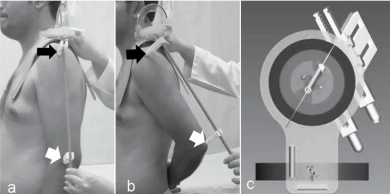
As a starting point, the patient was positioned standing upright with the upper limbs in anatomical position. The angular values obtained from the apparatus were always noted down by examiner E2, using the lateral edge of the acromion and the lateral epicondyle as the parameters, which corresponded to the axis of the humeral diaphysis in this position (Figure 2). Following this, the patient was asked to position the limb in medial rotation on his back, as high as possible, with the thumb outstretched. The apparatus was positioned using the lateral edge of the acromion and the tip of the olecranon as the parameters, which corresponded to the axis of the humeral diaphysis in this position. The angular measurements marked on the apparatus, the degree of elbow flexion and the vertebral level reached by
the outstretched thumb were noted down by examiner E2. This sequence was done firstly on the normal side and then on the affected side.
The angular value measured on the unaffected side positioned in medial rotation was used as a parameter for repositioning the affected limb. After alignment was done by examiner E1, a new measurement was made on the maximum vertebral level reached, using the visual scale, and was noted down by examiner E2 (Figure 3).
The angular variation observed between the limb with the limitation on medial rotation, with reposi-tioning of the humeral axis on this side, and the an-gular values from the normal side was analyzed and compared with the difference in maximum vertebral level reached by the affected member.
For the statistical analysis, the Minitab 16 statis-tical software was used. In all the hypothesis tests, descriptive levels (p values) were calculated, taking 95% confidence intervals to be statistically significant
and therefore rejecting hypotheses with p ≤ 0.05.
To compare means between pairs of variables, Student’s t test for paired data was used. Linear correlations between pairs of variables were also
calculated, such as the Δx variation, which quantitatively
represents the angular variation of the x axis (angle x on affected side minus x on unaffected side, in medial
rotation) and the Δy variation, quantitatively representing
Figure 2 – Positioning of the bi-goniometer on the shoulder, with patient standing upright. a) Anatomical position, with reference points on the lateral edge of the acromion (black arrow) and
angular variation of the y axis (angle y on affected side minus y on unaffected side, in medial rotation).
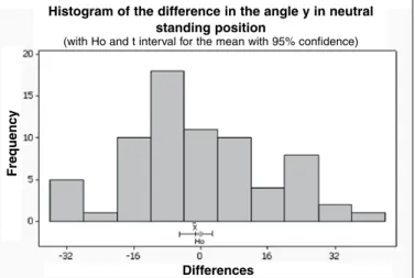
The mean, standard deviation (SD) and confidence interval (CI) were calculated for all the variables ana-lyzed. Regarding the angle x on the unaffected and affected sides, in the anatomical position, the mean difference in comparing the 70 patients was within the CI, with p > 0.005, and thus the hypothesis of equality between the measurements was accepted (p = 0.681) (Figure 4). The same was found in calcu-lating the difference for the angle y on the unaffected and affected sides, in the anatomical position, with p = 0.545 (Figure 5).
Figure 3 – Posterior view of the positioning of the bi-goniometer for measuring the medial rotation of the affected shoulder. a) Maximum vertebral level reached (T10). b)
Cor-rected vertebral level (L1). c) Axis of the angle x in the frontal plane, corresponding to abduction and medial rotation of the shoulder (Department of Mechanical Engineering, Mackenzie Presbyterian University).
Figure 4 – Distribution of the difference in x in the anatomical upright standing position
between the affected and unaffected sides.
Histogram of the difference in the angle x in neutral standing position
(with Ho and t interval for the mean with 95% confidence)
Differences
F
re
q
u
e
n
c
y
Figure 5 – Distribution of the difference in y in the anatomical upright standing position
between the affected and unaffected sides.
Histogram of the difference in the angle y in neutral standing position
(with Ho and t interval for the mean with 95% confidence)
Differences
F
re
q
u
e
n
c
y
REsULts
In evaluating the variable of the angle x for the 70 patients, comparing this angle between the unaffected and affected sides in medial rotation, the mean difference was within the CI, with p = 0.002. It was therefore concluded that the measurements were not equal for the unaffected and affected sides, such that they were greater on the affected side (Figure 6).
they were greater on the affected side (Figure 7). The maximum vertebral level reached in medial rotation with the thumb outstretched on the unaffected side ranged from T3 to T12, and 46% of the cases were between T8 and T9. On the affected side, the maximum vertebral level reached with the thumb outstretched ranged from T6 to the trochanter. When the affected limb was repositioned in medial rotation in accordance with the angular values of the normal side, the levels ranged from T6 to the trochanter, but with a different percentage distribution, such that 57.13% of the patients reached levels between the sacrum, gluteus and trochanter (Table 1).
The variable of maximum elbow flexion on the unaffected and affected sides was analyzed, comparing these measurements for the 70 patients. It was concluded that the maximum elbow flexion on the unaffected and affected sides was not equal, such that it was greater on the unaffected side (p = 0.000) (Figure 8). In the same way, in comparing the patients who reached vertebral levels higher than S1 with their
affected limb, with unaffected individuals, the elbow flexion was also greater on the unaffected side.
In evaluating the variables of difference in maximum
vertebral level reached and Δx variation (angle x on the
affected side minus x on the unaffected side, in medial ro-tation), it was concluded that there was a decreasing lin-ear relationship between these variables, i.e. the greater the angle of the diaphysis axis is, the smaller the variation in the vertebral level achieved is (p = 0.044) (Figure 9).
Following this, the linear correlation was calculated between the variables of difference in the maximum
vertebral level reached and Δy variation (angle y on the
table 1 – Incidence of vertebral levels for the unaffected, affected and
corrected affected sides.
Vertebral level
Unaffected
side (%) Affected side (%)
Corrected affected side (%)
T3 1.43 0 0
T4 2.86 0 0
T5 2.86 0 0
T6 5.71 1.43 1.43
T7 17.14 1.43 0 T8 24.29 4.29 1.43
T9 21.42 7.14 2.86
T10 14.29 7.14 7.14 T11 2.86 14.29 1.43
T12 7.14 8.57 5.71
L1 0 4.29 2.86
L2 0 10 8.57
L3 0 5.7 4.29
L4 0 0 5.72
L5 0 12.86 1.43
Sacrum 0 8.57 15.71
Gluteus 0 12.86 25.71
Trochanter 0 1.43 15.71
Total 100 100 100
Legend: T - thoracic; L - lumbar.
Source: medical files at Irmandade Santa Casa de Misericórdia de São Paulo.
Figure 8 – Distribution of the difference in elbow flexion in standing position between
the affected and unaffected sides.
Figura 6 – Distribuição da diferença entre a rotação medial x para o lado acometido
e o não acometido.
Figura 7 – Distribuição da diferença entre a rotação medial y para o lado acometido
e o não acometido.
Histogram of the difference in the angle x of medial rotation, in standing
(with Ho and t interval for the mean with 95% confidence)
Differences
F
re
q
u
e
n
c
y
Histogram of the difference in the angle y of medial rotation, in standing position
(with Ho and t interval for the mean with 95% confidence)
Differences
Histogram of the difference in elbow flexion in standing
(with Ho and t interval for the mean with 95% confidence)
Differences
F
re
q
u
e
n
c
y
15.0 12.5
10.0
7.5
5.0
2.5
0.0
F
re
q
u
e
n
c
affected side minus y on the unaffected side, in medial rotation), and the value obtained was –0.231. At the sig-nificance level of 5%, this value was zero (P = 0.054), and it was concluded that there was no well-established linear relationship between these variables (Figure 10).
Complaints of shoulder pain did not influence the ver-tebral level reached with the affected limb (p = 0.309). However, through correlating the elbow flexion on the side with medial rotation and the presence of pain, it was concluded that patients with pain performed greater compensatory flexion of the elbow (p = 0.004).
maintained the same measurement routine: while exa-miner E1 always positioned the instrument, E2 always made the measurements.
Although Armstrong et al(7) found that measurements
of medial rotation of the shoulder by means of visual estimation presented moderate reproducibility, Andrade
et al(1) showed through comparing this method with go-niometry that measurements using a visual scale were more reproducible. In our study, in testing the measu-rements of medial rotation according to the maximum vertebral level reached, we obtained high concordance between the two examiners, thus confirming from the sta-tistical analysis that the measurements were reproducible.
In accordance with the findings of Mallon et al(4),
who defined that hand positioning behind the back takes place in three distinct movements, we developed a numerical form of measurement of medial rotation of the shoulder that took into consideration the initial movement of abduction and medial rotation, measured using the angle of the x axis. This movement takes place mainly in the shoulder joint. Subsequently, there are ex-tension, adduction and medial rotation movements, per-formed by the scapulothoracic joint, which we measured using the angle of the y axis. Lastly, we measured the angle of elbow flexion using conventional goniometry: this joint is the main agent responsible for medial rota-tion from the midline of the sacrum, and it may move the hand 17 to 20 cm proximally, corresponding to a gain of seven to eight levels in normal individuals(14).
In our study, we concluded that the maximum el-bow flexion on the affected side was shown to be statistically less than on the unaffected side, for all the vertebral levels reached, and that patients who only reached levels lower than the lumbar level L5 had lo-wer degrees of elbow flexion. This may be explained
by the studies of Mallon et al(4), who demonstrated
that elbow flexion was the main agent responsible for reaching vertebral levels between S1 and T10.
In evaluating patients with complaints of pain in the affected shoulder, this variable was not correlated with lower vertebral levels. However, these patients’ elbow flexion was shown to be greater (p = 0.004), which may have been in an attempt to compensate for the medial rotation deficit. It should be noted that none of these individuals presented any pain or limita-tion of elbow movements.
Although Andrade et al(1) did not find any correlation between the angular measurement of medial rotation of the shoulder at 90° of abduction with the patient in a supine position and the vertebral level observed on the visual scale, given that these are different methods that cannot
Figure 9 – Dispersion between vertebral level and Δx (difference between angle x on
the affected side and x on the unaffected side, in medial rotation).
Figure 10 – Dispersion between vertebral level and Δy (difference between angle y on
the affected side and y on the unaffected side, in medial rotation).
Dispersion graph for difference in vertebral level versus difference in x, in standing position
Difference in x, in standing
D
iff
e
re
n
c
e
i
n
v
e
rte
b
ra
l
le
v
e
l
DIsCUssION
Measurement of medial rotation of the shoulder is very complex, and there is no standard for arm
posi-tioning, thus making such measurements imprecise(13).
In our study, we sought to standardize the reference points of the axis of the humeral diaphysis, so as to make measurements on medial rotation of the shoulder using the visual scale method less subjective. We also
Dispersion graph for difference in vertebral level versus difference in y, in standing position
Difference in y, in standing
D
iff
e
re
n
c
e
i
n
v
e
rte
b
ra
l
le
v
e
be compared, we were able to establish a decreasing linear relationship between the variation in vertebral level on the visual scale and the variation in angular values obtained using bi-goniometry in the x axis. Thus, the greater the compensatory angle of the affected upper limb was, in
relation to the normal side (Δx), the greater the variation
in the corrected vertebral level was (p = 0.044). On the
other hand, in comparing Δy with the variation in vertebral
level, the statistical value at the significance level of 5% was very close to 0.05 (p = 0.054), which suggests that we cannot affirm that there is a well-defined relationship. However, a strong association was shown between the variables, and thus the error may have been in the sample or in the measurement.
Our work clearly demonstrated that the value of the vertebral level measured in medical practice is overestimated, given that with repositioning of the affected upper limb according to the angular values found in the unaffected upper limb, there was a de-crease in these measurements by at least one level, for all the patients studied.
Greene and Heckman(11) cited the maximum
ver-tebral level reached as a measurement of interest to shoulder surgeons because of its functional importan-ce in activities of daily living, such as hygiene, clo-sure of bras by women and removal of wallets from back pockets of trousers/pants. Thus, although the value of medial rotation of the shoulder may be ove-restimated in the measurements, it is important to bear in mind that this measurement is a form of compen-sation of arm positioning for functional adaptation, thus making upper limbs with movement limitations functionally as close as possible to the normal side.
In conformity with the literature, in which the maximum vertebral level reached is cited as ranging
from T6 to T10 in normal individuals(11), 82.86% (58
cases) of our sample were within this range on the unaffected side. On the other hand, on the affected side, only 21.43% (15 cases) of the measurements were within this interval and, after correction of the angular positioning of the humeral diaphysis, this number went down to 12.86% (nine cases).
Taking into account that hand reach lower than ver-tebral level L5 is functionally very poor for performing activities of daily living, we can conclude that compen-sation for the trunk is very important, given that 16 pa-tients (22.86%) actively reached the sacrum, gluteus and trochanter levels with the upper limb, and after the correc-tion, the number of patients who were unable to reach le-vels above the sacrum increased to 40 patients (57.13%).
Although Andrade et al(1) suggested that a new device should be created for measuring medial rotation of the shoulder, given the difficulty in determining the center of rotation of the movements of this joint, we did not find any study developing such equipment in the litera-ture. Our study was a pioneer in attempting to develop a new measuring instrument and, although we tested its applicability, we suggest that it should be reevaluated in new studies in order to confirm its reliability.
Measurement of the medial rotation of the shoulder using a bi-goniometer is relatively fast and easy. However, because this instrument is not available on the market, there is some difficulty in conducting comparative studies, which makes its applicability limited, from a practical point of view.
CONCLUsION
In conclusion, we can affirm that measurements of medial rotation according to vertebral levels do not correspond to the real values, since they vary according to the positioning of the humeral diaphysis.
REFERENCEs
1. Andrade JA, Leite VM, Teixeira-Salmela LF, Araújo PMP, Juliano Y. Estudo comparativo entre os métodos de estimativa visual e goniometria para ava-liação das amplitudes de movimento da articulação do ombro. Acta Fisiátrica. 2003;10(1):12-6.
2. American Academy of Orthopaedic Surgeons. Joint motion: method of measu-rement and recording. Edinburgh: British Orthopaedic ASSn; 1965.
3. Kumar V, Satku S. Documenting rotation the glenohumeral joint. A technical note. Acta Orthop Scand. 1994;65:483-4.
4. Mallon WJ, Herring CL, Sallay PI, Moorman CT, Crim JR. Use of vertebral levels to measure presumed internal rotation at the shoulder: A radiographic analysis. J Shoulder Elbow Surg Am. 1996;5(4):299-306.
5. Edwards T, Bostick R, Greene C, Baratta R, Drez D. Interobserver and intra-observer reliability of the measurement of the shoulder internal rotation by vertebral level. J Shoulder Elbow Surg Am. 2002;11(1):40-2.
6. Ginn KA, Cohen ML, Herbert RD. Does hand-behind-back range of motion accura-tely reflect shoulder internal rotation? J Shoulder Elbow Surg Am. 2006;15(3):311-4. 7. Armstrong AD, MacDermid JC, Chinchalkar S, Stevens RS, King GJW. Rea-bility of range-of-motion measurement in the elbow and forearm. J Shoulder Elbow Surg Am. 1998;7(6):573-80.
8. Herrington L. Glenohumeral joint: internal e external rotation range of motion in javelin throwers. Br J Sports Med. 1998;32(3):226-8.
9. Green S, Buchbinder R, Forbes A, Bellamy N. A standardized protocol for measurement of range of movement of the shoulder using the Plurimeter-V inclinometer and assessment of its intrarater and interrater reliability.Arthritis Care Res. 1998;1(1):43-52.
10. Richards R, Bigliani L, Gartsman G, Iannotti J, Zuckerman J. A standardized method for the assessment of shoulder function. J Shoulder Elbow Surg Am. 1994;3:347-52.
11. Greene WB, Heckman JD. The shoulder. In: The clinical measurement of joint motion. Rosemont: Americam Academy Orthopaedic Surgeons; 1994. p. 15-26.
12. Wakabayashi I, Itoi E, Minagawa H, Kobayashi M, Seki N, Shimada Y, et al. Does reaching the back reflect the actual internal rotation of the shoulder? J Shoulder Elbow Surg Am. 2006;15(3):306-10.
13. Matsen FA. Presidential address: American shoulder and elbow surgeons, Tenth Anniversary, Annual Meeting. J Shoulder Elbow Surg.1992;1:63–4. 14. Hollinshead WH. Anatomy for surgeons. Back and Limbs. 3nded. Philadelphia: