Review
The role of key genes and pathways involved in the tumorigenesis of
Malignant Mesothelioma
Leonardo Vinícius Monteiro de Assis
b,c, Jamille Locatelli
b,d, Mauro César Isoldi
a,b,d,⁎
aDepartment of Biological Science, Institute of Exact and Biological Sciences, BrazilbLaboratory of Hypertension, Campus Universitário Morro do Cruzeiro, Brazil cPharmacy School, Federal University of Ouro Preto, Minas Gerais, Brazil
dNúcleo de Pesquisa em Ciências Biológicas (NUPEB), Universidade Federal de Ouro Preto, Ouro Preto, MG, Brazil
a b s t r a c t
a r t i c l e
i n f o
Article history:
Received 6 December 2013
Received in revised form 20 January 2014 Accepted 24 January 2014
Available online 31 January 2014
Keywords:
Malignant Mesothelioma Erionite/SV40
p16INK4a/p14ARF NF2
PI3K/AKT Genes and pathways
Malignant Mesothelioma (MM) is a very aggressive cancer with low survival rates and often diagnosed at an advanced stage. Several players have been implicated in the development of this cancer, such as asbestos, erionite and the simian virus 40 (SV40). Here, we have reviewed the involvement of erionite, SV40, as well as, the role of several genes (p16INK4a,p14ARF,NF2,LATS2,SAV,CTNNB1and among others), the pathways (RAS, PI3K, Wnt, BCL
and Hippo), and their respective roles in the development of MM.
© 2014 Elsevier B.V. All rights reserved.
Contents
1. An introduction to Malignant Mesothelioma . . . 233
1.1. An overview of molecular biology of MM . . . 233
1.2. Asbestos and MM . . . 234
1.3. SV40 and MM . . . 234
1.4. Erionite and MM . . . 235
2. Genes and MM . . . 235
2.1. p16INK4a/p14ARF . . . . 235
2.1.1. Role of p16INK4a/p14ARFand MM . . . . 236
2.1.2. p16INK4a/p14ARFgene therapy . . . . 236
2.2. NF2 . . . 236
2.2.1. NF2and MM . . . 237
2.2.2. NF2gene therapy . . . 238
2.3. BAP-1and MM . . . 238
2.4. LATS2 . . . 238
2.4.1. LATS2 and MM . . . 238
2.5. DNA methylation and MM . . . 238
2.6. MicroRNA and MM . . . 238
2.7. Other genes and MM . . . 239
3. Important pathways in MM . . . 239
3.1. Receptors tyrosine kinases . . . 239
3.2. Ras/Raf/MEK/MAPK pathway . . . 240
3.2.1. Ras/Raf/MEK/MAPK pathway in MM . . . 240
⁎ Corresponding author at: Departamento de Ciências Biológicas, Universidade Federal de Ouro Preto, Morro do Cruzeiro, 35400-000 Ouro Preto, MG, Brazil. Tel.: +55 31 3559 1693.
E-mail address:[email protected](M.C. Isoldi).
0304-419X/$–see front matter © 2014 Elsevier B.V. All rights reserved. http://dx.doi.org/10.1016/j.bbcan.2014.01.008
Contents lists available at
ScienceDirect
Biochimica et Biophysica Acta
3.3. PI3K/AKT/mTOR pathway . . . 240
3.3.1. PI3K/AKT/mTOR pathway and MM . . . 240
3.3.2. Therapeutic targets . . . 241
3.4. The BCL-family and MM . . . 241
3.4.1. Therapeutic targets . . . 241
3.5. The Merlin protein . . . 242
3.5.1. The hippo pathway . . . 242
3.5.2. Merlin-hippo pathway and MM . . . 242
3.6. Wnt pathway . . . 242
3.6.1. Wnt pathway and MM . . . 242
4. Conclusions . . . 242
Acknowledgements . . . 243
References . . . 243
1. An introduction to Malignant Mesothelioma
Malignant Mesothelioma (MM) is a slow-growing solid tumor
arising from mesothelial cells that can develop in the pleural space,
peri-cardium, peritoneum, tunica vaginalis testis and ovarian epithelium. In
its early stage, it is not common for these tumors to suffer metastasis
[1]. MM is divided into three categories according to its histological
morphology: epithelial, biphasic, and sarcomatoid with median survival
period of 18, 11, and 8 months, respectively
[2]. Moreover, MM displays
a long latency period that can take up to 40 years
[3]. During this period,
there is an accumulation of mutations on several key genes[4]. In the US
alone, it is expected 70,000 new cases of MM over the next 20 years
[5].
The Malignant Pleural Mesothelioma (MPM) is an aggressive form of
cancer that affects the pleura. MPM is known to be very aggressive, and
it is often diagnosed at a very advanced stage, which contributes to its
very poor prognosis with a median survival of 11 months
[6].
It has been more than 50 years since the
fi
rst study correlating
asbestos to the development of MM
[7]. Evidence has shown a strong
relationship between asbestos exposure and MM
[8]. Exposure to simian
virus 40 (SV40)
[9], erionite
[10], and genetic predisposition have been
also implicated in the development of MM
[11].
1.1. An overview of molecular biology of MM
MM frequently displays chromosomal loss involving the
chromo-somes 1, 3, 4, 6, 9, 13, 14 and 22
[12]. The most common genetic
alter-ations in MM are the homozygous deletion of
p16
INK4aand
p14
ARFgenes. It was found a homozygous deletion of
p16
INK4aand
p14
ARFin
72% of MM
[13]. In addition, homozygous deletion of the
p16
INK4awas
present in approximately 75% of MM, and it was associated with a
more aggressive cancer, and with a reduction on survival
[14].
The tumor suppressor Neuro
fi
bromatosis type II (
NF2
) has been
reported to be altered in MM.
NF2
inactivation is a frequent event in
MM with rates ranging from 20% to 60%
[15]. The
NF2
gene encodes
for the Merlin protein that is associated with the inhibition of several
mitogenic signaling pathways
[16].
The guardian of the DNA, the protein p53, is encoded by the
TP53
.
The p53 plays a crucial role in the cellular response to DNA damage,
and its expression is lost in many advanced cancers
[17]. However, in
MM only 20
–
25% of the tumors display mutations on
TP53
, a fairly low
rate when comparing to other cancers
[18]. In recent studies using
MM samples, it was reported an overexpression of p53 in 58.2%
[19],
and in 81% of the cases
[20].
The phosphatase and tensin homolog (PTEN), also known as MMAC
(mutated in multiple advanced cancers) is a dual lipid and protein
phos-phatase encoded by the
PTEN
, which is a tumor suppressor gene (TSG)
located on chromosome 10q23. PTEN is known to negatively regulate
the AKT pathway; thus the loss of
PTEN
expression increases AKT
path-way activation, which ultimately leads to an uncontrolled cell growth
[21–23].
New studies have demonstrated that other genes play important
roles in MM. Germline mutation of
BAP-1
has been identi
fi
ed in a cancer
syndrome predisposing individuals to cancer, including MM;
further-more, it has also been shown
BAP-1
somatic mutations in MM samples
[24]. Likewise, the
LATS2
has been recently implicated in the
develop-ment of MM
[25]. DNA methylation and MicroRNA (miRNA) expression
have exhibited signi
fi
cant roles in MM, as it is described later on.
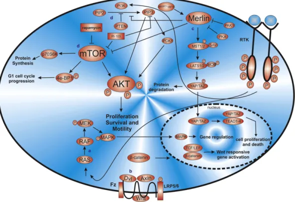
The PI3K/AKT/mTOR pathway is altered in MM and plays an
impor-tant role in cell proliferation, survival and motility in many cancers. In
62% of MM cell lines, AKT activation was reported
[26]. In another
study, it was shown that 65% of human MM species displayed elevated
levels of AKT activity
[22].
Furthermore, other pathways are dysregulated in MM. The Receptor
Tyrosine Kinases (RTKs) drive cell proliferation, survival, differentiation
and cell cycle control. Several mechanisms can activate this pathway in
cancer providing a good therapeutic option. The overexpression of the
epidermal growth factor receptor (EGFR) plays an important role in
the progression of several cancers
[27]. In a study, the EGFR was present
in 44% of MM samples; however, it is not found to be an independent
prognostic factor
[28].
The Vascular Endothelial Growth Factor Receptors (VEGF) are a
potent inducer of the angiogenesis, and its role in the cancer is well
established
[29]. High levels of VEGF in MM have been demonstrated,
being associated with a worse patient survival
[30]. Moreover, the
importance of this receptor in regulating the angiogenesis, and tumor
progression was established; thus making this pathway as a therapeutic
target in MM
[31].
The retinoblastoma protein (pRb) pathway plays an important role
in apoptosis and cell cycle regulation. Mutation on pRb is common in
many cancers, but not in MM
[32]. Nevertheless, the pRb and p53
path-ways play an important role in MM. The
p16
INK4aand
p14
ARFexert
effects on the pRb and on p53 pathways. The p16
INK4ainhibits the cyclin
dependent kinases (CDks), preventing the inactivation of pRb; on the
other hand, the p14
ARFpromotes degradation of MDM2, leading then
to the stabilization of p53
[33]. Indeed, mutations on
TP53
and on
RB
are not a common event in MM; however mutations and/or alterations
on
p16
INK4a/p14
ARFare very common. Thus, alterations and/or mutations
on
p16
INK4a/p14
ARFhave the potential to disrupt key cell cycle control
pathways.
The hippo pathway controls cell proliferation, growth,
differentia-tion and death
[37], and it has been implicated in the development of
MM
[38,39]. The Wnt pathway plays a fundamental role in the
determi-nation of cell fate, proliferation, polarity, and cell death during
embry-onic development
[40]. The Wnt signaling pathway has been reported
in MM
[41]. Therefore, it is clear that there are several players, genes,
and pathways involved in MM, all of which are described in depth in
the following sections.
1.2. Asbestos and MM
We have recently reviewed the role of asbestos in MM and its
carcinogenic mechanisms, which are summarized in
Fig. 1. In this
same work, we have also reviewed the roles of
PTEN
and
TP53
in the
development of MM
[42].
1.3. SV40 and MM
The Simian virus 40 (SV40) is a DNA monkey virus that was present
in contaminated polio vaccines produced from 1955 to 1978. It is
believed, that this is the most likely route of SV40 transmission into
humans
[43]. Furthermore, the SV40 has been implicated in MM
[44].
It was observed that speci
fi
c SV40 viral sequences were present in 57%
of epithelial invasive MM
[45]. In another study, initially, it was reported
the presence of SV40 in 60% of MM samples; then later on, it was shown
that these
fi
ndings were incorrect due to plasmid contamination, and
that only 6% of the positive samples had the presence of SV40 DNA
[46]. Recently, an Italian study in a hyperendemic area of MM has
detected SV40 DNA in 22% of the MM tumors with a low viral load
[47].
Negative results have also been published. In a study, the authors
have reported that SV40 was absent in 69 (100%) MM tumors
[9].
Another study has reached the similar conclusion
[48]. Recently, no
SV40 was detected in Korean MM samples
[49], and similar conclusions
has reached a recent study in Slovenia
[50]. Thus, these contradictions in
the literature have caused a huge controversy regarding the role of SV40
in MM. Until now, at least 50 laboratories have detected the presence of
SV40 in human tumors using a great variety of molecular biology
tech-niques; and thus raising even more the controversy about the role of
SV40 in MM
[51]. For instance, 100% of animals injected with SV40 in
the pleural tissue developed MM within 6 months
[52], thus showing
a relationship between SV40 and MM, at least in animal studies. Besides,
the relationship and the controversy between SV40 and MM have been
extensively addressed by Qi et al.
[51].
However, an animal study has shown that SV40 alone was not able
to cause MM, only asbestos exposure caused 20% of MM, and
remark-ably, asbestos and SV40 together caused 90% of MM in hamsters. This
study has shown that lower amounts of asbestos may cause MM in
animals infected by SV40
[43], and similar conclusions have been
found by another study
[53]. Therefore, these studies indicate that the
levels of asbestos exposure that are considered
“
safe
”
for the whole
population, may not be for those who were previously exposed to
SV40. Lastly, a recent study has shown that long-term exposure to
asbestos in SV40 infected cells generates resistance to
chemotherapy-induced apoptosis
[54].
The mechanisms behind SV40 carcinogenic activity are indeed
com-plex, and not fully understood. The SV40 oncogenic activity rests on the
production of two major proteins: the small t antigen (tag) and the large
T antigen (TAG). It is known that TAG is able to inactivate several tumor
suppressor genes, such as
TP53
and
RB
. These genes encode key proteins
to the cell cycle checkpoints, and the loss of these proteins leads to
uncontrolled cell proliferation
[53]. Furthermore, the tag protein
inhibits the cellular phosphatase 2A (PP2A), which is involved in the
dephosphorylation of many protein substrates, including elements of
the MAPK pathway (Fig. 1). Consequently, the loss of PP2A by tag may
alter the activity of several phosphoproteins
[55]. In addition to these
classical mechanisms, it was recently shown that TAG-p53-pRb-p300
complex regulates the transcription of the insulin-like growth factor I
(IGF-1) gene by binding to the IGF-1 promoter. In other words, there
is an increase of IGF-1 production, which leads to enhanced cell growth
[56]. Moreover, it has also been shown the involvement of SV40 in the
expression of VEGF
[57], and in the increase of telomerase activity in
MM cells
[58].
Taken altogether, it is still not clear the direct carcinogenic effects of
SV40 in MM in humans; however, it is widely accepted the role of SV40
as a co-carcinogenic player in association with asbestos in the
develop-ment of MM
[51,59].
1.4. Erionite and MM
It has been reported cases of MM without any previous known
contact with asbestos particles, in other words, not all MM cases are
eti-ologically related to asbestos exposure
[60]. Indeed, it is often wrongly
assumed that only asbestos causes MM, but in fact, other agents have
been implicated in the development of MM, among these agents, the
mineral erionite.
The erionite is a
fi
brous form of the zeolite group of minerals, which
is several times more carcinogenic than crocidolite asbestos in causing
MM
[10,61]. The relationship between erionite and MM was
fi
rst studied
in some villages in Turkey, where a strong correlation between erionite
exposure and MM incidence was found
[62,63]. The names of the
villages
fi
rst described were Karain, Tuzköy and Sarihidir, and at that
time, there were approximately 5000 people living in these villages.
The mortality rate due to MPM among Karain village was 8 deaths/
1000 inhabitants/year; in addition to 52% of deaths related to MM
from 1970 to 1994
[64]. Therefore, these studies have shown that
erionite is a strong inducer of MM in humans.
Until recently, erionite exposure was believed to be a health
prob-lem only in Turkey; however, this has changed dramatically with the
discovery of the
fi
rst erionite related MM case in the US
[65], and also
with the
fi
rst reported case of a patient with erionite-associated pleural
mesothelioma in North America
[66]. Astonishing evidence has shown
that over the past 30 years more than 300 miles of road was surfaced
with erionite-containing gravel in Dunn County, North Dakota, USA. In
this same study, it has been reported that the airborne concentration
of erionite in several places was equal or exceeded the concentrations
found in Turkish villages known to have a high incidence of MM
induced by erionite
[67].
Consequently, asbestos and erionite likely share the mechanisms of
toxicity and carcinogenesis
[68,69]; in addition, erionite is able to
induce the transformation of human mesothelial cells (MET5-A), but
on the other hand, asbestos is not able to cause such transformation
[70]. Furthermore, it has been speculated that the HMGB1 (High mobility
group box 1) (Fig. 1) is a critical initiator of the chronic in
fl
ammation in
erionite exposed individuals with the release of IL-1
β
and TNF-
α
[69]. It
has been reported that erionite activates NLRP3 in
fl
ammasome in
human mesothelial cells, which is associated with the release of IL-1
β
,
IL-6, IL-8 and VEGF, and with the activation of an autocrine feedback
loop modulated via the IL-1 receptor. Likewise, it has been shown that
IL-1 receptor blocking may play an important role in inhibiting MM
growth and progression
[71].
2. Genes and MM
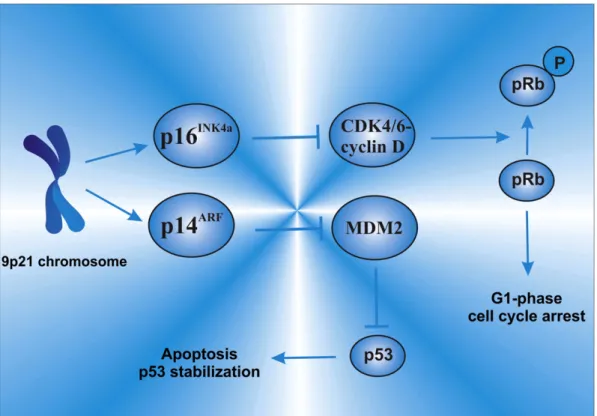
2.1. p16
INK4a/p14
ARFLocated at the 9p21 chromosome, the
p16
INK4a/p14
ARF(also known
as
CDKN2A/ARF
) are important tumor suppressor genes, which encode
two functionally unrelated proteins, the p16
INK4aand the p14
ARF(also
known as p19
ARFin mice). These proteins have unique
fi
rst exons (1
α
and 1
β
), but share exons 2 and 3, which are translated from an alternative
reading frame with no amino acid homology
[72]. The same locus harbors
another tumor suppressor gene (TSG) called
p15
INK4B(also known as
CDKN2B
), which encodes the protein p15
INK4B, a CDK (cyclin-dependent
kinase) inhibitor known to be induced by TGF
[73].
The p16
INK4ais a CDK inhibitor, and acts by inhibiting the
CDK-mediate hyperphosphorylation that leads to pRb inactivation, while
p14
ARFregulates p53 function by inhibiting p53 degradation through
MDM2 interaction
[27,74]. p16
INK4amaintains pRb in its active
hypophosphorylated form by disrupting the CDK4/6-cyclin D complex,
leading to G
1-phase cell cycle arrest (Fig. 2). Thus, both p16
INK4aand
p14
ARFare key cell cycle regulators due to their role in the p53 and the
pRb pathways
[75]. Therefore, genetic defects in the
p16
INK4a/p14
ARFare
able to lead to loss of function on both p53 and Rb pathways, which are
key players to a regulated cell cycle control (Fig. 3).
2.1.1. Role of p16
INK4a/p14
ARFand MM
The
p16
INK4a/p14
ARFhave been implicated in the development of
human cancers
[76–78]. This is no different in MM, in which these
genes have been extensively shown to be inactive. It has been shown
homozygous deletions of
p16
INK4ain 85% of mesothelioma cell lines
[79], abnormal p16
INK4aexpression in all primary mesothelioma
speci-mens and cell line
[32], and codeletion of
p16
INK4aand
p15
INK4Bin 72%
of primary mesotheliomas
[13]. Regarding the histological type, MM
epithelioid samples have shown approximately 70% of
p16
INK4a/
p14
ARFhomozygous deletions; the sarcomatoid and biphasic have shown
approximately 100% of homozygous deletions
[80–83]. Loss of the
9p21 locus was observed in 32% of MM cases. Furthermore, intermediate
methylation values were observed in the promoter region of the
p16
INK4a/p14
ARFin MM samples with no changes on the prognosis
[84].
Further studies are summarized in
Table 1.
Genetic engineering has been a great asset in better understanding
the functions of these genes. Thus, a knockout mouse for
p19
ARF, but
expressing
p16
INK4adevelops tumors early in life
[85]. Similar results
have been found using knockout mice for
p16
INK4a[86]. Not surprisingly,
knockout mice for both
p16
INK4a/
p19
ARFwere more prone to the
sponta-neous development of tumors at an early age, and highly sensitive to
carcinogenic treatments
[87].
Although, several studies have shown loss of
p16
INK4a/p14
ARFin MM,
only recently a knockout mice model for
p19
ARFhave been developed,
showing that the inactivation of this gene plays a signi
fi
cant role in
driving MM pathogenesis
[88]. Furthermore, an interesting study has
shed more light on the role of both genes in the development of MM
related to asbestos exposure. In this study, mice de
fi
cient for
p16
INK4a(+/−),
p19
ARF (+/−), and those with double de
fi
ciency
(
p16
INK4a(+/−)/
p19
ARF (+/−)) were exposed to asbestos. The mice
p16
INK4a(+/−)/
p19
ARF(+/−)displayed accelerated asbestos-induced
MM in comparison to
p16
INK4a(+/−)or
p19
ARF(+/−)mice alone. The
p16
INK4a(+/−)mice displayed bi-allelic inactivation of
p16
INK4a, loss of
the
p14
ARFor p53 expression, and frequent loss of
p15
INK4b; on the
other hand, mice
p19
ARF(+/−)exhibited loss of
p19
ARFexpression, but
no loss of
p16
INK4aor
p15
INK4b[89].
Thus, this study clearly shows that both genes play signi
fi
cant, and
not redundant roles in MM, and their inactivation increases the
tumor-igenesis caused by asbestos exposure. Another interesting
fi
nding of this
study was that p53 remained functional even in the absence of
p19
ARF,
thus showing that
p19
ARFloss contributes to MM progression via
p53-independent pathway(s)
[89], as it has been con
fi
rmed by other studies
[36,88].
2.1.2. p16
INK4a/p14
ARFgene therapy
Gene therapy has been growing considerably in the last decade
especially in Mesothelioma due to the poor response to the
tradi-tional chemotherapy. It has been shown previously, that loss of the
p16
INK4a/p14
ARFis the most common event in MM, and thus therapies
targeting the re-expression of these genes in mesothelioma cell lines
have shown interesting results. It has been demonstrated, that the
transduction of
p16
INK4aexpressing adenovirus in mesothelioma
cells resulted in cell cycle arrest, inhibition of pRb phosphorylation,
diminished cell growth, and eventual death of the transduced cells
[90]. Likewise, in a human mesothelioma xenografts study, the
re-expression of the
p16
INK4aled to: an increase in survival
[91]; an
in-crease in p53 protein levels; a reduction on the phosphorylation of
pRb, as well as G
1-phase cell cycle arrest and apoptotic cell death
[12].
The re-expression of
p16
INK4a/
p14
ARFusing different vectors did not
show better results in comparison to the single re-expression of the
p16
INK4a[92]. Taken altogether, gene therapy targeting both genes
seems to have promising results. The clinical gene therapy trials for
me-sothelioma has been recently revised elsewhere
[93].
2.2. NF2
The Neuro
fi
bromatosis type 2 (NF2) is a dominantly inherited tumor
predisposition syndrome characterized by the development of bilateral
vestibular schwannomas of the eighth cranial nerve, and by other brain
tumors, including meningiomas and ependymomas
[94]. This syndrome
is caused by mutations and lack of expression of the tumor suppressor
gene
NF2
(Fig. 3), which is located on chromosome 22q12, and encodes
the 595 amino acid protein called Merlin (Moesin-ezrin-radixin-like
protein)
[95].
2.2.1. NF2 and MM
In 1995, two groups
fi
rst demonstrated that this gene was mutated
in approximately 40
–
50% of MM, and its inactivation was important in
the tumorigenesis of MM
[95,96]. Follow-up studies have con
fi
rmed
the importance of this gene in the development of MM, and are
Table 1Methods in each study vary with different sensitivity/specificity rates, and definitions of mutations. The methodologies described in the table are the main methods used, but more methods may have been applied. Fish = Fluorescence in situ hybridization; MM = Malignant Mesothelioma; HMM = Human Malignant mesothelioma; SSCP = single-strand conformation polymorphism.
Evaluation ofp16INK4a/p14ARFexpression in MM
Study Methodology Main results
p16alterations and deletion mapping of 9p21–p22 in Malignant Mesothelioma[79]
Southern Blot and PCR.
40 cell lines and 23 primary tumors.
Homozygous deletion ofp16INK4ain 34 (85%) cell lines
and in 5 (22%) of primary tumors
p16deletion in sarcomatoid tumors of the lung and pleura[249] FISH
Sarcomatoid malignant mesotheliomas samples
Deletion of 9p21 in 26 of 32 (81%) in malignant mesotheliomas
Codeletion ofp15andp16in primary malignant mesothelioma[13]
FISH
Primary mesotheliomas samples
Codeletion ofp15INK4bandp16INK4ain 72% of
mesotheliomas Genomic profiling of malignant pleural mesothelioma
with array-based comparative genomic hybridization shows frequent non-random chromosomal alteration regions including JUN amplification on 1p32[247]
Genome-wide array-based CGH, RT-PCR 9 MPM cell lines and 17 MPM samples;
p16INK4a/p14ARFdeletion was found in 7 (41%) MPM
samples and in 9 (100%) MPM cell lines.
Diagnostic importance of 9p21 homozygous deletion in malignant mesotheliomas[83]
FISH
Pleural mesothelioma and Peritoneal mesothelioma samples
Homozygous deletion of the 9p21 in 35 of 52 cases (67%) of pleural mesothelioma and in 5 of 20 cases of peritoneal mesothelioma (25%)
Establishment and characterization of four malignant pleural mesothelioma cell lines from Japanese patients[248]
PCR, SSCP analysis and Western Blot 4 HMM cell lines
p16INK4a/p14ARFin all four HMM cell lines
9p21 deletion in the diagnosis of malignant mesothelioma in serous effusions additional to immunocytochemistry, DNA–ICM, and AgNOR analysis[81]
FISH
Malignant mesothelioma patient samples
9p21 homozygous deletion in 48.5%, heterozygous deletion in 36.4%
Promoter methylation of RASSF1A, RARÎ2and DAPK predict poor prognosis of patients with malignant mesothelioma[84]
Nested methylation-specific PCR DNA of mesothelioma patients
p16INK4aandp14ARFpromoter region methylation
in 28.2% and 44.2%, respectively Morphology of 9p21 homozygous deletion-positive pleural
mesothelioma cells analyzed usingfluorescence in situ hybridization and virtual microscope system in effusion cytology[250]
FISH
15 epithelioid MPM
12 positive for a homozygous deletion and 3 positive for both homozygous and heterozygous deletions with a predominantly heterozygous pattern
9p21 deletion in the diagnosis of malignant mesothelioma, usingfluorescence in situ hybridization analysis[82]
FISH
Malignant mesothelioma patient samples
9p21 deletion in 35 of 40 (88%) cases with MM
Genomic gains and losses in malignant mesothelioma demonstrated by FISH analysis of paraffin-embedded tissues[249]
FISH
Malignant mesothelioma patient samples
Loss p16INK4a/p14ARFin epithelioid 23/30 (77%) and
biphasic/sarcomatoid 12/12 (100%) mesotheliomas
Immunohistochemical analysis of thep16INK4
cyclin-dependent kinase inhibitor in malignant mesothelioma[32]
Immunohistochemical analysis and Immunoblot. Primary thoracic mesotheliomas and mesothelioma cell lines
Abnormal p16INK4aprotein expression in 12 of 12 primary mesothelioma specimens and in 15 of 15 mesothelioma cell lines
Homozygous deletion ofCDKN2A/ARF and codeletion of the methylthioadenosine phosphorylase gene in the majority of pleural mesotheliomas[247]
FISH
Pleural mesothelioma samples
p16INK4a/p14ARFhomozygous deletion in 70 samples
(74%). Homozygous loss ofp16INK4a/p14ARFin 49
of 71 epithelial (70%), 16 of 19 biphasic (89%) and 5 of 5 sarcomatous (100%) mesotheliomas.
Table 2
Methods in each study vary with different sensitivity/specificity rates, and definitions of mutations. The methodologies described in the table are the main methods used, but more methods may have been applied. MM = Malignant Mesothelioma; HMM = Human malignant mesothelioma; SSCP = single-strand conformation polymorphism; Comparative genomic hybridization (CGH).
Evaluation ofNF2expression in MM
Study Methodology Main results
Frequent mutations ofNF2and allelic loss from chromosome band 22q12 in malignant mesothelioma: Evidence for a two-hit mechanism ofNF2inactivation[251]
Western blot and DNA sequence analyses 25 MM cell lines
14 of 25 (56%) showed noNF2expression; 18 of 25 (72%) showed losses at one or both loci tested.
Heterogeneity of mesothelioma cell lines as defined by altered genomic structure and expression of theNF2gene[252]
Northern blot, RT-PCR and PCR. 18 HMM cell lines
NF2alterations were identified at a genomic level in 7 (39%) cell lines and were associated with a marked decrease in the concentration of the NF2 transcript.
Establishment and characterization of four malignant pleural mesothelioma cell lines from Japanese patients[248]
PCR, SSCP analysis and Western Blot 4 HMM cell lines
A point mutation ofNF2was observed in 1 cell line.
Genomic profiling of malignant pleural mesothelioma with array-based comparative genomic hybridization shows frequent non-random chromosomal alteration regions including JUN amplification on 1p32[247]
Genome-wide array-based CGH, RT-PCR 9 MPM cell lines and 17 MPM samples;