MORPHOLOGICAL CHANGES IN THE VAGINAL
EPITHELIUM DURING THE OESTROUS
CYCLE OF
Calomys callosus
(Rodentia, Cricetidae)
JEAN FÁBIO TORRES RODRIGUES and ELOISA AMÁLIA VIEIRA FERRO
Department of Morfhology, Federal University of Uberlândia, Uberlândia, Minas Gerais, Brazil Address correspondence to: Eloisa Amália Vieira Ferro, Departamento de Morfologia,
Universidade Federal de Uberlândia, Av. Pará,1720, Campus Umuarama, CEP 38400-902, Uberlândia, MG, Brazil
Received October 10, 1996 – Accepted May 05, 1998 – Distributed August 28, 1998
(With 22 figures)
ABSTRACT
This study describes changes in the pattern of microridges, keratinization, desquamation, secretion, leukocyte infiltration as well as the increasing number of mitotic cells in the vaginal epithelium of
Calomys callosus during the oestrous cycle. In proestrus, the epithelium is squamous and stratified
with a fine layer of keratin and it is overlain by secretory prismatic cells. In oestrous, the epithelium is squamous, stratified and keratinized. In metoestrus, the epithelium is squamous and stratified with loss of the keratin layer. A leukocyte infiltration, extending from the base to the surface of the epi-thelial layer is also present. At the end of this phase, the surface cells start to become PAS-positive. In dioestrus, the epithelium is stratified. The superficial cells are prismatic, exhibiting the structural and ultrastructural characteristics of glycoprotein secreting cells supported by a layer of squamous cells. At the end of this phase, kerato-hyaline granules appear in the granular layer of the epithelium, indicating the beginning of the keratinization process, present in the next proestrus.
Key words: oestrous cycle, vaginal epithelium, Calomys callosus, keratinization, secretion, leukocyte
infiltration.
RESUMO
Alterações morf ológicas do epitélio vaginal de Calomys callosus
(Rodentia, Cricetidae) durante o ciclo estral
Neste estudo são descritas mudanças que ocorrem nos padrões de microrrugas, queratinização, des-camação, secreção, infiltração leucocitária e aumento no número de células em mitose no epitélio vaginal de Calomys callosus durante o ciclo estral. No proestro, o epitélio é estratificado pavimentoso,
com uma fina camada de queratina e, sobre esta camada, há células prismáticas secretoras. No estro, o epitélio é estratificado pavimentoso e queratinizado. No metaestro, o epitélio é estratificado pa-vimentoso com poucas camadas de queratina. Infiltrados leucocitários estão presentes desde a base até as camadas superficiais do epitélio. No final desta fase as células da superficie começam a se tornar PAS-positivas. No diestro, o epitélio é estratificado. As células superficiais são prismáticas, exibindo características estruturais e ultra-estruturais de células secretoras de glicoproteínas, as quais estão apoiadas sobre camada de células pavimentosas. No final desta fase, aparecem grânulos de querato-hialina na camada granulosa do epitélio, indicando o começo do processo de queratinização, presente no proestro.
Palavras-chave: ciclo estral, epitélio vaginal, Calomys callosus, queratinização, secreção, infiltração
INTRODUCTION
Calomys callosus is a species of South
Ameri-can animal recently introduced in laboratory re-search. It is a cricetidae rodent widely found in Brazilian territory and easily adapted to laboratory conditions (Mello, 1981; Peter et al., 1967; Justines & Johnson, 1970). From a medical point of view C. callosus is the reservoir for Trypanosoma cr uzi, the
etiologic agent of Chagas’s disease (Ribeiro, 1973), and for the agent of Argentine hemorrhagic fever (Justines & Johnson, 1969). From the reproductive point of view, C. callosus is a polyestrus rodent and
has a postpartum oestrus. The species is character-ized by an oestrous cycle of 6.6 days duration, puberty occurs frequently with 40,1 days (± 7,6) in female and 19,6 days (± 6.6) in male (Mello, 1978). The adult animal has 12 cm long, weighs 30±8 g and the number of youngs born is 5±3. Although several studies have been published about this spe-cies, morphological changes in the vaginal epithe-lium during the oestrous cycle are not known. Cyclical changes in the vaginal epithelium during oestrous cycle have been described in many ani-mal species. In general, these changes are charac-terized by keratinization, acquisition of secretory activity by the epithelial surface, variation in the number of mitotic figures in the germinative stra-tum with consequent increase in thickness of the epithelium, leukocyte infiltration through the epi-thelial cells to the lumen of the organ, and an in-crease in the population of Langerhans cells among the keratinocytes.
The purpose of this study was to examine the morphological changes in vaginal epithelium of C. callosus during the oestrous cycle with light
micros-copy (LM), transmission electron microsmicros-copy (TEM) and scanning electron microscopy (SEM).
MATERIALS AND METHODS
Eighteen, twelve-week-old, female C. callo-sus were obtained from the Tropical Medicine
In-stitute of São Paulo. The animals were housed at 26 + 2o C under a 12-h light, 12-h dark light pe-riod and received water, granulated ration (Puri-na), sunflower seeds and corn ad libitum.
Every morning vaginal smears were taken, stained using Shorr’s technique (1945) and analyzed under the LM to define the phase of the cycle. Three females were sacrificed to characterize each cycle
phase. The animals were anaesthetized by ether inhalation. After laparotomy, the dissection was initiated in the median region of the vagina, di-viding it into 3 fragments. The first fragment was processed for LM, the second fragment for TEM and the last for SEM.
The first fragments were fixed in a solution of 95% ethanol, formalin, glacial acetic acid and distilled water (3:1:1:5 v/v) (Finn & McLaren, 1967) for 18 hours. The fragments were then rou-tinely processed for Glycol methacrylate embed-ding (Historesin, LKB). Sections of 2 µm thick-ness were stained with 0,25% toluidine blue in distilled water at 40oC. Some sections were treated using the Periodic-acid-Schiff (PAS) re-action according to McManus (1948).
Second fragments were fixed for about 3 hours in a mixture of equal parts of 2% glu-taraldehyde and 2% paraformaldehyde in 0.1 M phosphate buffer at pH 7.4.
They were then washed in 0.1 M phosphate buffer at pH 7.4, post-fixed in 1% osmium tetrox-ide in 0.1 M phosphate buffer at pH 7.4 during 1 h, embedded in Epon and analyzed in a Zeiss EM-109 Electron Microscope. The third fragments were fixed broadly similarly to the second frag-ments and were dehydrated in an ascending series of ethanol, dried with C02 in a critical-point dryer (Balzers), mounted on metal stubs with silver paint, coated with 9 nm gold in a sputter coated and analyzed in a Zeiss DSM 950 Scanning Elec-tron Microscope.
RESULTS
Proestrus
In this phase, the epithelium was squamous, stratified and keratinized, exhibiting about 20 lay-ers of cells. PAS-positive, prismatic cells (Fig. l) were observed on the keratinized layer.
approximately 10 nm diameter, either isolated or in bundles, and lipid droplets were also observed.
Fig. 1 — Vaginal epithelium of Calomys callosus in proestrus
stained with PAS and observed by LM. In this phase the epithelium is thick, presenting kerato hyaline granules (ar-rowheads) and a keratinized layer (K). In the vaginal lumen (L) many PAS-positi ve cells (asterisk) are supported on the keratinized layer. l70X.
In the middle third, most cells were globous or squamous; the other characteristics described for the basal layer were present, although the number of the desmosomes was greater (Fig. 2). In the apical portion, two distinct cellular populations were observed: an internal layer con-tained squamous cells, and an external layer exhibited prismatic secretory cells which secreted PAS-positive material. In the squamous cell layer, two further regions were distinguished: an infe-rior region, in contact with the cells of the middle third, and a superior region, in contact with the secretory cells layer. The inferior region was composed by cytoplasms of the cells which were filled with 10 nm of diameter filaments and kerato-hyaline granules. A reduction in the num-ber of mitochondria and cisternae of granular endoplasmic reticulum compared to the basal and middle layers was observed, although the large number of free polyribosomes still persisted. The nucleus presented the same characteristics as seen
in the previously described portions. The supe-rior region exhibited cells containing cytoplasm filled with an amorphous, homogeneous and electrondense substance. Nucleus and other or-ganelles were not observed. A linear accumula-tion of electron-dense material near the plasmatic membrane was observed (Fig. 3).
Fig. 2 — Electronmicrograph of vaginal epithelium of
Calomys callosus during proestrus. Middle region of the
vaginal epithelium. Nucleus (N) with prominent nucleolus (asterisk). In the cytoplasm, numerous keratin filaments (ar-rowheads) grouped in bundles or adhering to desmosomes. 6000X. Bar – 1µm.
The secretory cells presented predominantly heterochromatic nucleus. Their cytoplasm revealed organelles in different degrees of degeneration. There were numerous secretory granules of variable electron density in the cytoplasm (Fig. 4). Surface of epithelium exhibited high cells covered with microridges (Figs. 5 and 6). The vaginal lumen was characterized by the presence of many PAS-posi-tive, epithelial cells undergoing desquamation.
Oestrus
In the basal portion, the cells were similar to those in the same region in the proestrus phase. Mitotic figures were common. In the cytoplasm, well developed granular endoplasmic reticulum, free polyribosomes, 10 nm diameter filaments and mitochondria presenting either lamellar or tu-bular cristae (Fig. 8) were observed.
Fig. 3 — Vaginal epithelium of Calomys callosus in proestrus
observed by TEM. Interface between keratinized cells (as-terisk) and cells in the process of keratinization (K). In this cell there are numerous free polyribosomes (arrowheads), kerato-hyaline granules (Q) and bundles of intermediate filaments (arrow). 29000X. Bar – 1µm.
In the middle portion, the cells have already acquired a squamous form, presenting nuclei with loose chromatin. In the cytoplasm, few organelles were observed with the exception of free polyri-bosomes. There was an increase in the quantity of kerato-hyaline granules and 10 nm filament bundles. At some points in the layer of contact with the keratinized portion, these filament bundles adhered to the internal face of the desmosomes, which confered the coupling between the cells of this layer and the keratinized layer (Fig. 9).
In the apical portion, the cells were kerati-nized and their cytoplasms were completely filled with an amorphous, homogeneous, and electron-dense substance; residues of nuclei could be
observed at some points. The adherence of the adjoining keratinized cells was effected by des-mosomes. At these sites, an electron-dense ma-terial was deposited on the internal face of the plasma membrane (Fig. 9). The surface epithe-lium showed flattened cells in process of slough-ing (Fig. 10). At low magnification some surface cells possessed connected microridges (Fig. 11).
Fig. 4 — Vaginal epithelium of Calomys callosus in proestrus
observed by TEM. Interf ace between keratinized cells (K) and secretory cells (S). The plasma membrane of the secre-tory cells exhibits numerous microvilli. In the cytoplasm there are secretory granules (g); the nucleus (N) p resents an irregular profile and condensed chromatin. 6000X. Bar – 1µm.
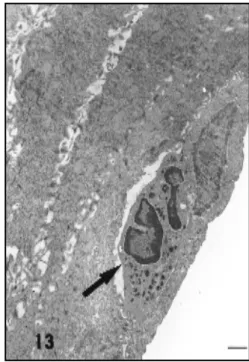
Metoestrus 1
Fig. 9 — Electronmicrographs of vaginal epithelium of Calomys callosus during oestrous. Interf ace between keratinized
(K) and non-keratinized layer (Q). The upper left insert demonstrates desmosomes seen in the keratinized layer. The two membranes forming each desmosome are in close juxtaposition and there are deposits of electron dense material on the in cytoplasmic f aces (arrows). The lower left insert shows the transition zone between the keratinized and non-keratinized layer. The non-keratinized layer is marked by the presence of numerous desmosomes (asterisk) into which intermediate filaments are inserted. 14000X. Upper left insert – 40000X. Lower left insert – 20000X. Bar – 1µm.
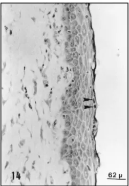
Fig. 7 — Vaginal epithelium during oestrous obseved by LM.
The epithelium is stratified, squamous and keratinized (K). Below this layer there are man y kerato-hyaline granules (as-terisk). 310X.
Fig. 8 — Electronmicrograph of vaginal epithelium of
Figs. 10 and 11— Scanning electronmicrographs of the vaginal epithelium of Calomys callosus in oestrus. Fig. 10 — Surface
Fig. 12 — Vaginal epithelium of Calomys callosus in me-toestrus 1 stained with PAS and observed by LM. The epi-thelium is stratified and squamous. Among the epithelial cells in the middle and upper epithelial layers, numerous infil-trated leukocytes are found (arrowheads). Leukocyte (arro w) infiltration is seen in the connective tissue (t). 180X.
Fig. 13 — Electronmicrographs of vaginal epithelium of
Calomys callosus during metoestrus l. In this
electronmicro-graph a leukocyte is located (arrow) among the epithelial cells. 5000X. Bar – 1µm.
Metoestrus 2
The epithelium was characterized by the presence of approximately 9 to 10 layers of cells and occasional mitotic figures. In this phase, the leukocytes accumulated in the apical layers of the epithelium (Fig. 14) and vaginal lumen charac-teristically presented desquamation of epithelial cells, together with a few leukocytes. The basal cells and those of the middle portion presented characteristics similar to those seen in the previ-ous phase. The apical portion differed from last phase in that it was PAS-positive (Fig. 14) and presented glycogen deposits (Fig. 15).
The surface epithelium similary to me-toestrus 1 and meme-toestrus 2, showed flattened cells with microridges in process of sloughing, very closely resembling that at oestrus (Fig. l6).
Dioestrus 1
In this phase, the epithelium were approxi-mately 5 cell layers thick with an accentuated in-filtration of leukocytes among the keratinocytes. In the basal third, the cells were prismatic and mitotic figures were rare. In the apical third, tall cubic and prismatic cells were present (Fig. 17). Ultrastructurally the cytoplasm of the cells pre-sented mitochondria, cisternae of granular endo-plasmic reticulum, Golgi complexes and secre-tion granules (Fig.18). Surface cells showed microridges. The number of the microridges was variable, numerous in some cells and few in oth-ers (Fig. l9).
Dioestrus 2
The epithelium was characterized by the presence of 15 to 20 layers of cells. Some mitotic figures were observed in the basal third of the epithelium. In the middle third, the cells were squamous and became progressively filled with kerato·hyaline granules towards the epithelial sur-face.
Fig. 14 — Vaginal epithelium of Calomys callosus in
me-toestrus 2 stained with PAS and observed by LM. In this phase, leukocytes predominate in the upper layer of the epi-thelium (arrowheads) which is PAS-positive. 160X.
Fig. 15 — Electronmicrograph of the upper re gion of the
epithelium seen in the previous figure. Observe the nucleus, mitochondria (arrow), desmosomes (arrowheads) and re gion of glycogen accumulation (star). 26000X. Bar – 1µm.
Fig. 16 — Scanning electron micrograph of the vaginal epithelium of Calomys callosus in metoestrus. The cells are in process
Fig. 19 — Scanning electron micrograph of the luminal surface of the vaginal epithelium in dioestrus l. Cell surface with few microridges (asterisk) or with many microridges (star). 12000X. Bar – 1µm.
Fig. 17 — Vaginal epithelium of Calomys callosus in
di-oestrus 1 stained with PAS and observ ed by LM. The epi-thelium comprises few cell layers and the superficial layer cells are cubic. In the connective tissue, leukocytes (arrow) also insinuate (arrowhead) among the epithelial cells. l50X.
Fig. 18 — Elect ronmicrograph of v aginal epithelium of
Calomys collosus during dioestrous l. Cubic cells seen in the
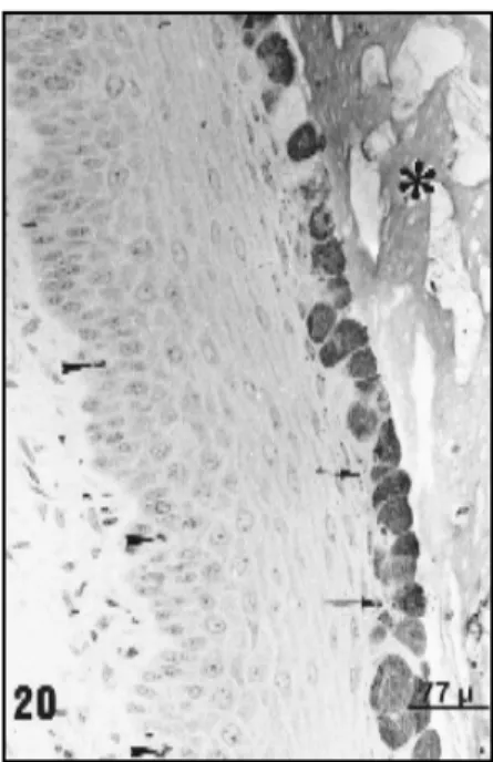
Fig. 20 — Vaginal epithelium of Calomys callosus in di-oestrous 2 stained with PAS an observed by L M. A thicker epithelium is observed; mitotic figures are common in the basal region (arrowhead). The superficial cells are PAS-posi-tive. Below this layer kerato-hyaline granules (arrow) are also found in the cytoplasm of the squamous cells. In the vagi-nal lumen (asterisk) a PAS-positive secretion is seen. l70X.
Fig 21 — Electronmicrograph of vaginal epithelium of Calomys callosus during dioestrous 2. Prismatic cells seen in Fig. 20.
Observe nucleus (n), many secretory granulea (g) are present in the cytoplasm of these cells. On the apical surface of the cells, certain expansions which may represent the process of gran-ule extrusion (asterisk) are seen. 6000X. Bar – 1µm.
Fig. 22 — Scanning electron micrograph from the vaginal epithelium of Calomys callosus in dioestrus 2. In cell surfaces
DISCUSSION
During the oestrous cycle, the vaginal epi-thelium of C. callosus undergoes profound
mor-phological changes. This phenomenon also oc-curs in rat (Parakkal, 1974; Centola, 1978; Vijayasaradhi & Gupta, 1987) and mouse (Allen, 1927; Young & Hosking, 1986; Nelson et al,
1991; Horvat et al, 1992). The acquisition of
secretory activity, changes in the pattern of kera-tinization, and an increase in the rate of mitosis were also seen. As in other animals, these changes probably occur due to oscillating levels of ova-rian hormones.
In the proestrus phase, the oestrogen level is maximal and influences the keratinocytes which present a greater fluidity of the plasma membrane (Reddy et al.,1989). In addition to this
indirect effect, oestrogen directly influences the enzyme glucose-6-phosphatase which has an important role in keratin synthesis (Kang & West, 1982). Roop (1987) has demonstrated that oestro-gen modulates the expression of the DNA seg-ment responsible for keratin synthesis. These data, obtained from studies in rats and mice, suggest that in Calomys callosus, the process of
keratinization, which starts in proestrus and be-comes maximal in oestrus, is related, either di-rectly or indidi-rectly, to high oestrogen levels. It is known that copulation occurs during oestrus; thus, greater protection of the vaginal mucous layer is necessary and this is provided by the keratin layer, by the increased thickness of the other epithelial layers and by the microridges at the surface of the cells, wich can hold mucus at the luminal surface of the vagina (Lamb et al.,
1978). The increased thickness of epithelial layers is the result of mitosis in a great number of basal cells. A similar fact was observed by Galand et al. (1971) who demonstrated that in mice,
dur-ing the oestrogenic phase, there is an increase in the number of mitotic figures due to a reduction of the S phase of the cellular cycle, thus reduc-ing the duplication time of DNA.
Morphologically, the end of oestrus is mar-ked by the liberation of the keratin to the vaginal lumen and a reduction in epithelial thickness. This moment marks the transition from oestrus to meto-estrus 1. As the cycle proceeds, the apical portion of the vaginal epithelium is characterized by gly-cogen deposits as noted by ultrastructural
evalu-ation and by the PAS reaction, characterizing metoestrus 2. During metoestrus, an intense leu-kocyte infiltration is observed among the epithe-lial cells as these migrate towards the vaginal lu-men. This leukocyte infiltration is a well known phenomenon and has already been described by various authors (Busch, 1966; Holtz et al., 1968).
Thus, by a still unclear mechanism, the leuko-cytes migrate through the epithelium towards the lumen, assisting in protection from infectious agents (Corbeil et al., 1985), or to accomplish the
phagocytosis of spermatozoa residues still present in the vaginal lumen (Branscheid & Holtz, 1988). In dioestrus l, the epithelium of the super-ficial layer is cubic and presents ultrastructural and histochemical features characteristic of secre-tory cells. This secresecre-tory activity may be related to increased progesterone levels, since in mice the maximum concentration of progesterone occurs in dioestrus 1 (Walmer et al., 1992). As the cycle
continues, the superficial layer cells become pris-matic or globous with a great number of secre-tory granules; this period is characterized as di-oestrus 2.
The secretion produced by the epithelial cells is discharged to the vaginal lumen where, together with the secretions from the cervix, seems to per-form an important role in the defense of the epi-thelium, since the leukocytes remain immersed in this gelatinous net, and thus may exert their ph-agocytic function (Branscheid & Holtz, 1988).
In diestrus 2, kerato-hyaline granules asso-ciated with intermediate filaments of 10 nm diam-eter accumulate below the layer of secretory cells forming the keratin layer present in the subsequent phase: the proestrus phase. In proestrus, secretory cells were observed in the keratinized layer, re-flecting an advanced degenerative condition which persisted since the last dioestrus.
In conclusion, the present results suggest that surface cells of vaginal epithelium could be secre-tory or keratinized, probably in response to ovarian hormones. Additionally, the present study gives evidence of wide cellular variety found in vaginal epithelium of C. callosus during oestrous cycle.
Acknowledgements — The authors thank Mr. Hélgio H.
instruction in the techniques and principles of scanning electron microscopy.
REFERENCES
ALLEN, E., 1927, The oestrus cycle in the mouse. Ameri-can Journal of Anatomy, 30(3): 297-371.
BRANSCHEID, W. & HOLTZ, W., 1988, Histochemical ex-amination of the vaginal epithelium of sows at varìous stages of the estrus c ycle. Anat. Histol. Embriol.,17:
12-26.
BUSCH, W., 1966, Die periodischen v eränderungen des vaginal epithels beim schwein und die möglichk eiten ihrer heranziehung zur graviditätsdiagnose. Wiss. Z. Humboldt-Uni v. Berlim, Math. Nat. R., 15: 833-865.
CENTOLA, G. M., 1978, Surface features of exfoliated vagi-nal epithelial cells during oestrous cycle of the rat exa-mined by scanning electron microscopy. J. Anat., 127: 553-561.
CORBEIL, L. B., CHATTERJEE, A., FORESMAN, L. & WESTFALL, J. A., 1985, Ultrastruture of cycle change in the murine uterus, cervix and v agina. Tissue & Cell, 17: 53-68.
FINN, C. A. & MC LAREN, A. , 1967, A study of the early stage of implantation in mice. J. Reprod. Fertil., 13: 259-267.
GALAND, P., LEROY, F. & CHRETIEN, J., 1971, Effect of vaginal on proliferation and histological changes in the uterus and vagina of mice. J. Endocr., 49: 243-252.
HOLTZ, W., SMIDT. D., THUME, O. & WESELOH, W., 1968, Veränderugen des scheiden-pH, der rektal und vaginal gemessenen Körpertemperatur und des scheidenepithels in abhängigkeit vom sexualzyklus beim gö ttinger zwergschwein. Zbl. Vet. Med, 15: 329-352.
HORVAT, B., VRCIC, H. & DAMJANOV, I., 1992, Trans-diferentiation of murine squamous vaginal epithelium in proestrus is associated with changes in the expres-sion of keratin polypeptides. Experimental Cell Re-sear ch, 199: 234-239.
JUSTINES, G. & JOHNSON, K. M., 1969, Imune tolerance in Calomys callosus infected with machupo virus. Natur e, 222: 1090-1091.
JUSTINES, G. & JOHNSON, K. M., 1970, Observations on the laboratory breeding of the cricetidae rodent
Calomys callosus. Lab. Anim. Core, 20: 57-60.
KANG, Y. & WEST, W. L., 1982, Ultrastructural localiza-tion of glucose-6-phosphatase and alkaline phos-phatase in the v aginal epithelium of rat. Journasl of Morpholo gy, 171: 1-10.
LAMB, J. C., NEWBOLD, R. R., STUMPF, W. E. & MCLACHLAN, J. A., 1978, Transiotional changes in the surf ace epithelium of the cycling mouse vagina, cervix and uterus: scanning electron microscopic stud-ies. Biol. reprod., 19: 701-711.
MCMANUS, J. F. A., 1948, Histological and histochemical use of periodic acid. Stain Tecnology, 23(3): 99-108.
MELLO, D. A., 1978. Biology of Calomys callosus under
laboratory conditions (Rodentia, Cricetidae). Rev. Bras. Biol., 38(4): 807-811.
MELLO, D. A., 1981, Studies on reproduction and longe vity of Calomys callosus (Renger, 1830) under laboratory
conditions ( Rodentia, Cricetidae ). Rev. Bras. Biol., 41(4): 841-843.
NELSON, K. G., TAKAHASHI, T., BOSSERT, N. L., WALMER, D. K. & MCLACHLAN, J. A.,1991, Epi-dermal growth factor replaces estrogen in the stimu-lation of female genital-tract growth and differentia-tion. Proc. Natl. Acad. Sci.,88: 21-25.
PARAKKAT, P. F., 1974, Cyclical changes in the v aginal epithelium of the sun by scanning electron micros-copy. Anat. Rec., 178: 529-538.
PETTER, F., KARIMI, Y. & ALMEIDA, C. R., 1967, Un nouvean ranger de laboratoire, lé cricetidé (Roden-tia, Cricetidae). C. R. Acad. Paris, 265: 1974-1976. RIBEIRO, R. D., 1973, Novo reservatório do Trypanosoma
cruzi. Rev. Bras. Biol., 33: 429·437.
REDDY, A. G., SHIVAJI, S. & GUPTA, P. D., 1989, Effect of estradiol on the membrane fluidity of the rat vaginal epithelial cells. J. Steroid. Biochem, 33(6): 1229-1233. ROOP, D. R., 1987, Regulation of k eratìn gene expression during differentiation of epidermal and vaginal epi-thelial cells. Current Topics in Developmental Biol-ogy,22: 195-207.
SHORR, E., 1945, A new technic for staining vaginal smears. A single differentive stain. Science,84: 545.
VIJAYASARADHI, S. & GIJPTA, P. D., 1987 Keratinization of rat vaginal epithelium. I - Cell surface study during natural and induced keratinization. J. Submicrosc. Cytol., 19: 595-603.
WALMER, D. K., WRONA, M. A., HLTGHES, C. L. & NELSON, K. G., 1992, Lactoferrin expression in the mouse reproductive tract during the natural estrous cycle: Correlation with circulating estradiol and progesterone. Endocrinodogy, 131(3): 458-1466. YOUNG, W. G. & HOSKING, A. R., 1986, Langerhans cells