13
Arq Neuropsiquiatr 2011;69(1):13-18
Article
Juvenile neuronal ceroid-lipofuscinosis
Clinical and molecular investigation in a large family in Brazil
Eugênia Ribeiro Valadares1,6, Mayara Xavier Pizarro2,
Luiz Roberto Oliveira3,6, Regina Helena Caldas de Amorim3,6,
Tarcísio Márcio Magalhães Pinheiro4, Ulrike Grieben5,
Helena Hollanda Santos6, Rachel Rabelo Queiroz2 ,
Guilherme de Castro Lopes2, Ana Lúcia Brunialti Godard2
ABSTRACT
Objective: Juvenile Neuronal Ceroid-Lipofuscinosis (JNCL, CLN 3, Batten Disease) (OMIM #204200) belongs to the most common group of neurodegenerative disorders of childhood. We report the clinical data and molecular analysis of a large Brazilian family. Method: Family composed of two consanguineous couples and thirty-two children. Clinical data of ten JNCL patients and molecular analyses on 13 participants were obtained. Results: The large 1.02 kb deletion was detected. The most severe phenotype, with autistic behavior, tics and parkinsonism was seen in a 12-year-old female and a milder phenotype in a 14-year-old male. Nyctalopia was the first symptom in one deceased child. The visual loss of six patients has been first observed in the school and not at home. Conclusion: The report highlights the phenotypical intrafamily variation in 10 affected children of this family. The molecular investigation of this large family in our metabolic center turned possible the diagnosis, right approach and genetic counseling.
Key words: Batten disease, neuronal ceroid-lipofuscinoses, polymerase chain reaction. Lipofuscinose ceróide neuronal juvenil: investigação clínica e molecular em uma família grande no Brasil
RESUMO
Objetivo: Lipofuscinose Ceróide Neuronal Juvenil (JNCL, CLN 3, Doença de Batten) (OMIM # 204200) pertence ao grupo mais comum de doenças neurodegenerativas na infância. É causada por mutações no gene CLN3, com padrão de herança recessiva. A deleção de 1,02 kb é a mutação mais comum. Relatamos os dados clínicos e análise molecular de uma família consanguínea numerosa. Método: Família composta por dois casais consanguíneos e trinta e duas crianças. Foram obtidos dados clínicos de dez pacientes e análises moleculares de 13 participantes. Resultados: Foi detectada deleção de 1,02 kb. O fenótipo mais grave, com comportamento autista, tiques e parkinsonismo foi visto em uma paciente do sexo feminino de 12 anos e o fenótipo mais leve em um paciente do sexo masculino de 14 anos. Nictalopia foi o primeiro sintoma de uma criança falecida. A perda visual de seis pacientes foi observada pela primeira vez na escola e não em casa. Conclusão: Destaca-se a variação fenotípica intrafamiliar em 10 pacientes. A investigação molecular desta família numerosa tornou possível o diagnóstico, a abordagem correta e aconselhamento genético.
Palavras-chave: doença de Batten, lipofuscinoses ceróides neuronais, reação em cadeia da polimerase.
Correspondence
Eugênia Ribeiro Valadares Av. Alfredo Balena 190
30130-100 Belo Horizonte MG - Brasil E-mail: [email protected]
Support
Research supported by a grant from Fundação de Amparo à Pesquisa do Estado de Minas Gerais - FAPEMIG/ SUS 005/2006 – EDT 3261-06
Received 22 July 2010
Received in final form 27 July 2010 Accepted 03 August 2010
1Departamento de Propedêutica Complementar, Faculdade de Medicina da UFMG, Belo Horizonte MG, Brazil; 2Departamento
de Biologia Geral, Instituto de Ciências Biológicas da UFMG, Belo Horizonte MG, Brazil; 3Departamento de Pediatria,
Faculdade de Medicina da UFMG, Belo Horizonte MG, Brazil; 4Departamento de Medicina Preventiva e Social, Faculdade de
Medicina da UFMG, Belo Horizonte MG, Brazil; 5Otto Heubner-Zentrum für Kinder und Jugendmedizin, SPZ, Neuropädiatrie,
he Neuronal Ceroid-Lipofuscinoses (NCL’s) are the most common group of neurodegenerative disorders of childhood, characterized by the lysosomal accumulation of autoluorescent material, which resembles ceroid and lipofuscin, in patient’s tissues1. his accumulation on
neu-ronal tissue leads to neocortical neurons death2. he
glob-al incidence of NCL’s is 1 to 8 in 100,000 births3. In the
Portuguese population, which is more similar to Brazil-ians from Minas Gerais State, the prevalence of overall NCL is estimated in 1.55 per 100,000 live births, distrib-uted in CLN3 (42.3%), CLN 2 (11.5%) and CLN 1 (3.8%)4.
he irst report of NCL was in 1826 by Stengal, who investigated four siblings presenting progressive blind-ness, epilepsy, cognitive decline and motor dysfunction. Later, in 1903, Batten reported the juvenile form of the disease (CLN 3)5. Until recently, there had been four main
forms of NCL’s classiied on age of onset and storage ma-terial at the ultrastructural level, i.e., infantile (CLN 1), classical late infantile (CLN 2), juvenile (CLN 3), and adult forms (CLN 4). However, due to recent advances in molecular and biochemical analysis, it is considered that the NCL’s are classiied into ten genetic forms (CLN1-10), only CLN 4 and CLN 9 not yet molecularly characterized (OMIM). he number of NCL forms can still grow, since syndromes with seizures and a variable degree of psy-chomotor deterioration will probably get in this group of disorders2. Most variants manifest cell death and
dys-regulated sphingolipid metabolism, suggesting that the proteins defective in these disorders may interact along a common pathway6.
NCLs are autosomal recessive diseases, except for the adult form (CLN 4) that can present both dominant and recessive autosomal patterns of inheritance2.
Juvenile-on-set Neuronal Ceroid-Lipofuscinoses (JNCL, CLN 3, Bat-ten Disease) (OMIM #204200) is caused by mutations in CLN3 gene. his gene is located at chromosome region
16p12. he gene is constituted by at least 15 exons7 and
CLN 3 can be caused by at least 25 diferent mutations, specially within exons 6 to 8 and 13. A common muta-tion achieving the rate of 81% worldwide is a delemuta-tion of 1.02kb8 that eliminates exons 7 and 8, resulting in a
trun-cated protein9. he CLN3 mutations cause only partial
loss of function of this lysosomal enzyme2.
he symptoms in classic or typical JNCL often begin after 4 years of age with progressive visual loss and other neurological dysfunction (dementia; seizures; pyramidal, extrapyramidal, and cerebellar signs). Before the recent progress in molecular genetic studies, diagnosis of NCLs was based on age at onset, clinical course, and
pathologi-cal indings of ingerprint proiles and/or vacuoles in the lymphocytes by electron microscopy10.
In 27 homozygous patients for the 1.02-kb deletion in Finland, visual failure was noticed at 4-10 years old (100%), onset of epilepsy at 8-13 years old (92%), par-kinsonian signs at 12-15 years of age (30%) and at 17-29 years of age (22%); and death occurred between 10 and 28 year11. In this study great clinical variability between
dif-ferent families and also within the families was noticed, and 22% of patients had good motor performance be-tween 14 and 22 years old. Problems related to behavior, emotions and thought have clinically been some of the main features of JNCL patients. he psychiatric symp-toms include anxiety, aggressive behavior, depression, hallucinations and psychotic symptoms. Female patients with JNCL have more diicult psychiatric problems than their male counterparts12.
We report the clinical data and molecular analysis of a large consanguineous Brazilian family from Minas Gerais State, highlighting the intrafamilial phenotypical variation in 10 afected children. We also focus in the importance of the molecular analysis to establish the diagnosis and to turn possible an accurate genetic counseling.
METHOD
Studied family
Patients were identiied by medical students in formal activity at a poor village in Minas Gerais State. heir fam-ily is composed of two consanguineous couples (broth-er and sist(broth-er who got married to their irst-cousins, also brothers) and their kindred of thirty-two children. he clinical evaluation of patients was performed by the re-search group in their hometown, without access to major clinical propedeutics other than those already done. We studied the clinical data of ten JNCL patients of this family.
he project was approved by the Ethics Committee on Research of Universidade Federal de Minas Gerais under the number ETIC 337/06, on November 29th (2006). he
study was performed in accordance with the ethical stan-dards laid down in the1964 Declaration of Helsinki. he written informed consent was given by all participants; parents gave it for their children.
Molecular analysis
Arq Neuropsiquiatr 2011;69(1)
15
Juvenile neuronal ceroid-lipofuscinosis Valadares et al.
genomic DNA was extracted and PCR ampliication of the exons 6, 7, 8 and 13 were conducted using previous-ly described primers13. Allele-speciic PCR ampliication
was conducted due to obtain diferential ampliication of a normal DNA fragment (729pb) and a mutated DNA fragment (426pb). he primers and the method were ex-tracted from the literature14. Sequencing was performed
to verify if there was any additional mutation in exons 6 and 13 of all patients. Exons 7 and 8 were sequenced in carriers only, using their normal allele, once we are deal-ing with a large deletion.
RESULTS
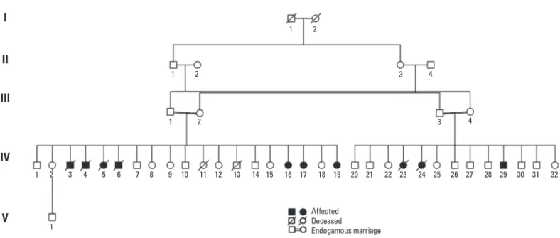
he family is composed by two related couples and their kindred (Fig 1). he irst couple is individuals III-1 and III-2, with their kindred of nineteen children. Seven of their sibs were afected by JNCL, four deceased (individu-als IV-3, IV-4, IV-5 and IV-6) and three alive (individu(individu-als IV-16, IV-17 and IV-19). he second couple is individuals III-3 and III-4 with thirteen children. hree of their sibs were afected by JNCL, only one alive (individual IV-29). Molecular analyses were performed on 3 parents and 10 children of the family, 4 symptomatic and 6
asymp-Fig 1. Family pedigree showing the consanguineous marriages and pointing the afected people in the family.
Control
Carrier
Affected
Weight pat tern
III-2 III-3 III-4 IV-12 IV-16 IV-15 IV-18 IV-30 IV-31 IV-17 IV-32 IV-19 IV-29
729pb 426pb
Fig 2. Agarose gel showing the genotypic results of afected, carriers and a con-trol individual. The 729pb band (the higher one) corresponds to the normal allele and the 426pb band (the lower one) indicates the 1.02kb deletion.
1 2
1 2
1
1
Affected Deceased Endogamous marriage
2 3 4 5 6 7 8 9 10 11 12 13 14 15 16 17 18 19 20 21 22 23 24 25 26 27 28 29 30 31 32
1 2 3 4
3 4
I
II
IV III
Arq Ne
uro
ps
iqu
ia
tr 2011;69(1)
n
eu
ro
na
l c
e
ro
id-li
pofu
sc
in
os
is
et
al.
Family Member IV-3 IV-4 IV-5 IV-6 IV-16 IV-17 IV-19 IV-23 IV-24 IV-29
First symptoms Visual loss
Visual loss*
Visual loss*
Visual loss
Visual loss*
Learning disorder (7y); visual loss (9y).
Visual loss
Visual loss*
Nyctalopia * Visual loss*
Sex and age of disease onset M, 7y M, 8y F, 7y M, 8y F, 9y F, 7y F, 6y F, 7y M, 7y M,7y
Age of latest evaluation NA NA NA NA 13y4m 12y3m 7y6m NA NA 14y2m
Dementia (age of onset) No Yes (8y)
Yes (10y)
Yes (10y)
Yes (13y)
Yes (7y)
Yes (7y) Mild learning disability.
No No (?) Yes (?). Learning dis-ability more related to visual loss.
Behavioral alterations ? Yes Yes No Temper tantrums. Tics (hands in the face).
Autistisc behavior, but understands and coop-erates. Tics (hands in the face and repetitive move-ment with the trunk).
No No ? Angry outbursts.
Temper tantrums.
Seizures (age of onset) 12y 12y 12y 10y Tonic-clonic (12y)
Tonic-clonic (8y) No No Myoclonic (9y)
12y
Inability to walk (age of onset) 17y 12y 12y 12y No Parkinsonism, ataxia, walk only with support since age 10.
No 9y No
Neurological evaluation (age of onset)
Spasticity (12y)
NP NP NP Normal tonus. Ran-dom eye movement Dysarthria (9y).
Normal tonus. Dysarthria (anarthria?). Equivocal Babinski sign.
Normal tonus
Normal tonus
Spasticity (9y)
Normal tonus. Good communication. Oc-ular apraxia. Mouth synkinesis.
EEG NP NP NP NP Abnormal Abnormal NP NP NP NP
ERG NP NP NP NP NP NP NP NP NP Retinal dystrophy
(8y)
CT/MRI NP NP NP NP Cortical
atrophy
Normal CT (8y) NP NP NP NP
Age of death 19y 14y 14y 14y Alive alive Alive 12y
(drown)
11y alive
Arq Neuropsiquiatr 2011;69(1)
17
Juvenile neuronal ceroid-lipofuscinosis Valadares et al.
tomatic (III-2, III-3, III-4, 12, 15, 16, 17, IV-18, IV-19, IV-29, IV-30, IV-31 e IV-32). he PCR ampli-ication of exons 6, 7, 8 and 13 were obtained from the patients who carried at least one normal allele. he ex-ons 7 and 8 of afected individuals could not be ampli-ied. It suggested a large deletion. hrough the allele-spe-ciic PCR the molecular diagnostic was obtained (Fig 2). he 1.02kb deletion in both of the mutated alleles was detected in all symptomatic children. All the others an-alyzed (parents and 6 asymptomatic children) were het-erozygote for this mutation. No additional mutation was detected through sequencing.
A total of 10 patients with JNCL were detected in this family, but only the four alive (individuals IV-16, IV-17, IV-19 and IV-29) were seen by the authors and had a mo-lecular diagnosis. Data of the six deceased ones was ob-tained from parents. The clinical findings from the 10 JNCL patients are shown in Table.
he irst symptom in 9 of 10 afected children was vi-sual failure (IV-3, IV-4, IV-5, IV-6, IV-16, IV-19, IV-23, IV-24 e IV-29) and learning disorder in early school years in one (IV-17). Visual loss of 6 patients have been irst ob-served in the school (4, 5, 16, 23, 24 e IV-29) and not at home. One child started with nyctalopia (IV-24) as irst symptom, diferent from the others that manifested visual loss both night and day since the be-ginning. As the disease advanced seven children showed progressive dementia, two temper tantrums and angry outbursts, and autistic behavior emerged in one. Within the group of alive patients, patient IV-17, a 12 year-old fe-male presented the most severe phenotype, with autistic behavior, tics and parkinsonism. Her brain CT was nor-mal. he less severe phenotype was observed in patient IV-19, a 7 year-old female with visual loss since she was six years-old and mild learning disability. he 14 year-old male (patient IV-29) with a milder phenotype, can walk and communicate well.
Eight of nine patients had seizures starting between 8 and 12 years of age; the youngest patient, the 7 year-old female had no seizure until now (IV-19). Death occurred between ages of 11 and 19 years-old (3, 4, 5, IV-6, IV-23 e IV-24). One blind 12 year-old girl drowned in a river (IV-23).
DISCUSSION
he molecular analysis demonstrated the 1.02kb dele-tion in the CLN3. he allele-speciic PCR gave us a very clear diagnostic. he sequencing was performed only to verify if there were additional mutations and we did not
ind any of those. As we are dealing with endogamous marriages this result was already expected. he lost part of the gene results in protein truncation. It disables the CLN3 protein capacity of localizing itself in the lyso-some9, afecting its correct function and resulting in
au-toluorescent material storage.
he clinical indings of ten patients, four males and six females, were described. Only four of them are alive and were seen by the authors. Data of the deceased ones were obtained from the parents. The age range of on-set of disease was 6 to 9 years-old, in agreement with lit-erature. Death occurred probably related to the natural course of the disease between ages of 11 and 19 years, ex-cept for one blind 12-year-old girl who drowned in a riv-er. An intrafamilial phenotypical variation was observed, as expected11.
Nyctalopia was the first symptom in one deceased child (IV-24), diferent from the others that manifested visual loss both night and day. We have not found in the literature any case of isolated nyctalopia. Vitamin A de-iciency as a cause of nyctalopia was not investigated in this patient. Severe visual loss was probably responsible by the chaotic eye movements presented by patient IV-16. In the group of the four alive patients, the only male patient has a milder phenotype, what might be explained by observations that female patients have more behavior problems than male patients12.
Patient IV-17 presented the most severe phenotype, with autistic behavior, tics and parkinsonism. Learning disorder before visual failure, an unusual inding, can be a predictor of a worsen outcome of the disease, as observed in this patient. It probably indicates a precocious onset of dementia, due to a more rapid degenerating process. In her case a normal brain CT at age of 8 had no correlation with the outcome. On her evaluations equivocal Babinski sign was observed. It was reported that the interobserver reliability and validity ofthe Babinski sign for identifying upper motor neuron weaknessare limited15.
he visual loss of six patients has been irst observed in the school and not at home. It can be a sign of negli-gence of observation by their parents for many reasons: poorness, excessive work, large kindred. he age of on-set of visual loss does not seem to be related to the prog-nosis of the disease in this family.
meta-bolic center turned possible the diagnosis, right approach and genetic counseling.
REFERENCES
1. IEM Digest. The function of CLN3P, the Batten disease protein. Mol Gen Metab 2008;93:269-274.
2. Peltonen L, Savukoski M, Vesa J. Genetics of the neuronal ceroid lipofusci-noses. Current Opinion in Gen Dev 2000;10:299-305.
3. Kovács AD, Pearce DA. Attenuation of AMPA receptor activity improves motor skills in a mouse model of juvenile Batten disease. Exp Neurol 2008; 209:288-291.
4. Teixeira C, Guimarães A, Bessa C, et al. Clinicopathological and molecular characterization of neuronal ceroid-lipofuscinoses in the Portuguese pop-ulation. J Neurol 2003;250:661-667.
5. Wisniewski KE, Kida E, Connell F, Zhong N. Neuronal ceroid lipofuscinoses: research update. Neurol Sci 2000;21(Suppl):S49-S56.
6. Persaud-Sawin DA, Mousallem T, Wang C, Zucker A, Kominami E, Bousta-ny RM. Neuronal ceroid-lipofuscinoses: a common pathway? Pediatr Res 2007; 61:146-152.
7. Mitchison HM, Munroe PB, O’Rawe AM, et al. Genomic structure and com-plete nucleotide sequence of the batten disease gene, CLN3. Genomics 1997;40:346-350.
8. Järvelä I, Mitchison HM, Munroe PB, O‘Rawe AM, Mole SE, Syvanen AC. Rap-id diagnostic test for the major mutation underlying Batten disease. J Med Genet 1996;33:1041-1042.
9. Kwon JM, Rothberg PG, Leman AR, Weimer JM, Mink JW, Pearce DA. Novel CLN3 mutation predicted to cause complete loss of protein func-tion does not modify the classical JNCL phenotype. Neuroscience Letters 2005; 387:111-114.
10. Wisniewski KE, Kaczmarski A, Kida E, et al. Reevaluation of neuronal ceroid lipofuscinoses: atypical juvenile onset may be the result of CLN2 mutations. Mol Gen Metab 1999;66:248-252.
11. Järvelä I, Autti T, Lamminranta S, Aberg L, Raininko R, Santavuori P. Clinical and magnetic resonance imaging indings in Batten disease: analysis of the major mutation (1.02-kb deletion). An Neurol 1997;42:799-802. 12. Bäckman ML, Santavuori PR, Åberg LE, Aronen ET. Psychiatric symptoms
of children and adolescents with juvenile neuronal ceroid-lipofuscinoses. J Intellec Disab Res 2005;49:25-32.
13. Munroe B, Mitchison HM, O’Rawe AM, et al. Spectrum of mutations in the Batten disease gene, CLN3. Am J Hum Genet 1997;61:310-316.
14. Taschner PEM, Vos N, Breuning MH. Rapid detection of the major dele-tion in the Batten disease gene CLN3 by allele speciic PCR. J Med Gen-et 1997; 34:955-956.