Clinical and genetic analysis of 29
Brazilian patients with Huntington’s
disease-like phenotype
Guilherme Riccioppo Rodrigues1, Ruth H. Walker2, Benedikt Bader3, Adrian Danek3, Alexis Brice4, Cécile Cazeneuve4, Odile Russaouen4, Iscia Lopes-Cendes5, Wilson Marques Jr.1, Vitor Tumas1
ABSTRACT
Huntington’s disease (HD) is a neurodegenerative disorder characterized by chorea, behavioral disturbances and dementia, caused by a pathological expansion of the CAG trinucleotide in the HTT gene. Several patients have been recognized with the typical HD phenotype without the expected mutation. The objective of this study was to assess the occurrence of diseases such as Huntington’s disease-like 2 (HDL2), spinocerebellar ataxia (SCA) 1, SCA2, SCA3, SCA7, dentatorubral-pallidoluysian atrophy (DRPLA) and chorea-acanthocytosis (ChAc) among 29 Brazilian patients with a HD-like phenotype. In the group analyzed, we found 3 patients with HDL2 and 2 patients with ChAc. The diagnosis was not reached in 79.3% of the patients. HDL2 was the main cause of the HD-like phenotype in the group analyzed, and is attributable to the African ancestry of this population. However, the etiology of the disease remains undetermined in the majority of the HD negative patients with HD-like phenotype.
Key words: Huntington’s disease, Huntington’s disease-like, chorea-acanthocytosis, Huntington’s disease-like 2.
Análise clínica e genética em 29 pacientes brasileiros com fenótipo doença de Huntington-símile
RESUMO
A doença de Huntington (DH) é uma doença neurodegenerativa caracterizada por coréia, alterações comportamentais e demência, causada por uma expansão patológica do trinucleotídeo CAG no gene HTT. Vários pacientes têm sido descritos com o fenótipo típico para a DH porém sem a mutação esperada. O objetivo deste estudo foi avaliar a ocorrência de doenças como doença de Huntington-símile 2 (DHS-2), ataxias espinocerebelares tipo 1, 2, 3 e 17, atrofia dentatorubral-palidoluisiana e coreo-acantocitose (CAc) entre 29 pacientes brasileiros com fenótipo doença de Huntington-símile. No grupo analisado, encontramos 3 pacientes com DHS-2 e 2 pacientes com CAc. O diagnóstico permaneceu obscuro em 79,3% dos pacientes. DHS-2 foi a principal causa do fenótipo DH-símile no grupo analisado, provavelmente devido a ancestralidade africana na população brasileira. Entretanto, a etiologia permaneceu indeterminada na maioria dos pacientes avaliados.
Palavras-chave: doença de Huntington, doença de Huntington-símile, coreo-acantocitose, doença de Huntington-símile 2.
Correspondence
Vitor Tumas
Department of Neuroscience and Behaviour Sciences Ribeirão Preto School of Medicine University of São Paulo Campus Universitário
14049-900 Ribeirão Preto SP - Brazil E-mail: [email protected]
Support
Dr. Danek serves on the editorial board of Zeitschrift für Gerontoneurologie and Translational Neuroscience; has received speaker honoraria from Merz, Pfizer, No-vartis, Janssen-Cilag and Neuro-Update; receives research suppor t as Principal Investigator from Bayerische Forschun-gsstiftung, German Federal Ministry of Research, Münchner Universitätsgesell-schaft, Deutsch-Französische Hochschul-stiftung, and Advocacy for Neuroacan-thocytosis Patients; and has served as an expert witness for German courts of justice in medicolegal cases
The other authors report no conflict of interest
Received 14 November 2010 Received in final form 23 February 2011 Accepted 11 March 2011
1Department of Neuroscience and Behaviour Sciences, Ribeirão Preto School of Medicine, University of São Paulo, Ribeirão
Preto SP, Brazil; 2Department of Neurology, James J. Peters Veterans Affairs Medical Center, Bronx, and Mount Sinai School
of Medicine, New York, USA; 3Department of Neurology, Ludwig-Maximilians-Universität, München, Germany; 4Department
of Genetics and Cytogenetics, Neurogenetics Unit, Hospital Pitié-Salpêtrière, Assistance Publique - Hôpitaux de Paris, France;
Huntington’s disease (HD) is a progressive, neurode-generative disorder characterized by chorea, dystonia or parkinsonism, cognitive impairment and behavioral ab-normalities, with typical onset in young adulthood. HD is caused by a pathological expansion of CAG trinucleo-tide repeats in the HTT gene.
Since the identiication of the causative mutation, it has been recognized that a number of patients with the classical HD phenotype have disease due to another eti-ology. hese include prion diseases such as HDL1; auto-somal dominant disorders due to inheritance of an ex-panded trinucleotide repeat sequence, such as HDL2, and DRPLA (dentatorubral-pallidoluysian atrophy); disorders afecting primarily the cerebellum, in which movements disorders can also be seen - FRDA (Fried-reich ataxia), SCA (spinocerebellar atrophy) 1, SCA 2, SCA 3 and SCA 17; the neuroacanthocytosis syndromes, chorea-acanthocytosis (ChAc) and McLeod syndrome; and the neurodegeneration with brain iron accumula-tion (NBIA) syndromes, neuroferritinopathy, acerulo-plasminemia and Pantothenate kinase-associated neu-rodegeneration (PKAN).
here have been several published studies addressing the etiological diagnosis of patients with an HD-like phe-notype1-14; however, these have been mainly drawn from
European and North American populations. We per-formed a transversal study of the genetic and clinical indings from a group of non-HD HD-like patients who attended a Brazilian movement disorders clinic.
METHOD
he medical records of 108 patients who were tested for HD between 1998 and 2006, were reviewed. Patients with an HD-like phenotype, deined as a progressive dis-order with chorea, dystonia, parkinsonism, ataxia or my-oclonus associated with a cognitive, psychiatric or be-havioral impairment who did not have a pathological expansion on HTT gene underwent further clinical ex-amination and testing for DRPLA, HDL2, SCA 1, 2, 3, 17 and ChAc. Although a true HD-like phenotype requires autosomal dominant inheritance, we did not use this cri-terion to select our patients, as an absent family history is non-informative.
DNA analysis of ATN1 (DRPLA), JPH3 (HDL2), ATX1 (SCA1), ATX2 (SCA2), ATX3 (SCA3) and TBP (SCA17) were performed by sizing of luorescent PCR products encompassing the CTG/CAG expansion site. he PCR products were loaded on 3730 DNA Analyzer or Megabace 1000 (GE) and analyzed using GeneMapper software [AppliedBiosystems] or Fragment Proiler (GE). As the standard PCR protocol does not allow detection of alleles carrying very large expansions as described for the juvenile, severe form of HD, triplet repeat primed PCR15, which permits detection of large expansions, was
performed for all patients who were not heterozygous at the HTT locus and/or the JPH3 locus.
Patients who were found to have acanthocytes on peripheral blood smear, elevation of serum CK levels, or evidence of myopathy or peripheral neuropathy, had
Table 1. Clinical and molecular indings in HD and HDL patients.
HD HDL p
Number of cases 37 29
Age of onset 35.1±12.8 30.7±17.6 0.25†
Male:Female ratio 16:21 14:15 0.68‡
Chorea 37 (100%) 22 (75%) 0.002#
Ataxia 7 (18.9%) 8 (27.5%) 0.40‡
Myoclonus 0 2 (6.8%) 0.19#
Dystonia* 0 2 (6.8%) 0.19#
Tremor 0 1 (3.4%) 0.43#
Parkinsonism* 0 0 –
Psychiatric disturbance 17 (45.9%) 15 (51.7%) 0.64‡
Dementia 25 (65.7%) 20 (68.9%) 0.90‡
Epilepsy 4 (10.8%) 4 (13.7%) 0.72#
Familiar history 34 (91.8%)
AD:34 AD: 14 (48.2%)19 (65.5%) non-AD: 5 (17.2%)
0.007‡
htt CAG range 37-87 15-27
chorein levels in peripheral blood qualitatively analyzed by Western blot16 (n=2).
Statistical analysis was performed with SPSS version 10.0 (SPSS, Inc., Chicago, IL, USA). his study was ap-proved by the ethical board of the Hospital das Clínicas de Ribeirão Preto and all patients have signed an in-formed consent.
RESULTS
Of the 108 patients tested for HD, seven were ex-cluded due to insuicient data in their medical records. From the remaining subjects, 37 were diagnosed with HD, 35 were not classiied as having an HD-like pheno-type, and 29 were classiied as a HD-like phenotype. As
a result, considering the patients with a typical or com-patible HD phenotype, we found 29/66 patients (43%) without pathological CAG expansion on HTT gene.
Table 1 summarizes the main clinical features of HD and HD-like patients. MRI exams were performed in 24 patients and brain CT in 3. he most common inding was brain atrophy in 23 (85%) patients. We found no ev-idence of iron deposition on those patients who under-went MRI exams. All 29 HDL patients had their DNA analyzed for trinucleotide repeat expansions of HTT, JPH3, ATX1, ATX2, ATX3, TBP and ATN1. Two pa-tients met criteria for chorein testing and had absent or markedly reduced levels in peripheral blood. From these tests, we diagnosed 3 (10.3%) cases of HDL2 and 2 (6.8%)
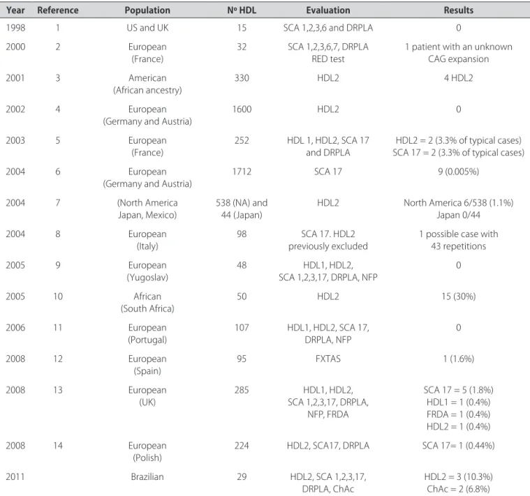
Table 2. Review of HD-like screening studies.
Year Reference Population Nº HDL Evaluation Results
1998 1 US and UK 15 SCA 1,2,3,6 and DRPLA 0
2000 2 European
(France) 32 SCA 1,2,3,6,7, DRPLARED test 1 patient with an unknown CAG expansion
2001 3 American
(African ancestry) 330 HDL2 4 HDL2
2002 4 European
(Germany and Austria)
1600 HDL2 0
2003 5 European
(France)
252 HDL 1, HDL2, SCA 17
and DRPLA
HDL2 = 2 (3.3% of typical cases) SCA 17 = 2 (3.3% of typical cases)
2004 6 European
(Germany and Austria)
1712 SCA 17 9 (0.005%)
2004 7 (North America
Japan, Mexico) 538 (NA) and 44 (Japan) HDL2 North America 6/538 (1.1%)Japan 0/44
2004 8 European
(Italy) 98 previously excludedSCA 17. HDL2 1 possible case with 43 repetitions
2005 9 European
(Yugoslav)
48 HDL1, HDL2,
SCA 1,2,3,17, DRPLA, NFP
0
2005 10 African
(South Africa)
50 HDL2 15 (30%)
2006 11 European
(Portugal) 107 HDL1, HDL2, SCA 17, DRPLA, NFP 0
2008 12 European
(Spain) 95 FXTAS 1 (1.6%)
2008 13 European
(UK)
285 HDL1, HDL2,
SCA 1,2,3,17, DRPLA, NFP, FRDA
SCA 17 = 5 (1.8%) HDL1 = 1 (0.4%) FRDA = 1 (0.4%) HDL2 = 1 (0.4%)
2008 14 European
(Polish)
224 HDL2, SCA17, DRPLA SCA 17= 1 (0.44%)
2011 Brazilian 29 HDL2, SCA 1,2,3,17,
DRPLA, ChAc HDL2 = 3 (10.3%)ChAc = 2 (6.8%)
cases of ChAc. he patients with HDL2 were clinically indistinguishable from those with typical HD pheno-type. However, patients with ChAc presented clear clin-ical diferences as absence of autosomal dominant his-tory, presence of peripheral involvement and epilepsy in both cases. he patients with HDL2 and ChAc have been previously reported in detail elsewhere17,18.
One patient of this study was a deceased sister of one ChAc patient, who presented with chorea and cog-nitive abnormalities, but whose diagnosis could not be conirmed by Western blot due to the non-availability of required biomaterial. he remaining 23 (79.3%) pa-tients tested negative for HD, HDL2, SCA1, 2, 3, 17, and DRPLA.
DISCUSSION
his is the irst study to address the diagnosis of dis-eases responsible for the HD-phenotype in Brazilian pa-tients. Compared with previous studies (Table 2), we found a higher frequency of ChAc and HDL2 among our patients. Considering the 3 unrelated HDL2 cases re-ported here, together with the patients rere-ported by Teive et al.19 and Santos et al.20, it is possible that HDL2 is the
most common cause of HD-like phenotype in Brazil. A probable explanation is that 44% of Brazilian population is of African descent21, even though this ancestry may be
occult19,20. his regional diference contrasts with the
ar-ticle of Wild and colleagues13, in which SCA 17 was
con-sidered the most important cause of the HD-like pheno-type in patients from the UK.
Based upon previous studies, we initially decided to test for disorders whose frequencies were higher than 0.5% of all HD phenocopies. Consequently, we included SCA 17 and HDL2 and excluded HDL1 and FRDA13. We
have not included neuroferritinopathy because it appears to be even rarer in HD-like patients, with a very small number of families being reported to date, and it is un-likely to occur in patients without evidence of iron de-position in the basal ganglia on MRI22.
Screening for ChAc was performed as the clinical characteristics of our patients suggested the diagnosis. It was important to include the investigation for ChAc because Brazil represents the largest population of Jap-anese and descendants outside Japan21, and ChAc
ap-pears to be particularly prevalent in Japanese subjects23.
DRPLA was included for the same reason. In addition, studies have demonstrated that the number of DRPLA cases in non-Asian populations may be higher than pre-viously considered24,25. Finally, chorea has been reported
as an occasional non-ataxic symptom on SCAs, occur-ring in approximately 7% of SCA 1 and 2 and 10% of SCA 3 patients26.
Our data showed that almost 43% of the patients
who had a phenotype compatible with HD tested nega-tive for CAG expansions at the HTT gene. his data di-verges from reference studies in this ield, which suggest that the frequency of HD-like phenocopies is about 1% of HD cases27,28. We believe that this discrepancy cannot
be addressed only as an ascertainment bias, because our sample was selected based on restrictive criteria. In ad-dition, other studies have found proportions of HD-like patients in 33%10, 35.5%11, and 36.3%9 of their study
pop-ulations. Krause and colleagues10 provided an insight in
this problem, showing that, in their population, only 16% of white patients were HD phenocopies, in contrast to 64% of the black patients. herefore, the percentage of HD-like patients may difer according to the ethnicity of the study population.
Our clinical data shows that the absence of an auto-somal-dominant history and signs of peripheral involve-ment or seizures may help diferentiate between ChAc and HD. Although the presence of acanthocytes may im-prove the diagnostic accuracy, their absence does not rule out the diagnosis of ChAc29. In addition to screening
for acanthocytes, we considered the diagnosis of ChAc in patients with elevated CK levels and clinical signs of myopathy or peripheral neuropathy.
he patients with HDL2 were clinically very similar to the classical HD phenotype, and this diagnosis should be suspected in the presence of typical HD phenotype with absent HTT mutation, especially if an autosomal dominant inheritance and African ancestry is present.
However, this survey has some limitations. First, the small number of patients identiied as having an HD-like phenotype, what can be explained by the rarity of these disorders and because we have enrolled patients from only one clinical center. Second, referral bias is unavoid-able when including patients from a referral movement disorders clinic, therefore, it is possible that the relative frequency of HD-like disorders was overestimated. hird, in spite of screening systematically for seven diseases, 79.3% of our patients remained undiagnosed, conirming a considerable etiologic heterogeneity among HD-like disorders and indicate the need for additional studies, using a broader panel of diagnostic tests. Considering the discrepancy between the high percentage of cryptic cases and the rareness of the remaining diseases to be tested for, it is likely that among the HD-like phenotypes some causalities have not been described yet.
REFERENCES
1. Rosenblatt A, Ranen NG, Rubinsztein DC, et al. Patients with features similar to Huntington’s disease, without CAG expansion in huntingtin. Neurology 1998;51:215-220.
3. Holmes SE, O’Hearn E, Rosenblatt A, et al. A repeat expansion in the gene encoding junctophilin-3 is associated with Huntington disease-like 2. Nat Genet 2001;4:377-378.
4. Bauer I, Gencik M, Laccone F, et al. Trinucleotide repeat expansions in the junctophilin-3 gene are not found in Caucasian patients with a Hunting-ton’s disease-like phenotype. Ann Neurol 2002;51:662.
5. Stevanin G, Fujigasaki H, Lebre AS, et al. Huntington’s disease-like pheno-type due to trinucleotide repeat expansions in the TBP and JPH3 genes. Brain 2003;126:1599-1603.
6. Bauer P, Laccone F, Rolfs A, et al. Trinucleotide repeat expansion in SCA17/ TBP in white patients with Huntington’s disease-like phenotype. J Med Genet 2004;41:230-232.
7. Margolis RL, Holmes SE, Rosenblatt A, et al. Huntington’s disease-like 2 (HDL2) in North America and Japan. Ann Neurol 2004;56:670-674. 8. Cellini E, Forleo P, Nacmias B, et al. Spinocerebellar ataxia type 17 repeat in
patients with Huntington’s disease-like and ataxia. Ann Neurol 2004;56:163. 9. Keckarević M, Savić D, Svetel M, Kostić V, Vukosavić S, Romac S. Yugoslav HD phenocopies analyzed on the presence of mutations in PrP, ferritin, and Jp-3 genes. Int J Neurosci 2005;115:299-301.
10. Krause A, Hetem C, Holmes SE, Margolis RL. HDL2 mutations are an im-portant cause of Huntington’s disease in patients with African ancestry [abstract #A17]. J Neurol Neurosurg Psychiatr 2005;76(Suppl 4):S17. 11. Costa MC, Teixeira-Castro A, Constante M, et al. Exclusion of mutations in
the PRNP, JPH3, TBP, ATN1, CREBBP, POU3F2 and FTL genes as a cause of disease in Portuguese patients with a Huntington-like phenotype. J Hum Genet 2006;51:645-651.
12. Rodriguez-Revenga L, Santos MM, Sánchez A, et al. Screening for FXTAS in 95 Spanish patients negative for Huntington disease. Genet Test 2008; 12:135-138.
13. Wild EJ, Mudanohwo EE, Sweeney MG, et al. Huntington’s disease pheno-copies are clinically and genetically heterogeneous. Mov Disord 2008;23: 716-720.
14. Sułek-Piatkowska A, Krysa W, Zdzienicka E, et al. Searching for mutation in the JPH3, ATN1 and TBP genes in Polish patients suspected of Hunting-ton’s disease and without mutation in the IT15 gene. Neurol Neurochir Pol 2008;42:203-209.
15. Warner JP, Barron LH, Goudie D, et al. A general method for the detection of large CAG repeat expansions by luorescent PCR. J Med Genet 1996; 33:1022-1026.
16. Dobson-Stone C, Velayos-Baeza A, Filippone LA, et al. Chorein detection for the diagnosis of chorea-acanthocytosis. Ann Neurol 2004;56:299-302. 17. Rodrigues GR, Walker RH, Bader B, Danek A, Marques W Jr, Tumas V.
Chorea-acanthocytosis: report of two Brazilian cases. Mov Disord 2008;23: 2090-2093.
18. Rodrigues GR, Walker RH, Brice A, et al. Huntington’s disease-like 2 in Brazil: report of 4 patients. Mov Disord 2008;23:2244-2255.
19. Teive HAG, Becker N, Munhoz RP, et al. Huntington’s disease-like 2: the irst case report in Latin America in a patient without African ethnic origin. Mov Disord 2007;22(Suppl 16):S27.
20. Santos C, Wanderley H, Vedolin L, Pena SD, Jardim L, Sequeiros J. Hun-tington disease-like 2: the irst patient with apparent European ancestry. Clin Genet 2008;73:480-485.
21. IBGE. Características gerais da população: censo demográfico 2000. Rio de Janeiro, Brasil. 2003:37-48.
22. McNeill A, Birchall D, Haylick SJ, et al. T2* and FSE MRI distinguishes four subtypes of neurodegeneration with brain iron accumulation. Neurology 2008;70:1614-1619.
23. Hirose G. Neuroacanthocytosis in Japan: review of the literature and cases. In: Walker RH, Saiki S, Danek A (Eds). Neuro-acanthocytosis syndromes II. Berlin: Springer; 2008:75-86.
24. Martins S, Matamá T, Guimarães L, et al. Portuguese families with dentato-rubropallidoluysian atrophy (DRPLA) share a common haplotype of Asian origin. Eur J Hum Genet 2003:808-811.
25. Wardle M, Majounie E, Williams NM, Rosser AE, Morris HR, Robertson NP. Dentatorubral pallidoluysian atrophy in South Wales. J Neurol Neurosurg Psychiatry 2008;79:804-807.
26. Schmitz-Hübsch T, Coudert M, Bauer P, et al. Spinocerebellar ataxia types 1, 2, 3, and 6: disease severity and nonataxia symptoms. Neurology 2008; 71:982-989.
27. Kremer B, Goldberg P, Andrew SE, et al. A worldwide study of the Hunting-ton’s disease mutation. The sensitivity and peciicity of measuring CAG repeats. N Engl J Med 1994;330:1401-1406.
28. Andrew SE, Goldberg YP, Kremer B, et al. Huntington disease without CAG expansion: phenocopies or errors in assignment? Am J Hum Genet 1994;54:852-863.