Significance of Background
activity and PoSitive SharP WaveS in
neonatal electroencePhalogram aS
PrognoStic of cereBral PalSy
Márcia de Oliveira Nicolini Nosralla
1, Délrio Façanha Silva
2, Ricardo Vieira Botelho
3abstract – Objective: To study the significance of electroencephalographic background activity and positive sharp waves in neonatal electroencephalogram as prognostic of cerebral palsy. Method: We studied prospectively and sequentially 73 newborns who had severe neonatal complications (neonatal anoxia, seizures, respiratory distress, sepsis, and meningitis). Nineteen newborns were excluded and 54 children formed the object of our study and were followed for 2 years. We analyzed gestational age, conceptional age, electroencephalographic background activity and positive sharp waves, which were correlated with cerebral palsy. Results: There were no statistically significant correlation between gestational age and conceptional age and cerebral palsy; the electroencephalographic background activity was correlated with cerebral palsy as well as the positive sharp waves. Conclusion: Children with electroencephalographic background activity markedly abnormal and accompanied by positive sharp waves were associated with a worse prognosis. Key Words: neonatal, cerebral palsy, newborn, anoxia, seizures, sepsis, meningitis, children, gestacional age, prognosis.
valor do ritmo de base e da onda aguda positiva no eletrencefalograma neonatal como prognóstico da paralisia cerebral
resumo – Objetivo: estudar o valor do ritmo de base e das ondas agudas positivas no eletrencefalograma neonatal como prognóstico da paralisia cerebral. Método: Nós estudamos, prospectiva e sequencialmente, 73 recém-nascidos que apresentaram complicações neonatais graves (anoxia neonatal, crises convulsivas, desconforto respiratório, septicemia e meningite). dezenove recém-nascidos foram excluídos e 54 crianças formaram o objeto do nosso estudo e foram seguidas por 2 anos. Nós analisamos a idade gestacional, a idade corrigida, o ritmo de base e as ondas agudas positivas, que foram correlacionadas com a paralisia cerebral. Resultados: Não houve correlação estatisticamente significante entre as idades gestacional e corrigida com a paralisia cerebral; o ritmo de base foi correlacionado com a paralisia cerebral, tanto quanto as ondas agudas positivas. Conclusão: Crianças com o eletrencefalograma com o ritmo de base marcadamente anormal e ondas agudas positivas estão associadas com pior prognóstico neurológico.
PAlAvrAs-ChAve: neonatal, paralisia cerebral, recém-nascido, anoxia, crises, sepsis, meningite, crianças, idade gestacional, prognóstico.
hospital e Maternidade Cruz Azul de são Paulo e hospital do servidor Público estadual “Francisco Morato de oliveira” – IAMsPe, são Paulo sP, Brazil:
1Médica Neuroisiologista do hospital e Maternidade Cruz Azul de são Paulo; 2Neurologista, Unidade de vídeo-eeG do CITe (Centro de Investigação
e Tratamento das epilepsias) do hospital Beneiciência Portuguesa; 3doutor em Ciências-Professor do Programa de Pós-graduação em Ciências da
saúde – IAMsPe.
received 6 october 2008, received in inal form 19 March 2009. Accepted 18 May 2009.
Dra. Márcia de Oliveira Nicolini Nosralla – Avenida Brigadeiro Luiz Antônio 3333 / 24 - 01401-001 São Paulo SP - Brasil. E-mail: [email protected] The mortality and morbidity of high risk neonates,
de-ined as those with severe neonatal complications (neona-tal anoxia, seizures, respiratory distress, sepsis, and menin-gitis), has declined due to advances in obstetrics and
pro-fessionals alike. This has led to a number of researchers establishing criteria for determining neurologic progno-sis in these neonates1-11.
Interest in this subject grew when new concepts re-garding the neonatal electroencephalogram (NeeG) were formulated in 193812 and stimulated many researchers in
the observation of the electroencephalophic background activity (BA) of newborn normal and pathological in dif-ferent age2-3,6-7,9-12. The presence of positive sharp waves
(PsW) on the central and temporal areas of the NeeG in preterm infants (deined as gestational age less than 37 weeks) with intraventricular haemorrhage was reported for the first time in 19725. Further associations of PsW
were subsequently described including periventricular leukomalacia, other types of intracranial haemorrhage, hy-drocephaly, meningitis, various amino acid deiciency dis-eases and midline infarcts with electrographic crises1, 9,13-20.
This study was designed to study the signiicance of BA and PsW in eeGN as prognostic of cerebral palsy.
method
This was a prospective sequential study of 73 high risk ne-onates with a gestational age of between 28 to 42 weeks, who were admitted to the Neonatal Intensive Care Unit of the hos-pital and Maternity Cruz Azul in são Paulo, in the period from 2001 to 2005 . Were included in this study, newborns with se-vere neonatal complications (neonatal anoxia, seizures, respi-ratory distress, sepsis, and meningitis) and excluding newborns who show genetic syndromes.
The following variables were described for all infants: sex, gestational age (it was determined by calculation from the mother’s last menstrual period and the result of the dubowitz and New Ballard examination for maturity), conceptional age (the gestational age plus the chronological age from birth), Ap-gar score at one and ive minute, birth weight, neonatal comor-bidities and maternal complications.
All newborns held the first month of life, neuroimaging study, mainly of cranial ultrasound scan and when necessary, cranial computed tomographic.
The NeeG was done in the first week of life and was re-corded at the patient’s bedside with 20 channel Neurotec NeU-roMAP eQsA 240 electroencephalograph, through the digi-tal method. Twelve surface disc electrodes were applied to the scalp with electrode paste according to the International sys-tem 10–20 modiied for newborns and recorded with a the tech-nique previously described21-23. The set up used in our service
was in-line with internationally accepted standards21-22,24.
Four-teen channels were devoted to NeeG and four recorded poly-graphic parameters including oculogram (eoG), electro-myogram (eMG), electrocardiogram (eCG) and chest respirations: F3-C3, C3-o1, F3-T3, T3-o1, F4-C4, C4-o2, F4-T4, T4-o2, C3-Cz, Cz-C4, T3-Cz, Cz-T4, T3-C3, T4-C4, eoG, eMG, eCG and respira-tion. Tracing were typically recorded for 60 minutes.
The criteria for analysis applied were those established in the literature8,11,15,21,23-25:
A – The BA was classiied into three groups: (0) normal; (1) mildly altered; (2) markedly abnormal.
The records were considered normal if: (1) temporal orga-nization discontinuous NeeG with attenuated periods lasting up to 3 minutes in preterm; (2) the number of delta-brushes decrease with increasing gestational age and conceptional age delta-brush is a rhythmic activity with slow waves, character-ized by outbreaks of 8 to 20 cycles / second, 25 to 200 micro-voltz in duration from 0.8 to 1.5 cycles / second, with overlap-ping waves of fast, with a range from 10 to 100 microvoltz; (3) the percentage of interhemispheric synchrony increases with gestational age and conceptional age; (4) the presence of fron-tal wave acute transitional and fronfron-tal rhythmic delta activity in the fullterm (gestational age greater or equal to 37 weeks); (5) sharp transients in all states and all location in the preterm; (6) continuous activity in wakefulness and active sleep and inter-mittent activity in portions of quiet sleep in fullterm
The records were considered mildly altered when: (1) inter-hemispheric synchrony less than 25%; (2) poor correlation be-tween clinical parameters and NeeG according to the stage of sleep-wake cycle; (3) mild focal abnormality (excessive sharp wave temporal and / or central, focal attenuation); (4) imma-turity for conceptional age - these patterns are speciic to chil-dren with corrected age less than 2 weeks (Fig 1); (5) low voltage with BA normal; (6) asymmetry <50%.
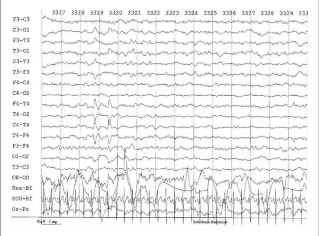
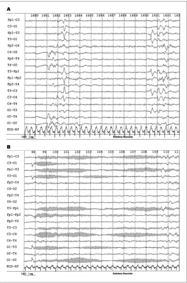
It was markedly abnormal when shown: (1) excessive disconti-nuity for gestational age (the attenuated period exceeds 60 sec-onds); (2) excessive asynchronous for age (asynchronous persis-tent, more than 25% in children older than 30 weeks conceptional age); (3) excessively low voltage (below 20 microvoltz) without the characteristic patterns for gestational age; (4) interhemispher-ic asymmetry more than 50%; (5) absence of differentiation be-tween sleep stages; (6) excessively slow cerebral electrical activity, without the presence of characteristic patterns for the concep-tional age, which persisted in all stages and are not reactive to stimuli (Fig 2); (7) the presence of focal or multifocal discharg-es (Fig 2); (8) electrographic seizurdischarg-es with or without clinical sei-zures (Fig 3B); (9) burst-suppression pattern (Fig 3A); (10) inactive. B – The PsW were classiied into two groups: (0) absence of PsW; (1) presence of PsW.
PsW were deined as sharp transient with a duration lon-ger than 400 msec, clearly distinguishable from the BA, maxi-mally expressed in the mid-temporal areas (central or tempo-ral), with an initial and predominant surface positive polarity23
and with a frequency of greater than 1 per minute15. These PsW
appear on the second day after birth and steadily increase over subsequent days and weeks, reaching a peak between the sec-ond and fourth weeks after birth (mean of 20 days). Generally, they disappear from the registers at 3–4 weeks of age24. In full
Fig 1. Newborn to 39 4 / 7 weeks gestational age and 40 weeks of conceptional age. Presented a EEGN with a BA with immature pattern corrected for age and PSW temporal. Evolved with a left hemiparesis.
The newborns included in the study were followed up to two years of age. At the end of this period, were tested in rela-tion to the proporrela-tions existing in two age groups, one younger and one older, divided by their median, were classiied accord-ing to the standard neurological exam26 like normal (without
ce-rebral palsy) and abnormal (with cece-rebral palsy) and then were determined the proportions of variables: occurrence of cerebral palsy, change of BA and the presence of PsW.
At the end of the study, we correlatede the variables gesta-tional age, concepgesta-tional age, BA and PsW with cerebral palsy25.
Statistical analysis
The gestational age and conceptional age were described by mean, standard deviation, median and the distribution differenc-es were differenc-estimated by non-parametric tdifferenc-ests (Mann-Whitney). The correlation between cerebral palsy and the other variables (ges-tational age, conceptional age, BA and PsW) were performed by the spearman test. The association between these variables were studied by logist regression test, between BA and cerebral palsy and was calculated by Chi square, and the association be-tween the PsW and cerebral palsy was calculated using the Fis-cher exact test. The sensitivity, speciicity and positive and neg-ative predictive values were determined from cerebral palsy.
The adopted level of signiicance was 5% (α=0.05). This study was authorized after the appreciation of the Commission on ethics in research of the hospital and Materni-ty Cruz Azul de são Paulo with free and informed consent signed by parents and guardians.
reSultS
Among 73 initially selected newborns for study, 3 (4.11%) died of clinical complications, mainly septicemia, during the neonatal period. Two subjects (2.74%) were ex-cluded as they had a genetic syndrome, and 14 (19.18%) abandoned treatment before study end point of two years of age. The remaining 54 neonates were included in the study.
of the 54 infants in this study, 31 (57.40%) were boys and 23 (42.60%) were girls; 38 (70.37%) of the neonates were preterm and 16 (29.63%) were fullterm (deined as gestational age greater than or equal to 37 weeks at birth). Apgar score at one minute ranged from 1 to 9 (mean of 5±3) and 5 minutes varied from 2 to 10 (mean 7±2). Weight of the preterm ranged from 0.790 g (mean 1475±0.64g) and of fullterm from 2850g to 3070g (mean 2952±0.10g).
The most common maternal complications in the pre-term included urinary tract infections, prolonged labor and placenta previa, while in the fullterm, the most fre-quent maternal complication was prolonged labor. In the neonatal period, the most common complications were respiratory distress, anoxia and convulsions.
of the 54 newborns (100%) who underwent the neu-roimaging study, 38 (70.37%) were normal and 14 (25.93%)
showed signiicant changes (intraventricular hemorrhage grade III or Iv with or without periventricular leukomalacia).
At the end of the study, the 54 children were divided into two groups according to neurological examination and 29 (53.7%) had cerebral palsy and 25 (46.3%) did not develop with cerebral palsy.
The gestational age of 54 newborns ranged from a minimum of 24 and a maximum of 42 weeks (X=33.65+4.31 weeks and a median of 34 weeks).
The group of 29 newborns with cerebral palsy present-ed with gestational age and mean±33.83+4.60 weeks and the group of 25 newborns without cerebral palsy of 33.44+ 4.02 weeks (p=0.76, Mann-Whitney).
The gestational age of 54 cases was divided by its me-dian, of 34 weeks, in two groups, the youngest with 26 cases of gestational age <34 weeks and older with 28 cas-es ≥34 weeks. Then, the proportions were tested accord-ing with occurrence of cerebral palsy, changes in BA and presence of PsW. When confronted the groups there were no statistically signiicant differences between the pro-portions calculated (age group × cerebral palsy – Fisher: p=0.78; age groups × BA – c2=0.28:p=0.86; Age groups ×
PsW – Fisher: p=0.39).
Conceptional age followed the gestational age, with slightly higher values. A total of 54 newborns had the min-imum 25 and maxmin-imum of 42 weeks (X=34.30+4.15 weeks and a median of 34 weeks).
The group of 29 babies born with cerebral palsy had conceptional age of 34.48+4.43 weeks and the group of 25 newborns without cerebral palsy of 34.08+3.88 weeks (p=0.70, Mann-Whitney).
of the 25 newborns (100%) that progressed with the normal neurological examination at 2 years of age, 19 (76%) had the BA normal. of the 29 children (100%) who had cerebral palsy to 2 years of age, 20 (69%) showed the BA markedly abnormal in NeeG (Table 1).
There were no difference in proportions between the three groups of BA in the two groups, with and without cerebral palsy (c2=20.69, p=0.00003*).
The PsW group was associated with signiicantly differ-ent with the presence of cerebral palsy (Fisher: p=0.00963) (Table 2).
Table 1. Distribution of the 54 newborns in the 3 groups do background activity (0=normal; 1=mildly altered; 2=markedly abnormal), according to the absent or presence of cerebral palsy.
Background activity
Cerebral palsy
Total
No yes
0 19 (76%) 5 (17.2%) 24
1 3 (12%) 4 (13.8%) 7
2 3 (12%) 20 (69.0%) 23
The BA markedly abnormal and the presence of the PsW were significantly correlated with cerebral palsy (spearman, p=0.000001 and p=0.0054 respectively). Ges-tational age and conceptional age were not correlated (spearman, p=0.76 and p=0.71, respectively).
Performed logistic regression with cerebral palsy as a dependent variable, there were signiicant correlations between cerebral palsy and BA PsW (r=0.66 and r=0.40, respectively).
diScuSSion
Both the families of patients and the medical pro-fession and society eager for prognostic factors to guide the development likely neuropsychomotor of newborns at high risk, ie those with severe neonatal complications (neonatal anoxia, seizures, respiratory distress, sepsis, and meningitis).
According to the literature, examination of the im-age, the eeGN, the gestational age and conceptional age are the methods used to predict the neurological prog-nosis of newborns11.
The types and location of brain injury in newborns, especially in response to hypoxia-ischemia are related to the time of appearance of the lesion and the nature of the insult23. In the fullterm, the main pathology of
hypox-ia-ischemia is the lesion of the gray matter and the sub-strate is the pathological lesion of the gray matter. In pre-term, the two main consequences of hypoxic-ischemic in-jury are haemorrhage of germinal matrix and periventric-ular leukomalacia23.
In the neurological development of children, there is spastic diplegia due to damage of pyramidal ibers in the cortex, which passes through the central white substance, or spastic quadriplegia, when the area involved is larger27-29.
observed in our patients structural lesions, particularly intraventricular haemorrhage grades III or Iv with or with-out periventricular leukomalacia in 25.93% of cases, which is similar to published data.
data from eeGN are designed to bring prognostic infor-mation. The BA altered and the presence of PsW is the chang-es disclosed to eeGN potential correlation with prognosis.
There is evidence suggesting that the BA, showing its maturity by agreeing to gestational age, indicating a good prognosis for normal neurological development. More-over, when the eeGN presents a BA abnormal, indicates a poor prognosis for children with 1 to 2 years of age, with the possibility of developing cerebral palsy2,14,18,20,23,29.
our study conirmed these data. We observed a pre-dominance of BA normal in the group without cerebral palsy and BA markedly abnormal in patients with cere-bral palsy. The disorganization BA was signiicantly corre-lated with the presence of cerebral palsy.
The PsW central and PsW temporal are morpholog-ically similar and both are associated with structural brain injury, including intraventricular haemorrhage and periventricular leukomalacia, and other types of intrac-ranial haemorrhage, hydrocephalus, meningitis, and some aminoacidopatias infarction of the middle line with elec-trographic seizures 1,9,13-20,23. In the fullterm, the PsW is
lo-cated in the temporal region.
The PsW seen in children showed the morphological characteristics described above. The PsW were consid-ered pathological by its association with high incidence of abnormalities of BA, both in preterm, newborn and in the end, which were exposed to attacks the central nervous system. Moreover, were associated with structural lesions, mainly intraventricular haemorrhage grades III and Iv.
A relevant question in the study of eeGN refers to the different presentations of the routing related to the age of the newborn, because there could be changes in presentation. separate the group of newborns with and without cerebral palsy checking whether the gestation-al age and conceptiongestation-al age were confounding factors in the work. There was no difference in distribution between the ages of the groups with and without cerebral palsy (x=33.83+4.60 × X weeks=33.44+4.02 weeks, Mann-Whit-ney, p=0.76). The conceptional ages were not different be-tween groups (x=34.48+4.43 × weeks=34.08+3.88 weeks, Mann-Whitney, p=0.70).
We conclude that the presence of the BA changed and PsW in eeGN were signiicantly associated with cerebral palsy in infants at high risk, and the change of BA were more strongly associated with prognosis. Prematurity was not isolated factor related to neurological prognosis.
ACKNOWLEDGMENTS – The authors would like to thank dr.
ro-berto Flores Guevara for his help in the discussion of the manuscript ( Université Pierre et Marie Curie – Paris, France) and dr. Arnaldo Zanoti, of Faculadade of Medicina da Universidade de são Paulo, Brazil.
referenceS
1. Banker BG, Larroche J. Periventricular leukomalacia of infancy, a form of neonatal anoxic encephalopathy. Arch Neurol 1962;7:386-410.
Table 2. Distribution of the 54 newborns in the 2 groups of positive sharp waves (0=absent of positive sharp wave; 1=presence of positive sharp waves), according to the absent or presence of cerebral palsy.
Positive sharp waves
Cerebral palsy
Total
No yes
0 21 (84%) 14 (48.3%) 35
1 4 (16%) 15 (51.7%) 19
2. Torres F, Anderson C. The normal E.E.G. of human newborn. Clin Neu-rophysiol 1985;2:89-103.
3. Biagioni E, Bartelena L, Boldini A, et al. Background EEG activity in preterm infants: correlation of outcome with selected maturational fea-ture. Electroencephalogr Clin Neurophysiol 1994;91:154-161. 4. Blume WT, Dreyfus-Brisac, C. Positive rolandic sharp wave in
neona-tal EEG: type and signiicance. Electroencephalogr Clin Neurophysiol
1982;53:277-282.
5. Cukier F, André M, Monod N, Dreyfus-Brisac C. Apport de le EEG au diagnostic des hémorragies intra-ventriculaires du prématuré. Rev EEG Neurophysioll Clin 1972;2:318-322.
6. Lamblin MD, Andre M, Auzoux M, et al. Indications of electroenceph-alogram in the newborn. Arch Pediatr 2004;11:829-833.
7. Lombroso CT. Neonatal EEG polygraphy in normal and abnormal new-borns. In: Niedermeyer E, Lopes da Silva F (Eds).
Electroencephalogra-phy. 3rd Edition. Baltimore: Urban and Schwarzenberg, 1993:803-875.
8. Murat I. Intérêt discriminatif des pointes positives rolandiques. Con-tribution au diagnostic des hémorragies intraventriculaires. Thesis for the Doctor of Medicine, Academy of Paris, University René Descartes, Faculty of Medicine Cochin Port Royal, Paris, 1978.
9. Scher MS. Midline electrographic abnormalities and cerebral lesions in the newborn brain. J Clin Neurol 1988;3:135-146.
10. Tharp BR. Neonatal and pediatric electroencephalography. In: Aminoff MJ (Ed). Electrodiagnosis in clinical neurology. Edinburgh: Churchill-Livingstone, 1980:67-117.
11. Tharp BR, Cukier F, Monod N. The prognostic value of the electro-encephalogram in premature infants. Electroencephalogr Clin Neuro-physiol 1981;51:219-236.
12. Smith JR. The electroencephalogram during normal infancy and child-hood. In Rhythmic activities present in the neonate and their subse-quent development. J Genet Psychol 1938;53:431-453.
13. Bejar R, Coen RW, Merritt TA, Vaucher Y, Trice J, Centeno RGF. Focal necrose of white matter (periventricular leukomalacia): sonographic, pathologic and electroencephalographic features. AJNR Am J Neuro-radiol 1986;60:1073-1079.
14. Marret, S, Parain, D, Samson-Dollfus, D. Positive rolandic sharp waves and periventricular leukomalacia in the newborn. Neuropediatrics 1986;17:199-202.
15. Novotny EJ, Tharp BR, Coen RW, Bejar R, Enzmann D, Vaucher YE.
Posi-tive rolandic sharp waves in the EEG of the premature infant. Neurology 1987;37:1481-1486.
16. Okumura A, Hayakawa F, Kato T, Kuno K, Watanabe K. Positive ro-landic sharp waves in preterm infants with periventricular leukomal-acia: their relation to background electroencephalographic abnormali-ties. Neuropediatrics 1999;30:278-282.
17. Okumura A, Hayakawa F, Maruyama K, et al. Abnormal sharp tran-sients on electroencephalograms in preterm infants with periventricu-lar leukomalacia. J Pediatr 2003;143:26-30.
18. Clancy RR, Tharp BR. Positive rolandic sharp waves in the electroen-cephalograms of premature neonates with intraventricular hemorrhage. Electroencephalogr Clin Neurophysiol 1984;57:395-404.
19. Clancy RR, Tharp BR, Enzman D. EEG in premature infants with intra-ventricular hemorrhage. Neurology 1984;34:583-590.
20. Biagioni E, Bartalena L, Boldrini A, Pieri R, Cioni G. Electroencepha-lography in infants with periventricular leukomalacia: prognostic fea-turest at preterm and term age. J Child Neurol 2000;15:1-6.
21. Werner S, Stockard J, Bickford R. Atlas of neonatal electroencephalog-raphy. New York: Raven Press, 1977.
22. American EEG Society. Guidelines in EEG and evoked potentials. J Clin Neurophysiol 1986;3(Suppl):S1-S37.
23. Chung HJ, Clancy RR. Signiicance of positive temporal sharp waves
in the neonatal electroencephalogram. Electroencephalogr Clin Neuro-physiol 1991;79:256-263.
24. Nunes ML, Costa JC. Manual de EEG e polissonograia neonatal Atlas
e traçados. Porto Alegre: EDIPUCRS, 2003.
25. Marret S, Parain D, Jeannot E, Eurin D, Fessard C. Positive
rolandan-dic sharp waves in the EEG of the premature newborn: a ive year pro -spective study. Arch Dis Child 1992;67:948-951.
26. Diament A, Cypel S. Neurologia infantil. 2ª Ed. São Paulo: Livraria Ath-eneu, 1989.
27. Volpe JJ. Intraventricular hemorrhage in the premature infant. Current concepts. Part II. Ann Neurol 1989;25:109-116.
28. Van Bor M, Van Dijk G, Van Belf, Brouwer OF, Van Sweden B. Electri-cal brain activity in preterm infants at risk for intracranial hemorrhage. Acta Paediatr 1994;83:588-595.