DOI: 10.1590/0004-282X20160070
ARTICLE
Impaired executive functions in experimental
model of temporal lobe epilepsy
Prejuízo das funções executivas no modelo experimental de epilepsia do lobo temporal
Fabiane Ochai Ramos1,2, Luiz Renato Rodrigues Carreiro1, Fulvio Alexandre Scorza 2,
Roberta Monterazzo Cysneiros1
with approximately 65 million people affected worldwide, epilepsy is one of the most common, chron-ic and serious neurologchron-ical disease1,2,3,4. temporal lobe epilepsy (tLE) is the commonest form of human epilep-sy, affecting approximately 40% of patients and often re-sistant to antiepileptic drugs5,6,7. From the structural and functional points of view, tLE is often associated with spe-cific structural abnormalities that affect limbic structures as well as frontal lobe, which is associated with cognitive processes, including executive functions, working memory (wM), decision making, planning, cognitive flexibility and sustained attention8,9. Epilepsy is frequently associated
with some psychiatry comorbities, such as attention def-icit hyperactivity disorder (ADhD) and autism spectrum disorder (ASD). the prevalence of ADhD in children with epilepsy is higher than in general population, being esti-mated in 30-40%10,11,12. Although there is a well-established relationship between the two disorders, the underlying mechanisms are still unclear and more research needs to be performed. thus, the animal models allow investigate many issues related to epilepsy in the absence of iatro-genic neurobehavioral abnormalities. Based on these, the present study aimed to investigate ADhD-like behaviour in male rats with pilocarpine-induced tLE.
1Universidade Presbiteriana Mackenzie, Laboratório de Neurobiologia, Programa de Pós-Graduação em Distúrbios do Desenvolvimento, São Paulo SP, Brasil; 2Universidade Federal de São Paulo, Escola Paulista de Medicina, Departamento de Neurologia e Neurocirurgia, São Paulo SP, Brasil.
Correspondence: Roberta Monterazzo Cysneiros; Rua da Consolação, 930 / Prédio 28; 01302-907 . São Paulo SP, Brasil. E-mail: [email protected]
Conflict of interest: There is no conflict of interes to declare.
Supported: This work was sponsored by grants from Conselho Nacional de Desenvolvimento Científico e Tecnológico (CNPq-302528/2011-3) and Fundação de Amparo à Pesquisa do Estado de São Paulo (FAPESP-2011/50680-2). Fabiane Ochai Ramos was fellow from CNPq.
Received 11 November 2015; Received in final form 16 February 2016; Accepted 15 March 2016.
ABSTRACT
Objective: The present study aimed to investigate cognitive and behavioural changes consistent with attention deficit hyperactivity disorder (ADHD -like behavior in male Wistar rats with temporal lobe epilepsy (TLE). Method: Male Wistar rats at 25 day of age were submitted to animal model of TLE by pilocarpine injection (350 mg/kg, ip) and a control group received saline 0.9%. The animals were continuously video monitored up to the end of experiments. The behavioural tests (open field, elevated plus maze and operant conditioning box) started from 60 days postnatal. Results: Animals with TLE exhibited elevated locomotor activity, reduced level of anxiety-related behavior, impulsivity and impaired visuospatial working memory. Conclusion: Taken as a whole, we concluded that animals with TLE exhibited some cognitive and behavioural changes consistent with ADHD-like behavior.
Keywords: Epilepsy, temporal lobe; executive function; pilocarpine; memory, short-term.
RESUMO
Objetivo: O presente estudo teve como objetivo investigar as alterações cognitivas e comportamentais consistentes com o comportament de transtorno de deficit de atenção e hiperatividade (TDAH) -like em ratos Wistar machos com epilepsia do lobo temporal (ELT). Método: Ratos Wistar machos com 25 dias de vida foram submetidos a modelo animal de ELT pela injeção de pilocarpina (350 mg / kg, ip) e grupo controle recebeu salina 0,9%. Os animais foram monitorados continuamente por vídeo até ao final dos experimentos. Os testes comportamentais (campo aberto, labirinto em cruz elevado e caixa de condicionamento operante) começaram a partir de 60 dias pós-natal. Resultados: Os animais com ELT exibiram aumento da atividade locomotora, redução do comportamento relacionado com a ansiedade, impulsividade e prejuízo da memória de trabalho visuospatial. Conclusão:
Em conjunto, concluímos que os animais com ELT apresentaram algumas alterações cognitivas e comportamentais consistentes com o comportamento TDAH-like.
METHOD
All procedures were approved by Universidade Presbite-riana Mackenzie Ethical Committee (CEUA 093/08/2012). Male wistar rats were maintained under controlled conditions (07:00–19:00 hours, light/dark cycle; 22–24°C).
SE induction
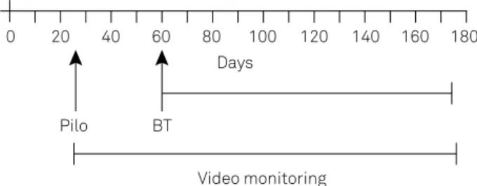
At 25 days of age the experimental animals were sub-mitted to pilocarpine-induced epilepsy13. Experimental group (15 animals) received methyl scopolamine (1 mg/kg, s.c.) 30 minutes before pilocarpine adminis-tration (350 mg/kg, ip) and control group (11 animals) received saline 0.9%. three hours after the onset of the status epilepticus both groups received diazepam (7.5 mg/kg, sc) (Figure 1). Epilepsy induced experimen-tally exhibits high and variable mortality rates, and thus, the following groups of animals were designed:
Experimental group: 6 animals; Control group: 11 animals
he animals were monitored 24 h/day starting at the SE induction up to end of experiments. he behavioural tests
(Bt) started from 60 days postnatal after all animals have been evolved to epilepsy.
Behavioural Tests (BT)
the animals were transferred to the testing room 60 min before each day session. All apparatus were cleaned with a 5% alcohol solution after each behavioral proce-dure. At the end of behavioral tests, the rats were anaes-thetized with urethane 1,200 mg/kg (ip). the half animals of each group were decapitated, the brains were dissected and frozen to -80C and the other half was submitted to transcardial perfusion.
Open field
he apparatus consisted of a circular arena (100 cm di
-ameter) enclosed by plain white walls and a loor divided
into 12 zones, being 8 peripheral and 4 central (insight Ltda, Brazil). Each animal was placed into the central area and ob-served for 10 min. During this time, the locomotor activity was expressed as the number of peripheral, central or total lines crossed. in addition, the time spent on central zone was
measured. he test was repeated 7 and 15 days later.
Elevated plus maze (EPM)
he apparatus had two closed arms with walls 45 cm
in height and two open arms 50 cm long (insight Ltda,
Brazil) and was elevated 50 cm from the loor. he ani -mals were placed in the central zone of the maze with their nose pointing towards an open arm and explored
the maze for 5 minutes. he number of entries and the
time spent in both arms were recorded and expressed as percent of entries (EOA) or time (tOA) in open arms: ([Open arm /(Open arm + Closed arm)] * 100).he test was
repeated 7 and 15 days later.
Operant conditioning box (OCB)
he OCB aimed to analyse attention, impulsivity and vi -suospatial working memory in a visual discrimination task.
he apparatus (Habitest Coulbourn Instruments) consisted
of two responder levers, two cue lights located above the
le-vers and a water dispenser. he apparatus’ activities were
controlled by Graphic State and the data stored to posterior
analysis. he apparatus was enclosed in a sound-attenuating box outitted with an exhaust fans.
the animals were evaluated using three experimental protocols, each one with fifteen sessions and lasting thirty minutes. Before that, the animals were habituated to the apparatus followed by acquisition sessions. the animals were deprived of water 21 hours before the procedures. For habituation, the animals remained 30 minutes in the apparatus; the reinforcement was not delivered and the cue lights above the levers were off. After that, the ani-mals were subject for two acquisitions sessions, lasting 30 and 15 min, respectively. the cue light located inside the apparatus was on, but the cue lights above the levers were not lit. the reinforcement was delivered every 10s
independently of the rat’s behavior. During subsequent
sessions, the learning was acquired by the method of suc-cessive approximations. During the initial sessions, the rats learned to press the left lever in order to receive re-inforcement immediately after every correct response. the cue light above the left lever was now lit the entire session and the lights above the right lever were off. it was considered that the animals learned to press the left lever when it was pressed at least eight consecutively times in order to obtain the reinforcement. After that, the animals were trained to press the right lever following the same shaping procedure.
in the first and second protocols the light above the levers stayed lit for 5 or 1s, respectively, shifted randomly and the reinforcement was delivery immediately after the correct lever being pressed. in the third protocol, the light stayed lit for 1 s, but the reinforcement was delivery after 10 s. it was analysed the number lever presses and the per-centage of correct responses. Correct responses were con-sidered when the animal pressed the lever while the light points were lit.
Figure 1. Timeline of the procedures. Pilo: picocalpine; BT: behavioural test.
0 20 40 60 80 100 120 140 160 180
Days
Pilo BT
Statistical analyses
he data were expressed as mean ± standard error and
analysed by Mixed ANOVA, using Bonferroni for post-hoc
testing. p-values of 0.05 or less were considered signiicant. he analyses were efectuated using commercial program
(Prism 5.03 for windows).
RESULTS
Frequency of seizures
he irst spontaneous seizure started between 4–15 days
after SE onset with an average frequency of 1.34 + 0.27 per week. Most seizures were recorded during the day light (67.74%).
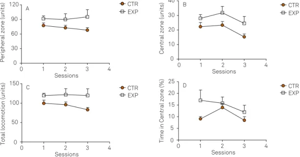
Open field
For peripheral locomotion, it was noted a signiicant dif -ference between groups (F2,30 = 5.365, p = 0.035), with no dif-ference between sessions (F2,30 = 0.167, p = 0.84) nor efect of
interaction between factors (F2,30 = 0.634, p = 0.53). he pe -ripheral locomotion was higher in experimental group as compared to control (Figure 2A).
Central locomotion was a signiicantly diferent between
groups (F2,30 = 6.43, p = 0.022) and the sessions (F2,30 = 3.81,
p = 0.033), with no efect of interaction between the factors
(F2,30 = 0.20, p = 0.81). he experimental group showed high -er locomotor activity in the central zone as compared to control (Figure 2B).
he total locomotion was signiicantly higher in the ex -perimental group as compared to control (F1,30 = 6.24, p = 0.02),
with no efect of interaction between the factors (F2,30 = 0.64,
p = 0.52). Only control group exhibited reduction on locomo-tor activity over time (Figure 2C).
the time spent in the central zone was significantly different between groups (F1,30 = 6.032, p = 0.026), with no effect of sessions (F2,30 = 2.23, p = 0.12) nor interaction be-tween the factors (F2,30 = 0.94, p = 0.39). the time in the central zone was higher in the experimental group as compared to control (Figure 2D).
in a non-aversive context, animals with ELt exhibited hy-peractivity and reduced level of anxiety-related behaviour.
Elevated plus maze
Total number of entries was signiicantly diferent between
groups (F1,30 = 5.25, p = 0.036), with no diference between ses -sions (F2,30 = 1.29, p = 0.28) nor efect of interaction between fac -tors (F2,30 = 0.91, p = 0.41). he locomotor activity was higher in the experimental group as compared to control group (Figure 3A).
For the percentage of entries and the time spent on the
open arms no diferences was noted for groups (F1,30 = 1.12, p = 0.30; F1,30 = 0.27, p = 0.60, respectively), neither for sessions (F2,30 = 1.67, p = 0.20; F2,30 = 0.14, p = 0.86, respectively), nor for interaction between factors (F2,30 = 0.87, p = 0.42; F2,30 = 0.05, p = 0.94, respectively (Figure 3B and 3C).
Operant conditioning box
Both groups learned to press the levers in the irst ses -sion. in the following sessions, both bars were used to obtain the reinforcement. Cue lights located above the levers
indi-cated which bar should be pressed. he cue lights light up
randomly and remained lit for periods of time varying in ac-cordance with the protocol (5 or 1 second).
Figure 2. Peripheral (A), central (B), total locomotion (C) and time spent in central zone (D) on the open field in 3 sessions with 7 days apart were expressed as mean ± standard error of CTR group (n = 11) and EXP group (n = 6). The experimental animals exhibited higher total and central locomotor activities and time spent in the central zone as compared to control, suggesting the presence of hyperactivity and reduced anxiety-related behavior. CTR: control; EXP: experimental.
0 1 2 3 4
0 30 60 90
120 CTR
EXP
A
Sessions
Peripheral zone (units)
0 1 2 3 4
0 10 20 30 40
CTR EXP
B
Sessions
Central zone (units)
0 1 2 3 4
0 50 100 150
CTR EXP
C
Sessions
Total locomotion (units) 0 0 1 2 3 4
5 10 15 20 25
CTR EXP
D
Sessions
Protocol 1
it was analysed the percentage of the correct response
and the number of bar presses. he percentage of cor
-rect responses was signiicantly diferent between session
(F14,210 = 36.75, p < 0.0001), with efect of interaction between factors (F14,21 = 3.57, p < 0.0001) and no diference between
groups (F 1,210 = 0.379, p = 0.54, Figure 4A). Both groups im-proved their performance over time.
he number of the lever presses was signiicantly difer -ent between session (F14,21 = 7.79, p < 0.0001), with no dif-ference between groups (F[1,210] = 0.49, p = 0.07) and nor in-teraction between factors (F14,210 = 1.63, p = 0.07, Figure 4B).
he number of lever presses reduced to almost half concomi -tantly with the increase of the percentage of correct response which stabilized approximately in the twelfth session around
of 60%. he data suggests that the attention to the task was
similar between groups.
Protocol 2
In order to increase the degree of diiculty in the subse -quent 15 sessions, the cue lights remained lit for 1 second.
he percentage of correct responses was signiicantly difer -ent across the sessions (F 14,210 = 3.32, p < 0.0001), with no ef-fect of interaction (F14,210 = 1.28, p = 0.21) nor diference be -tween groups (F1,210 = 3.43, p = 0.083, Figure 5A).
he number of lever presses was signiicantly diferent
across the sessions (F14,210 = 4.02, p < 0.0001), with no difer -ence between groups (F1,210 = 0.14, p = 0.71) nor interaction between factors (F14,210 = 1.53, p = 0.099, Figure 5B).
Protocol 3
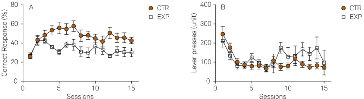
in order to assay the delay aversion and wM, the luminous spots remained lit for 1 second and the rein-forcement was released 10 seconds after the correct le-ver being pressed.
For the percentage of correct response, it was not-ed a significant effect of interaction between factors (F14,210 = 3.12, p = 0.0002) and effect of session (F14,210 = 5.77, p < 0.0001) with no difference between groups (F 1,210 = 4.39, p = 0.053, Figure 6A). the percentage of correct response, in the initial sessions, was similar between groups, fol-lowed by improvement in control group and a moder-ate reduction in experimental group. in the 5th and the 12th session the percentage of correct response in the experimental group was significantly lower as compared to control group (t = 3.29, p < 0.05 and t = 3.15, p < 0.05, respectively). the number of lever presses was significant-ly different over time (F14,210 = 7.44, p < 0.0001) with signifi-cant effect of interaction between factors (F14,210 = 2.62, p < 0.0016), with no difference between groups (F1,210 = 0.73,
Figure 3. Total entries (A), percentage of entries in open arms (EOA%, B), percentage of time in open arms (TOA %, C) on the elevated plus maze in 3 sessions with 7 days apart were expressed as mean ± standard error of the CTR group (n = 11) and EXP group (n = 6). The experimental group exhibited higher locomotor activity as compared to control with no differences between groups for the percentage of entries, nor time spent in open arms. In an aversive context, the results showed that animals with epilepsy exhibited hyperactivity. CTR: control; EXP: experimental.
0 1 2 3 4
15 20 25 30
A
Sessions
To
ta
l en
tr
ies
(num
be
r)
0 1 2 3 4
40 45 50 55 60 65 70 75
B
Sessions
EO
A
(%
)
0 1 2 3 4
35 40 45 50 55
C
Sessions
TO
A
(%
)
CTR EXP
CTR EXP
CTR EXP
Figure 4. Operant Conditioning Box. The light above the levers stayed lit for 5 s, shifted randomly and the reinforcement was delivery immediately after the correct lever being pressed. Percentage of correct answers (A) and total lever presses (B) expressed as mean ± standard error of CTR (n = 11) and EXP (n = 6) groups. No significant differences were found between the groups. CTR: control; EXP: experimental.
0 5 10 15
20 40 60 80
A
Sessions
Correct Response (%)
0 5 10 15
200 400 600 800
B
Sessions
Lever presses (unit)
CTR EXP
p = 0.40, Figure 6B). the number of presses gradually de-creased in both groups over time, stabilizing in a higher level in the experimental group.
the results of the three protocols were plotted on the same graph to better visualization and comparison the performance of both groups. when the difficulty of the challenge increases, the percentage of correct responses decreased. in the third protocol, note a sudden reduction in the percentage of correct response in both groups as compared to protocol 2, followed by recovery of the con-trol group. For the experimental group, the percentage of
correct response stabilized in a lower level as compared to previous protocol, and also when compared to control group within the same protocol (Figure 7A). Regarding the number of lever presses, note a drastic reduction in both groups when compared to the previous proto-col. For the control group, the number of bar presses re-mained reduced with a concomitant increase in the per-centage of correct response. For the experimental group, the number of lever presses stabilized in a higher level as compared to control, while the percentage of response stabilized in a lower level (Figure 7B).
Figure 6. Operant Conditioning Box.The light above the levers stayed lit for 1s, shifted randomly and the reinforcement was delivery 10 s after the correct lever being pressed.Percentage of correct answers (A) and lever presses (B) expressed as mean ± standard error of the CTR (n = 11) and EXP (n = 6) groups. The percentage of correct answers stabilized at a lower level and the lever presses stabilized at a higher level in the experimental group compared to the control group. CTR: control; EXP: experimental.
0 5 10 15
0 20 40 60 80
Sessions
Co
rr
ect
R
esponse
(%
)
0 5 10 15
0 100 200 300 400
Sessions
L
ever
pr
esses
(uni
t)
A CTR B
EXP
CTR EXP
Figure 7. Operant Conditioning Box. Data of the three protocols were represented in the same graph. Percentage of correct answers (A) and number of lever presses (B). CTR: control; EXP: experimental.
0 5 10 15 20 25 30 35 40 45 20
30 40 50 60 70
80 Aphase 1 phase 2 phase 3
Sessions
Correct Response (%)
0 5 10 15 20 25 30 35 40 45 0
100 200 300 400 500 600 700
B
phase 1 phase 2 phase 3
Sessions
Lever presses (unit)
CTR EXP
CTR EXP Figure 5. Operant Conditioning Box. The light above the levers stayed lit for 1 s, shifted randomly and the reinforcement was delivery immediately after the correct lever being pressed. Percentage of correct answers (A) and total lever press (B) expressed as mean ± standard error of CTR (n = 11) and EXP (n = 6) group. No significant differences were found between the groups. CTR: control; EXP: experimental.
0 5 10 15
30 40 50 60 70
A
Sessions
Correct Response (%)
0 5 10 15
0 200 400 600 800
B
Sessions
Lever presses (unit)
CTR EXP
he results observed in protocols 1 and 2 suggest that an
-imals with epilepsy showed no attention deicit in a visual
discrimination task. in the protocol with the highest degree
of diiculty, the diference between groups became evident. he performance of experimental animals reduced as com -pared to control, as result of the lower percentage of correct response and greater number of lever presses.
DISCUSSION
we investigated the presence of cognitive and behav-ioral dysfunctions in experimental tLE suggestive of con-comitant presence of ADhD. Animals showed reduced anxiety-related behavior, hyperactive behavior, impulsivity and deficit in visuospatial working memory. hyperactivity was observed over time in a familiar environment with neutral context (open field) and in a threatening environ-ment. it is interesting to mention that the locomotor hy-peractivity in children with ADhD as well as animal model of ADhD (e.g spontaneous hypertensive rats) tend to be less pronounced in novel environments than in familiar ones14, and for that reason the test was repeated 3 times with 7 days apart. in addition, our results are in agreement with Kubova et al.15, which reported increased locomo-tor activity in rats submitted to kainic acid (KA) model at PN25 (25 day of life).
in a no aversive environment, experimental group ex-hibited reduced anxiety-related behavior with no chang-es in an aversive environment, suggchang-esting that the state of anxiety is context dependent. in the lithium-pilocarpine (LiP) model, Detour et al.16 found increased number of en-tries and more time spent into the open arms of the aver-sive environment. Using the same paradigm, we did not
ob-serve diference between the groups, but a trend towards to increased activity in the open arms. he small sample size
may have contributed to the discrepant results between studies. inostroza et al.17 compared the cognitive and be-havioral performance and anatomic changes between LiP
model and KA model animals, which difered signiicantly
in the pattern and extent of tLE-associated brain lesions. LiP-treated rats showed reduced state of anxiety against a slight decrease in the KA-treated rats. LiP-treated animals also exhibited increased motivation to consume sucrose, and both showed reduced motivation for social contact,
being particularly afected LIP model.
After all behavioral tests, plasma corticosterone levels were increased only in LiP model, suggesting that altered emo-tional behaviors were not related to the epileptic condition;
instead of probably relect deregulation, model-dependent of
the hPA axis. to the best of our knowledge, no study com-pared cognition, behavioral performance, anatomic changes and the pattern of seizure frequency between LiP model and pilocarpine model.
the OCB´s results suggest that learning, attention, and associative memory in a visual discrimination task were preserved in animals with epilepsy. however, when a de-lay for reinforcement was used, the performance was re-duced, with increased impulsivity and visuospatial work-ing memory impairment, which is prominent in ADhD. interestingly, Pineda et al.18 observed in the LiP model that half the animals exhibited increased impulsivity and diminished attention in the lateralized reaction time test and the other half exhibited depressive related behav-ior. the seizure frequency ranging from 1 to 5 per week, but any correlation was investigated between the seizures frequency and behavioral changes. Faure et al.19 demon-strated in LiP model that animals displayed attention def-icit with a tendency toward impulsivity and compulsivi-ty in a five-choice serial reaction time task. the seizures frequency was not reported. in our study, animals did not display attention deficit, rather than, they exhibited in-creased impulsivity and visuospatial working memory im-pairment. Some issues may be underlying the differences
in the animals’ performance among studies.
An important issue to be considered regards the seizure frequency and its impact on the cognitive and behavioral changes. As seen in human condition, seizure activity is highly variable among animals and occurs in clusters with seizures free-intervals20,21. Bajorat et al.21 using video-EEG in
pilocarpine model identiied three patterns of seizure distri -bution during the course of recording: a) > 3/day and evenly distributed seizures, b) > 2/day and again evenly distribut-ed seizures and c) > 3/day and seizures clusterdistribut-ed with sei-zure-free intervals. in our study, the average of seizures was 1.34 per week, being lower than observed by Bajorat et al.21. we do not rule that the low accuracy of the video monitoring may have contributed to underestimate the seizure activity. Despite of the well-established concept that epilepsy is a progressive disease and that the hippocampal/neocortical atrophy increases over time22,23,24, remains the controversy if seizure frequency has substantial impact on brain damage and cognition. Fuerst et al.24 and Briellmann et al.25 showed a correlation between the seizures frequency and ipsilat-eral hippocampal volume loss. On the contrary, Liu et al.26 did not reported that the brain volume reduction was likely
to be related to an initial brain insult and being inluenced
by age. Pacagnella et al.27 reported that the memory perfor-mance and the degree of hippocampal atrophy did not dif-fer between patients with frequent and infrequent seizures, rather than, a positive correlation was found between age of onset and degree of hippocampal atrophy. in this sense, we
argue that the lack of attentional deicits in our experimen -tal animals could be related to the low seizure frequency and or the newly onset of epilepsy.
Another issue regards the degree of challenge of the task.
lever presses and a stimulus presentation of 1 s or 5 sec. in
the Faure and colleagues’ study, ive holes were used with a
stimulus presentation of 0.5 s or 5 sec. with a stimulus pre-sentation of 0.5 s, which require a high attentional demand,
the percentage of correct response was signiicantly difer -ent between groups, but not when the stimulus pres-enta- presenta-tion was set at 5 sec. we argue that our protocol condipresenta-tion
was not enough sensitive to detect attentional deicits.
in regarding the visuospatial working memory, experi-mental15 and clinical evidences9,28,29,30,31 have been shown that it is impaired in tLE. we showed evidences that animals with tLE exhibited hyperactivity, reduced level of anxiety-related behavior, increased mild impulsivity and impaired visuospa-tial working memory, suggesting that the pilocarpine mod-el of epilepsy is appropriate to investigate the interplay be-tween epilepsy and ADhD.
References
1. Sander JW. The epidemiology of epilepsy revisited. Curr Opin Neurol. 2003;16(2):165-70. doi:10.1097/00019052-200304000-00008 2. Boer HM, Mula M, Sander JW. The global burden and
stigma of epilepsy. Epilepsy Behavior. 2008;12(4):540-6. doi:10.1016/j.yebeh.2007.12.019
3. Thurman DJ, Beghi E, Begley CE, Berg AT, Buchhalter JR, Ding D. Standards for epidemiologic studies and surveillance of epilepsy. Epilepsia. 2011;52(suppl 7):2-26. doi:10.1111/j.1528-1167.2011.03121.x
4. Laxer KD, Trinka E, Hirsch LJ, Cendes F, Langfitt J, Delanty N et al. The consequences of refractory epilepsy and its treatment. Epilepsy Behav. 2014;37:59-70. doi:10.1016/j.yebeh.2014.05.031
5. Duncan JS, Sander JW, Sisodiya SM, Walker MC. Adult epilepsy. Lancet. 2006;367(9516):14. doi:10.1016/S0140-6736(06)68477-8 6. Kwan P, Sander JW. The natural history of epilepsy:
an epidemiological view. J Neurol Neurosurg Psychiatry. 2004;75(10):1376-81. doi:10.1136/jnnp.2004.045690 7. Sander JW. Some aspects of prognosis in the
epilepsies: a review. Epilepsia. 1993;34(6):1007-16. doi:10.1111/j.1528-1157.1993.tb02126.x
8. Zamarian L, Trinka E, Bonatti E, Kuchukhidze G, Bodner T, Benke T et al. Executive functions in chronic mesial temporal lobe epilepsy. Epilepsy Res Treat. 2011;2011:ID596174. doi:10.1155/2011/596174
9. Stretton J, Winston GP, Sidhu M, Bonelli S, Centeno M,
Vollmar C et al. Disrupted segregation of working memory networks in temporal lobe epilepsy. Neuroimage Clin. 2013;2:273-81. doi:10.1016/j.nicl.2013.01.009
10. Cohen R, Senecky Y, Shuper A, Inbar D, Chodick G, Shalev V et al. Prevalence of epilepsy and attention-deficit hyperactivity (ADHD) disorder: a population-based study. J Child Neurol. 2013;28(1):120-3. doi:10.1177/0883073812440327
11. Dunn DW, Austin JK, Harezlak J, Ambrosius WT. ADHD and epilepsy in childhood. Develop Med Child Neurol. 2003;45(1):50-4. doi:10.1111/j.1469-8749.2003.tb00859.x
12. Loutfi KS, Carvalho AM. Possíveis interfaces entre TDAH e epilepsia. J Bras Psiquiatr. 2010;59(2):146-55. doi:10.1590/S0047-20852010000200011
13. Turski WA, Cavalheiro EA, Schwarz M, Czuczwar SJ, Kleinrok Z, Turski L. Limbic seizures produced by pilocarpine in rats: behavioural, electroencephalographic and
neuropathological study. Behav Brain Res. 1983;9(3):315-35. doi:10.1016/0166-4328(83)90136-5
14. Levant B, Zarcone TJ, Davis PF, Ozias MK, Fowler SC. Differences in methylphenidate dose response between periadolescent and adult rats in the familiar arena-novel alcove task. J Pharmacol Exp Ther. 2011;337(1):83-91. doi:10.1124/jpet.110.174425
15. Kubová H, Haugvicová R, Suchomelová L, Mares P. Does status epilepticus influence the motor development of immature rats? Epilepsia. 2000;41(Suppl 6):S64-9. doi:10.1111/j.1528-1157.2000.tb01559.x
16. Detour J, Schroeder H, Desor D, Nehlig A. A 5-month period of epilepsy impairs spatial memory, decreases anxiety, but spares object recognition in the lithium-pilocarpine model in adult rats. Epilepsia. 2005;46(4):499-508. doi:10.1111/j.0013-9580.2005.38704.x
17. Inostroza M, Cid E, Menendez de la Prida L, Sandi C. Different emotional disturbances in two experimental models of temporal lobe epilepsy in rats. PLoS One. 2012;7(6):e38959. doi:10.1371/journal.pone.0038959
18. Pineda E, Jentsch JD, Shin D, Griesbach G, Sankar R, Mazarati A. Behavioral impairments in rats with chronic epilepsy suggest comorbidity between epilepsy and attention deficit/hyperactivity disorder. Epilepsy Behav. 2014;31:267-75. doi:10.1016/j.yebeh.2013.10.004
19. Faure JB, Marques-Carneiro JE, Akimana G, Cosquer B, Ferrandon A, Herbeaux K et al. Attention and executive functions in a rat model of chronic epilepsy. Epilepsia. 2014;55(5):644-53. doi:10.1111/epi.12549
20. Arida RM, Scorza FA, Peres CA, Cavalheiro EA. The course of untreated seizures in the pilocarpine model of epilepsy. Epilepsy Res. 1999;34(2-3):99-107. doi:10.1016/S0920-1211(98)00092-8 21. Bajorat R, Wilde M, Sellmann T, Kirschstein T, Köhling R.
Seizure frequency in pilocarpine-treated rats is independent of circadian rhythm. Epilepsia. 2011;52(9):e118-22. doi:10.1111/j.1528-1167.2011.03200.x
22. Liu RS, Lemieux L, Bell GS, Hammers A, Sisodiya SM, Bartlett PA et al. Progressive neocortical damage in epilepsy. Ann Neurol. 2003;53(3):312-24. doi:10.1002/ana.10463 23. Cascino GD. Progressive damage in epilepsy. Epilepsy Curr.
2003;3(6):214-5. doi:10.1046/j.1535-7597.2003.03615.x 24. Fuerst D, Shah J, Shah A, Watson C. Hippocampal sclerosis is
a progressive disorder: a longitudinal volumetric MRI study. Ann Neurol. 2003;53(3):413-6. doi:10.1002/ana.10509
25. Briellmann RS, Berkovic SF, Syngeniotis A, King MA, Jackson GD. Seizure-associated hippocampal volume loss: a longitudinal magnetic resonance study of temporal lobe epilepsy. Ann Neurol. 2002;51(5):641-4. doi:10.1002/ana.10171
26. Liu RS, Lemieux L, Bell GS, Sisodiya SM, Bartlett PA, Shorvon SD et al. Cerebral damage in epilepsy: a population-based longitudinal quantitative MRI study. Epilepsia. 2005;46(9):1482-94. doi:10.1111/j.1528-1167.2005.51603.x
27. Pacagnella D, Lopes TM, Morita ME, Yasuda CL, Cappabianco FA, Bergo F et al. Memory impairment is not necessarily related to seizure frequency in mesial temporal lobe epilepsy with hippocampal sclerosis. Epilepsia. 2014;55(8):1197-204. doi:10.1111/epi.12691
29. Krauss GL, Summerfield M, Brandt J, Breiter S, Ruchkin D. Mesial temporal spikes interfere with working memory. Neurology. 1997;49(4):975-80. doi:10.1212/WNL.49.4.975
30. Owen AM, Morris RG, Sahakian BJ, Polkey CE, Robbins TW. Double dissociations of memory and executive functions in working memory tasks following frontal lobe excisions, temporal lobe excisions or
amygdalo-hippocampectomy in man. Brain. 1996;119(5):1597-615. doi:10.1093/brain/119.5.1597