953
DOI: 10.1590/0004-282X20160155
ARTICLE
Neurological outcomes after hematopoietic
stem cell transplantation for cerebral X-linked
adrenoleukodystrophy, late onset metachromatic
leukodystrophy and Hurler syndrome
Desfechos neurológicos após transplante de células tronco hematopoiéticas na
adrenoleucodistrofia ligada ao X, forma cerebral, na leucodistrofia metacromática de
início tardio e na síndrome de Hurler
Jonas Alex Morales Saute1,2,5, Carolina Fischinger Moura de Souza1, Fabiano de Oliveira Poswar 7, Karina
Carvalho Donis1,3, Lillian Gonçalves Campos4,5, Adriana Vanessa Santini Deyl6, Maira Graeff Burin1, Carmen
Regla Vargas1,13, Ursula da Silveira Matte7,8,9, Roberto Giugliani1,3,5,7,8,9, Maria Luiza Saraiva-Pereira1,2,7,10,
Leonardo Modesti Vedolin4,5,, Lauro José Gregianin6,12, Laura Bannach Jardim1,2,3,5,7, 11
1Hospital de Clínicas de Porto Alegre, Serviço de Genética Médica, Porto Alegre RS, Brasil; 2Hospital de Clínicas de Porto Alegre, Laboratório de Identiicação Genética, Porto Alegre RS, Brasil;
3Universidade Federal do Rio Grande do Sul, Programa de Pós-Graduação em Saúde da Criança e do Adolescente, Porto Alegre RS, Brasil; 4Hospital de Clínicas de Porto Alegre, Serviço de Radiologia, Porto Alegre RS, Brasil;
5Universidade Federal do Rio Grande do Sul, Programa de Pós-Graduação em Ciências Médicas, Porto Alegre RS, Brasil; 6Hospital de Clínicas de Porto Alegre, Serviço de Oncologia Pediátrica, Porto Alegre, Brasil;
7Universidade Federal do Rio Grande do Sul, Programa de Pós-Graduação em Genética e Biologia Molecular; Porto Alegre RS, Brasil; 8Hospital de Clínicas de Porto Alegre, Laboratório de Terapia Gênica, Porto Alegre RS, Brasil;
9Universidade Federal do Rio Grande do Sul, Departamento de Genética e Biologia Molecular, Porto Alegre RS, Brasil; 10Universidade Federal do Rio Grande do Sul, Departamento de Bioquímica, Porto Alegre RS, Brasil;
11Universidade Federal do Rio Grande do Sul, Departamento de Medicina Interna, Porto Alegre RS, Brasil; 12Universidade Federal do Rio Grande do Sul, Departamento de Pediatria, Porto Alegre RS, Brasil; 13Universidade Federal do Rio Grande do Sul, Faculdade de Farmacia, Porto Alegre, Brasil
Correspondence: Laura Bannach Jardim; Serviço de Genética Médica do Hospital de Clinicas de Porto Alegre; Rua Ramiro Barcelos, 2350; 90035-903 Porto Alegre RS, Brasil; Email: [email protected]
Conflict of interest: There is no conlict of interest to declare.
Support: C.F.M. de Souza received travel grants and speaker honoraria from Genzyme, which has products for MPS-I treatment. R. Giugliani is an investigator in clinical trials sponsored by Genzyme and Shire, and has received travel grants and speaker honoraria from these companies, which have products for MPS-I and MLD treatment, respectively.
C.R.Vargas, U.S. Matte, R. Giugliani, M.L. Saraiva-Pereira and L.B. Jardim received research fellowships from Conselho Nacional de Desenvolvimento Cientíico e Tecnológico (CNPq), Brazil.
Received 17 June 2016 ; Received in inal form 01 August 2016 ; Accepted 24 August 2016.
ABSTRACT
Hematopoietic stem cell transplantation (HSCT) is the only available treatment for the neurological involvement of disorders such as late-onset metachromatic leukodystrophy (MLD), mucopolysaccharidosis type I-Hurler (MPS-IH), and X-linked cerebral adrenoleukodystrophy (CALD). Objective: To describe survival and neurological outcomes after HSCT for these disorders. Methods:
Seven CALD, 2 MLD and 2 MPS-IH patients underwent HSCT between 2007 and 2014. Neurological examinations, magnetic resonance imaging, molecular and biochemical studies were obtained at baseline and repeated when appropriated. Results: Favorable outcomes were obtained with 4/5 related and 3/6 unrelated donors. Two patients died from procedure-related complications. Nine transplanted patients were alive after a median of 3.7 years: neurological stabilization was obtained in 5/6 CALD, 1/2 MLD, and one MPS-IH patient. Brain lesions of the MPS-IH patient were reduced four years after HSCT. Conclusion: Good outcomes were obtained when HSCT was performed before adulthood, early in the clinical course, and/or from a related donor.
954 Arq Neuropsiquiatr 2016;74(12):953-966
Allogeneic hematopoietic stem cell transplantation (HSCT) was proposed as a treatment for inherited leukoencephalopa-thies in the eighties, based on the assumption that transplanted macrophages migrate through the blood-brain barrier, and dif-ferentiate into microglia that would replace or support native microglia with the functional enzyme through cross-correction1.
Since then, HSCT has been the only available treatment for the neurological involvement of some lysosomal storage disorders
that afect the central nervous system (CNS), such as juvenile
and adult metachromatic leukodystrophy (MLD), mucopoly-saccharidosis type I Hurler (MPS-IH), and others. In addition, HSCT has been mostly indicated for the cerebral form (CALD) of the peroxisomal disorder X-linked adrenoleukodystrophy (X-ALD)2,3,4,5,6,7,8,9,10. Considering that transplant-related mortality
has declined to 10% and the rate of engraftment has substantial-ly improved in recent years11,12, risk of transplant is worthwhile in
contrast to the certainty that the natural history of these disor-ders will lead to dementia, decorticate or vegetative states and death, some years after onset.
here is consensus about the eicacy of HSCT when
performed early in life for MPS-IH9,13,14 and at early stages in
CALD6,7,12,15. Case reports suggested that HSCT might reduce
demyelination in juvenile (onset between two and 14 years)
and adult forms of MLD when performed as early as possi-ble3,4,5,16. A recent case control study conirmed that HSCT,
at a presymptomatic or early symptomatic stage of juvenile
MLD, is associated with disease stabilization10.
he aim of the present study was to describe a seven-year
experience of a university hospital with HSCT for these dis-orders, focusing on survival and on neurological outcomes.
METHODS
Patients and study procedures
his is a retrospective study of a population consisting of seven CALD, one juvenile and one adult MLD and two MPS-IH patients,
who underwent HSCT at Hospital de Clínicas de Porto Alegre be-tween 2007 and 2014. All procedures were covered by the Brazilian
Uniied Health System, and received the same level of health in
-surance. hree CALD patients have been reported previously17.
Biochemical diagnosis was obtained for all patients and
was conirmed by molecular analysis of the appropriate gene. Neurological examinations, neuropsychological tests, magnetic resonance imaging (MRI), and speciic biochemi -cal markers were performed before HSCT, and six months to seven years after the procedure.
An HSCT was ofered as early as possible when an
HLA-matched donor or cord blood was available. Additional
clinical criteria for ofering HSCT were applied, according to each speciic disorder. International recommendations were
followed for CALD patients: the presence of white matter
lesions in the CNS and a Loes score lower than 10 points7.
An HSCT was also ofered as early as possible following diag
-nosis of juvenile or adult forms of MLD, provided that some
gross motor functions such as walking (with or without aid) were still present. Finally, HSCT was indicated for MPS-IH patients under 2.5 years of age if the clinical status, especially pulmonary function, allowed the procedure9.
Biochemical and molecular analyses
Plasma docosanoic (C22:0), tetracosanoic (C24:0) and hexacosanoic (C26:0) acids were obtained from CALD pa-tients, and analyzed as described previously18. Arylsulfatase
A (ARSA) activity in leukocyte and a thin-layer chromatogra-phy of urinary sulfatides were studied in the MLD patients. In the MPS-IH patients, urinary glycosaminoglycans (GAGs), colorimetric assay (DMB test) and electrophoresis were per-formed, and α-L-iduronidase (IDUA) activity in leukocytes was measured. All biochemical studies were performed at the time of diagnosis: their values were subsequently com-pared to those obtained after HSCTs.
Molecular analyses of IDUA, ABCD1 and ARSA were
per-formed in DNA isolated from peripheral blood prior to the HSCT. In all patients, individual exons and lanking regions ampliied by PCR were submitted for Sanger sequencing.
For ARSA analysis we also evaluated regions associated with
pseudodeiciency (PD) variants using a similar approach19.
Brain MRI
Brain MRI data were obtained with a 1.0 or 1.5 T sys-tems equipped with a standard circularly polarized head coil.
RESUMO
O transplante de células tronco hematopoiéticas (TCTH) é o único tratamento disponível para o envolvimento neurológico de doenças como a leucodistroia metacromática (MLD), a mucopolissacaridose tipo I-Hurler (MPS-IH) e a adrenoleucodistroia (CALD). Objetivos: Descrever a sobrevida e os desfechos neurológicos após o TCTH nessas doenças. Métodos: Sete pacientes CALD, 2 MLD e 2 MPS-IH realizaram TCTH entre 2007 e 2014. Avaliações neurológicas, ressonância nuclear magnética e estudos bioquímicos e moleculares foram feitos no baseline e repetidos quando apropriado. Resultados: Desfechos favoráveis foram obtidos em 4/5 TCTH de doadores relacionados e em 3/6 não relacionados. Dois pacientes faleceram de complicações do procedimento. Nove transplantados sobreviveram após uma mediana de 3,7 anos: estabilização neurológica foi obtida em 5/6 CALD, ½ MLD e em um caso MPS-IH. As lesões encefálicas de um caso MPS-IH reduziram-se quatro anos após o TCTH. Conclusão: Bons desfechos foram obtidos quando o TCTH foi feito antes da vida adulta, cedo no curso clínico e/ou a partir de um doador relacionado.
955 Saute JAM et al. Outcomes after HSCT for X-ALD, MLD and MPS IH
Axial luid-attenuated inversion recovery (FLAIR), axial and
coronal T2 and sagittal T1 weighted images were obtained in
all patients. he MRI was performed at baseline and in the
follow-up visits after HSCT. Disease-related MRI scores20,21
were reviewed for each patient at the time of the present re-port. Images were analyzed by two independent neuroradi-ologists (LMV and LC) blind at the time of the given study. In case of discordant scores, both neuroradiologists conferred
together to ind a consensus. Any 0.5 T or low-quality MRI
images were excluded from the analysis. An independent re-searcher (JAMS) rebuilt the time frame later.
HSCT procedures
The HSCT was performed according to institutional protocol recommendations, and all patients received the myeloablative conditioning regimen consisting of an com-bination of busulfan and cyclophosphamide, as previously published22. Details of the HSCT procedures are described
in Table 1.
Ethics, consent and permissions. Consent to publish.
Consents for the transplant procedure and for follow up visits and ancillary tests were obtained from all individ-uals or their representatives at the time of the procedure, and according to ethical requirements of our institution.
his report summarizes results from studies registered as
GPPG 13-0390 ( for X-ALD), 07-599 ( for MLD) and 03-066 ( for MPS-IH), all approved by the Ethics Committee (EC) of Hospital de Clínicas de Porto Alegre, following the World Medical Association International Code of Medical Ethics (Declaration of Helsinki).
RESULTS
he HSCT were performed in 11 patients (six with un -related donors), in the last seven years. Patients and donor characteristics, conditioning, graft source and engraftment, are shown in Table 1.
Baseline results of the neurological findings, intel -ligence quotient (IQ), MRI and biochemical markers, as well as the follow-up findings after HSCT, are present-ed in Table 2. Two patients (one with CALD and one with MPS-IH) died from HSCT complications. Seven of the re-maining nine individuals had stabilization of symptoms one year or more following HSCT.
Seven CALD patients underwent HSCT; preliminary data on three of them were reported previously20. Case
re-ports are presented in the Supplemental Material. In sum-mary, HSCTs were done in five CALD patient younger than 12 years, in one who was 19.4 years old and in one who was 28.2 years old. The oldest of them also presented with the highest Loes score at the time of his HSCT (8 points): he died nine months after the procedure. Five out of the six
CALD patients who survived HSCT were clinically stable at the time of this report (in one patient, the Loes score had even improved). The progression of Loes scores of all seven individuals is shown in Figure 1.
Two MPS-IH underwent HSCT: their data are depicted in Tables 1 and 2, and also on Supplemental Material. Of note,
a clear improvement of MRI indings of MPS IH-10 case ap
-peared three years after her HSCT, with signiicant reduction of periventricular and deep WM lesions. he mild ventricular
enlargement also improved (Figure 2).
Due to data scarcity on HSCT eicacy in MLD, individual
case reports will be reported bellow.
Patient MLD-8b
Patient MLD-8b, female, eight years old, was the young-est sister of MLD-8a, the male index patient. A gait distur-bance started when MLD-8a was nine years old. He was brought for evaluation at 11 years old: there was loss of cog-nitive abilities, spastic tetraparesis (wheelchair-bound) and
dysarthria. MRIs showed difuse leukodystrophy and brain
atrophy (Eichler score of 21) (Figures 3 A, B, C). ARSA ac-tivity in leukocytes was 0.6 nmol/h/mg prot (normal range: 5-20) and thin-layer chromatography detected increased sulfatides in urine. ARSA sequencing revealed a
homozygo-sis p.P426L mutation. he family and staf team agreed to
not perform HSCT. At the time of the genetic counseling of this family, MLD-8b was eight years old, her school per-formance was normal and, on clinical examination, only an intermittent strabismus (exotropia) was noted. ARSA activ-ity, urinary sulfatides and ARSA analysis conirmed the di
-agnosis of MLD. Her Eichler score on MRI was 6 (Figure 3D, Table 2). Patient MLD-8b was transplanted when she was nine years, two months old; her oldest sister did not car-ry p.P426L mutation at ARSA and was the donor. MRI
le-sions worsened at the time of HSCT (Eichler score of 10, Figure 3E), but she remained asymptomatic. She started with complex partial seizures at 12 years old, thee years af-ter HSCT. An EEG showed left temporal paroxysmal spikes.
Seizures were well controlled with valproic acid. he MRI lesions stabilized until the last observation, ive years after
HSCT (Eichler score of 12, Figures 3F-G). Her last follow-up was at 16 years of age: MLD-8b showed normal perfor-mance at school and normal neurological examination.
Neurophysiological studies depicted subclinical abnormali -ties (Table 2).
Patient MLD-9
Patient MLD-9, male, presented with personality chang-es and cognitive decline at 19 years of age. An MRI scan
revealed a difuse leukodystrophy (Eichler score of 20). No ARSA activity was detectable in his leukocytes and was
956
Ar
q Neur
opsiquia
tr 2016;
74(12):953-966
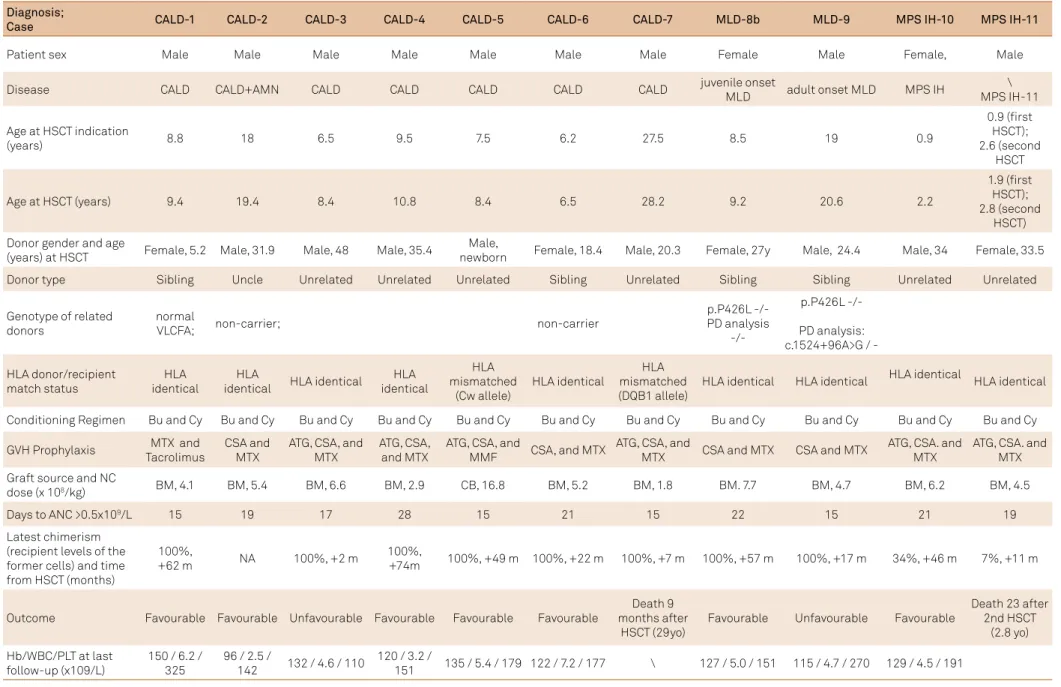
Table 1. Characteristics of the patients and donors, conditioning, composition of the graft, and engraftment.
Diagnosis;
Case CALD-1 CALD-2 CALD-3 CALD-4 CALD-5 CALD-6 CALD-7 MLD-8b MLD-9 MPS IH-10 MPS IH-11
Patient sex Male Male Male Male Male Male Male Female Male Female, Male
Disease CALD CALD+AMN CALD CALD CALD CALD CALD juvenile onset
MLD adult onset MLD MPS IH
\ MPS IH-11
Age at HSCT indication
(years) 8.8 18 6.5 9.5 7.5 6.2 27.5 8.5 19 0.9
0.9 (irst HSCT); 2.6 (second
HSCT
Age at HSCT (years) 9.4 19.4 8.4 10.8 8.4 6.5 28.2 9.2 20.6 2.2
1.9 (irst HSCT); 2.8 (second
HSCT) Donor gender and age
(years) at HSCT Female, 5.2 Male, 31.9 Male, 48 Male, 35.4
Male,
newborn Female, 18.4 Male, 20.3 Female, 27y Male, 24.4 Male, 34 Female, 33.5
Donor type Sibling Uncle Unrelated Unrelated Unrelated Sibling Unrelated Sibling Sibling Unrelated Unrelated
Genotype of related donors
normal
VLCFA; non-carrier; non-carrier
p.P426L -/-PD analysis
p.P426L
-/-PD analysis: c.1524+96A>G /
-HLA donor/recipient match status
HLA identical
HLA
identical HLA identical
HLA identical
HLA mismatched
(Cw allele)
HLA identical
HLA mismatched (DQB1 allele)
HLA identical HLA identical HLA identical HLA identical
Conditioning Regimen Bu and Cy Bu and Cy Bu and Cy Bu and Cy Bu and Cy Bu and Cy Bu and Cy Bu and Cy Bu and Cy Bu and Cy Bu and Cy
GVH Prophylaxis MTX and Tacrolimus
CSA and MTX
ATG, CSA, and MTX
ATG, CSA, and MTX
ATG, CSA, and
MMF CSA, and MTX
ATG, CSA, and
MTX CSA and MTX CSA and MTX
ATG, CSA. and MTX
ATG, CSA. and MTX
Graft source and NC
dose (x 108/kg) BM, 4.1 BM, 5.4 BM, 6.6 BM, 2.9 CB, 16.8 BM, 5.2 BM, 1.8 BM. 7.7 BM, 4.7 BM, 6.2 BM, 4.5
Days to ANC >0.5x10⁹/L 15 19 17 28 15 21 15 22 15 21 19
Latest chimerism (recipient levels of the former cells) and time from HSCT (months)
100%,
+62 m NA 100%, +2 m
100%,
+74m 100%, +49 m 100%, +22 m 100%, +7 m 100%, +57 m 100%, +17 m 34%, +46 m 7%, +11 m
Outcome Favourable Favourable Unfavourable Favourable Favourable Favourable
Death 9 months after
HSCT (29yo)
Favourable Unfavourable Favourable
Death 23 after 2nd HSCT
(2.8 yo)
Hb/WBC/PLT at last follow-up (x109/L)
150 / 6.2 / 325
96 / 2.5 /
142 132 / 4.6 / 110
120 / 3.2 /
151 135 / 5.4 / 179 122 / 7.2 / 177 \ 127 / 5.0 / 151 115 / 4.7 / 270 129 / 4.5 / 191
957 Sa ut e JAM e t al . Out comes aft er HSC T f or X -ALD
, MLD and MPS IH
Table 2. Baseline and follow-up results after HSCT: neurological and clinical indings, IQ, MRI, and biochemical markers.
Variable CALD-1 CALD-2 CALD-3 CALD-4 CALD-5 CALD-6 CALD-7 MLD-8b MLD-9 MPS IH-10 MPS IH-11
Genotype p.L628Q p.A232fsX64 p.A232fsX64 p.R518Q p.S358X p.A401W p.P560L
p.P426L/p.P426L; PD negative at
ARSA gene
p.P426L/ p.P426L; PD
negative at ARSA gene
p.W402X/ p.W402X p.W402X/ p.W402X
Age at onset of cerebral signs (years) 8.9 Addison-only at 7yo 18 6.5 9.5 Addison-only at 7yo
7.5 6.4 27.5 NA 19 0.8 0.9
Age at HSCT
(years) 9.4 19.4 8 10.9 8.4 6.5 28.2 9.1 20.6 2.1
1.9 (irst HSCT) and 2.8 (second HSCT)
Neurological and clinical examination indings at the time of HSCT Asymptomatic AMN without encephalic manifestations Ataxia, ankle clonus and attention deicit to auditory stimuli Asymptomatic Asymptomatic Brisk relexes on the left hemibody Hemiparesis, dysarthria and a conduction aphasia Intermittent strabismus Personality changes, seizures and cognitive loss. Babinski sign on right side
Recurrent upper air infections, hepatomegaly,
failure to thrive, developmental delay, and coarse facies. Severe PMD retardation: unable to walk
or talk.
Moderate PMD retardation:
at the time of irst HSCT, still starting to
walk and talk.
Age and status at last follow-up (time elapsed)
16yo (7 years after HSCT): No complains
Mild signs of neuropathy on
neurological examination
27yo (8 years and 9 months after HSCT) ): AMN without encephalic manifestations.
Progressive worsening of gait since 18yo
(needs canes for walking since 25yo). 9yo (12 months after HSCT) Severe GVHD. Marked worsening: aphasia, tetraparesis 16yo (5 years after HSCT): Stable
(clinically normal). NCS disclosed peripheral neuropathy. 13y10m (5 years after HSCT): Stable (asymptomatic) 8y8m (2 years after HSCT): Stable (brisk relexes on the left hemibody) 29yo (9 months after HSCT): Death due to Chronic GVH + opportunistic infection 9 months after HSCT
16yo (7 years and 6 months
after HSCT) Stable. Normal neurological examination. Focal seizures controlled with valproic acid. Subclinical
demyelinating polyneuropathy on nerve conduction studies, and slowed P40 latencies at somatosensory evoked responses. Normal visual evoked responses.
22 yo (18 months after HSCT)
Akinetic mutism
5y11m (3 years and 8months after HSCT) Stable. No further cognitive losses, Communication was nonverbal, though enough to her daily needs. Coarse facies, weight and height on percentile 3, gibbosity, and joint restriction in hands, arms and legs still present. No hepatomegaly on ultrasound. Thickening of the lealets of the aortic and mitral valves without
changes in transvalvular low on echocardiogram.
3 years: Death due opportunistic infection at 23 days after 2nd
HSCT
Baseline IQ Performance 103 Total 96
Performance
105 Total 91 NA
Performance 141 Total 140
Performance
99 Total 86 NA NA NA NA NA NA
Last IQ (time elapsed)
Performance 106 Total 105 (5 years later)
NA NA
Performance 136 Total 123 (2 years later)
Performance 108 Total 105 (2 years later)
NA NA NA NA NA NA
Baseline MRI: (CALD-Loes score, MLD-Eichler score)
Loes of 2 Loes of 6.5 Loes of 4.5 Loes of 4.5 Loes of 4 Loes of 3 Loes of 8 Eichler score of 10 Eichler score of 20
WM lesions in the periventricular and deep WM associated with mild ventricular enlargement
(Figure 4A-B)
958 Arq Neuropsiquiatr 2016;74(12):953-966
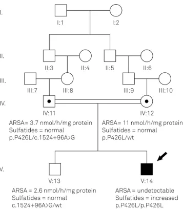
he appears as individual V-14 in Figure 4. Born from con-sanguineous parents (IV-11 and IV-12, Figure 4), MLD-9 was genotyped as p.P426L/p.P426L; PD - / - at ARSA
(pseudode-iciency variants not present). Mother was p.P426L/ - ; PD - /
-, and presented with normal ARSA activity and absent uri-nary sulfatides. Father was p.P426L/-; PD c.1524+96A>G/ -:
urinary sulfatides were absent, and ARSA activity was
com-patible with pseudodeiciency (Figures 3 and 4). he old -er broth-er of MLD-9, individual V-13 in Figure 4, was HLA identical to the patient, but was formerly excluded as a can-didate donor due to low ARSA activity. He was genotyped as p.P426L - / -; PD - c.1524+96A > G / -: absent urinary
sul-fatides, and ARSA activity were compatible with
pseudode-iciency. He was chosen to be the HSCT donor, performed
when the patient was 20 years, seven months old. Patient MLD-9 was discharged 79 days after HSCT. He present-ed with acute graft-versus-host disease (GVHD) with skin
and conjunctival involvement and no other major compli -cations. At the follow-up six months after HSCT, MLD-9 presented with akinetic mutism, being able to walk when forced to, and daily vomiting associated with cyclosporine use. In the last follow-up, 18 months after HSCT (22 years of age), the clinical picture had not changed. His family did not agree to perform MRI and nerve conduction follow-ups.
DISCUSSION
In the present study, we reported the neurologic pro-gression of Brazilian patients with CALD, MLD, and MPS-IH following HSCT.
hese diseases are associated with untreatable and pro -gressive neurological impairment, leading to vegetative states and death11,17,23,24. Pathophysiology varies according to
gene defect, and notable variability in age at onset and pro-gression rate occurs in all them. To date, HSCT is a unique therapy with potential to bring long-term survival and neu-rological stabilization for CALD and for some lysosomal stor-age disorders with brain involvement. Around 500 children with MPS-IH, 114 with MLD, and 465 with CALD underwent HSCT worldwide before 201211,12. In the same period, South
American HSCT procedures were described in seven CALD patients (the follow-ups of three of them were reported by Jardim et al.17), six MPS and one Gaucher disease type 125. Our
present report adds information about four new and three already-reported CALD patients, two new MLD, and two new MPS-IH patients, focusing on their neurological outcomes.
Nine out of our 11 patients (81%) were alive after a me -dian of 3.7 years after procedure: 6/7 CALD, 2/2 MLD and 1/2 MPS-IH. According to the literature, survival rates improved from 55-65% in early series6,8,25,26 to 85–95% in the most
re-cent series7,11. Our rates were comparable to the latter. A
28-year-old CALD man (CALD-7), and a three-year-old MPS-IH boy (MPS-MPS-IH-11) died 23 and 270 days, respectively, after
Las t MRI: (C ALD-L oes scor e, MLD-Eic hl er scor e) L oes o
f 1 (7y
aft
er HSC
T)
L
oes o
f 4 (6
year
s and 9
months aft er HSC T) L oes o
f 7 (12
months aft er HSC T) L oes o
f 6.5 (5
year
s and 6
monts aft er HSC T) L oes o
f 4 (5
year s aft er HSC T) L oes o
f 7 (2
year s aft er HSC T) L oes o
f 8 (2
months aft er HSC T) Eic hl er scor e o f 12 (5 y ear s aft er HSC T) NA Signiicant r educ tion o f periv
entricular and deep
WM l esions . Mil d v entricular enlar
gement also impr
o
ved
(Fig
ur
e 4C and D
) (3 y
ear s aft er HSC T) NA Baseline bioc hemical mark er s C26
: 2.2 µmol/L
C26 /22 r a tio 0 .0 4 C26 :3.47 µmol/L C26 /22 ra tio 0 .10 C26 :2.90 µmol/L C26 /22 r a tio 0 .1 5 C26 : 1 .94 µmol/L C26 /22 ra tio 0 .06 C26 : 4.33 µmol/L C26 /22 ra tio 0 .049 C26 : 3.54 µmol/L C26 /22 ra tio 0 .08 C26 : 4.53 µmol/L C26 /22 r a tio 0 .0 7 ARS A ac tivity in l euk oc yt es: 1 nmol/h/ mg pr o t Urinary sul fa tides ++ ARS A ac tivity in l euk oc yt es: unde tec tabl e Urinary sul fa tides: ++ α -L -idur onidase ac tivity in leuk oc yt es: 0 .2 nmol/h/ mg pr o
t Urinary GA
Gs: 532 ug / mg cr ea tinine α -L - idur onidase ac tivity in leuk oc yt es: 4. 0 nmol/h/ mg pr o
t Urinary
GA Gs: 638 ug / mg cr ea tinine Las t bioc hemical mark er s(time elapsed) C26 :2.9 µmol/L C26 /22 r a tio 0
.05 ( 6 y
ear s) NA C26 :5. 7 µmol/L C26 /22 r a tio 0
.16 (1 month)
C26 :1 .9 µmol/L C26 /22 r a tio 0
.13 (6 y
ear s) C26 : 1 .8 µmol/L C26 /22 r a tio 0
.17 (5 y
ear s) C26 : 0 .7 µmol/L C26 /22 ra tio 0 .12 (2 year s) NA ARS A ac tivity in leuk oc yt es: 9.5 ml/ mg o f pr o t/h Urinary sul fa tides
++ (6 y
ear s) ARS A ac tivity in l euk oc yt es:
5.3 nmol/h/ mg
pr o t α -L - idur onidase ac tivity in leuk oc yt es: 22 nmol/h/ mg pr o
t Urinary GA
Gs: 185 ug / mg cr ea tinine α -L - idur onidase ac tivity in leuk oc yt es: 12 nmol/h/ mg pr o
t Urinary
GA Gs:289 ug / mg cr ea tinine AMN: adr enomy el oneur opa th y; ARS A: arylsul fa tase A; C ALD: cer ebr al adr enol euk ody s tr oph y; HSC T: hema topoie tic s
tem cell tr
ansplanta tion; IQ: int ellig ence quo tient; GA G: gl ycosaminogl ycan; G VHD: gr aft-ver sus-hos t disease; MLD: me tac hr oma tic leuk ody s tr oph y; MPS: mucopol ysacc haridosis; MRI: magne tic resonance imaging; NA: no t a vailabl e; NCS: nerv e conduc tion s tudies; PMD: ps yc homo tor d; VL CF A: v ery l ong-c hain f a tty acid; WM: whit e ma tt er . nor mal l euk oc yt e ARS A ac
tivity =
5-20 nmol/h/ mg pr o t, nor mal l euk oc yt es α -L -idur onidase ac
tivity = 32-52 nmol/h/
mg pr
o
t,
nor
mal urinary GA
Gs l
e
vels =
79-256 ug
/mg cr
ea
tinine
959 Saute JAM et al. Outcomes after HSCT for X-ALD, MLD and MPS IH
Figure 1 . Loes score progression of seven CALD patients before and after HSCT. Individual dots indicate when brain magnetic resonance imaging was performed. The irst MRI was the image obtained at the time of HSCT indication. Time zero (continuous line) indicates when HSCT was performed. The 12-month interval (dotted line) indicates the time window for eficacy.
ALD7 ALD6 ALD5 ALD4 ALD3 ALD2 ALD1
Months
Loes score
84 72
60 48
36 24
12 0
-12 -24
10
9
8
7
6
5
4
3
2
1
0
A
B
D
C
960 Arq Neuropsiquiatr 2016;74(12):953-966
HSCT, from opportunistic infection, with or without GVHD. Multiple reasons probably contributed to their deaths. In common, both deceased patients received grafts from un-related donors, an already-known factor associated with in-creased risk of mortality26,27. Moreover, the CALD-7 patient
started with symptoms in adulthood, another factor asso-ciated with higher HSCT morbidity and mortality risks27.
Although mortality rates after HSCT in CALD of less than 5% have been reported7, none of the studies on HSCT
out-comes published so far included adult forms15. A report on
a 36-year-old CALD man showing cognitive deterioration in the previous year described the patient’s death from GVHD three months after HSCT28.
he remaining nine individuals of the present report had suicient follow-up time to be described. hey were six
CALD, two MLD and one MPS-IH patients.
One out of the six CALD patients surviving HSCT pre-sented with several complications from HSCT and worsen-ing of Loes score up to 12 months after HSCT. In contrast,
the other ive patients showed neurological stabilization
after transplantation: 1, 2, 4, CALD-5 and CALD-6 (Table 2 and Figure 1). Mean age and Loes scores at the procedure were 10.9 years and 4 points. Mean Loes scores changed to 5.9 after a mean time of four years
after their HSCTs; their neurological status was mostly sta-ble. One adolescent patient (CALD-2) presented
simulta-neously with adrenomyeloneuropathy (AMN) and CALD at the time of HSCT. Nine years later, AMN indings wors
-ened while CALD indings stabilized. All the other subjects
showed normal functioning at school and in daily life
activi-ties. Signs of peripheral neuropathy appeared ive and sev -en years after HSCT in two 16-year-old males (CALD-1 and
CALD-4), probably related to AMN evolution. hese results are in agreement with data showing that HSCT is efective in stabilizing CALD, but not AMN7,15.
herapeutic results in MLD transplanted patients were
completely divergent. While HSCT in patient MLD-8b seemed to be a success, it did not halt the disease progression in the adult form of patient MLD-9. Both patients shared the same
genotype: the main diference was the clinical picture at the
time of HSCT. Patient MLD-8b was a completely asymptom-atic girl (Eichler 10) transplanted at nine years old, and her neurological examination remained normal until the last
fol-low-up at 16 years of age. Although other juvenile onset pa -tients remained stable after HSCT29,30, the lack of association
of genotype p.P426L/p.P426L with age at onset complicates the evaluation of HSCT success. For instance, two untreated patients homozygous for p.P426L were still asymptomatic at
Figure 3. Brain FLAIR axial magnetic resonance images (MRI) of the transplanted patient MLD-8b and her non-transplanted brother MLD-8a. A, B and C. FLAIR axial MRIs from MLD-8a (non-tranplanted) patient. Images show hyperintense lesions diffusely distributed in the frontal, temporal, parietal and occipital white matter (WM) and in genu and splenium of the corpus callosum at 9 (A), 10 (B) and 12 years of age (C). There was a progressive worsening of brain atrophy during the follow up. White arrows highlight the progressive atrophy of the splenium of corpus callosum. D, E, F and G. FLAIR axial MRIs from MLD-8b patient show WM lesions distributed in the frontal, parietal and occipital lobes at 8 (D), 9 (E) (immediately before HSCT), 11 (F) and 13 years of age (G). Hyperintensities on the splenium of corpus callosum worsened until HSCT, with stabilization thereafter. The Eichler score was the same at 11 (F), 13 (G) and 14 years of age (image not shown).
8 years
9 years 10 years
11 years 9 years: HSCT
12 years of age
13 years of age Case MLD-8b
Case MLD-8a
A
B
C
961 Saute JAM et al. Outcomes after HSCT for X-ALD, MLD and MPS IH
ages 14 and 322; our patient MLD-9 was also homozygous for
p.P426L and had a disease onset at 19 years old. Late onset among p.P426L homozygotes points to longer follow-up
ob-servations to conirm the efectiveness of HSCT in these pa -tients. It is important to note that evolution of patient MLD8a
cannot be used as an untreated control for HSCT eicacy in
patient MLD8b. His report emphasizes the importance of ge-netic counseling for the early, presymptomatic diagnosis of MLD. Patient MLD8a came to our attention too late, when an
HSCT would not ofer a favorable outcome. However, his diag -nosis allowed the detection of patient MLD8b before disease onset. And indeed, two recent studies – a prospective cohort and case-control study – have raised evidence that HSCT is associated with a reasonable chance of disease stabilization if performed pre-symptomatically10,16.
Patient MLD-9 came for HSCT evaluation one year after the
beginning of cognitive losses. An HSCT was ofered to this patient
based on the assumption that adult forms progress slower than early-onset forms and that there was a chance that microglial cross correction could happen before the development of more se-vere neurological deterioration. However, follow-up revealed that this was not the case. Soon after HSCT, he developed an
akinet-ic mutism. Since therapeutakinet-ic efects are expected some months
(usually 12) after HSCT, MLD-9’s deterioration was probably re-lated to a rapid disease progression in an individual with already-extensive leukodystrophy on MRI at presentation. Recently, HSCT performed in four adult MLD patients was associated with stabili-zation four to 18 years after the procedure6. hree of them showed
high Eichler scores, from 18 to 26, before transplantation. Patient series have reported either stabilization31 or failure after HSCT32 in
juvenile and adult MLD patients. Better evidence favoring HSCT in juvenile MLD was recently obtained, when the neurological
follow-up of 24 transplanted patients was better than those of 41 non-transplanted patients10. Several factors were associated with
a better prognosis after HSCT: gross motor function preserved, IQ of at least 85, and age at onset older than four years.
Table 3. Factors potentially associated with unfavorable outcomes in the present patient series.
Factors All Favorable outcome
Unfavorable outcome
Death due to HSCT or worsening of clinical picture
11 7 4 (2 CALD, 1 MLD, 1 MPS-IH)
HSCT related donor 5 4 1
HSCT unrelated donor 6 3 3 (two deaths)
Adult-onset disease 2 0 2 (one MLD and one CALD)
Loes score at the time of HSCT, in
CALD 7 (2 to 8) 4 (2 to 6.5) 6.25 (4.5 and 8)
Mean (range)
Disease duration before HSCT *,
in years 0.94 (0 to 1.6) 0.8 (0 to 1.4) 1.2 (0.7 to 1.6)
Mean (range)
*Disease duration = the difference between age at onset of cerebral signs and age at HSCT (see Table 2). HSCT: Hematopoietic stem cell transplantation; CALD: Cerebral from of X-linked adrenoleukodystrophy ; MLD: Metechromatic Leukodystorphy ; MPS-IH: mucopolysaccharidosis type I-Hurler.
Figure 4. Pedigree of MLD-9 family. Proband is indicated with a black arrow. Squares indicate males and circle, females; dark ill indicates affected individuals. Arylsulfatase A (ARSA) activity in leukocytes (normal range: 5-20 nmol/hg/mg protein), urinary sulfatides, and ARSA genotypes are depicted for MLD-9 patient (V:14 individual), his parents IV:11 and IV:12, and his brother and bone marrow donor, V:13. Gene sequencing detected p.P426L as the pathogenic mutation in homozygosis in MLD-9 (V:14); both parents (IV:11 and IV:12) carried this mutation (were heterozygous), as expected. In addition, father (IV:11) and brother (V:13) were carriers of the variant associated with ARSA pseudodeiciency, c.1524+96A>G.
I.
II.
III.
IV.
V.
I:1 I:2
II:3 II:4 II:5 II:6
III:7 III:8 III:9 III:10
IV:11 IV:12
V:13 V:14
ARSA= 3.7 nmol/h/mg protein Sulfatides = normal
p.P426L/c.1524+96A>G
ARSA= 11 nmol/h/mg protein Sulfatides = normal p.P426L/wt
ARSA = 2.6 nmol/h/mg protein Sulfatides = normal
c.1524+96A>G/wt
962 Arq Neuropsiquiatr 2016;74(12):953-966
Early transplantation has been related to optimal long-term cognitive and language outcomes in MPS-IH9,14.
Although our MPS-IH survivor (patient MPS-IH-10) was a late transplanted patient, her follow-up four years later was very encouraging. Clinical improvement, despite relatively
low engraftment, should not be surprising. Studies in ibro
-blasts from MPS-I patients with diferent phenotypes and dif
-ferent genetic backgrounds conirmed that tiny diferences
in residual enzyme activity are related to important clinical
diferences33. Several studies on HSCT in MPS-IH reported
improvements in cardiopulmonary function, hearing and vi-sion, and preservation of neurocognitive function9,14,34. Some
of them documented that subsequent cognitive deteriora-tion might occur after HSCT, mostly if it is performed late35.
None of these studies, however, reported a reduction of brain
lesions on MRI after HSCT, such as happened in this patient (Figure 2). Since this patient was not receiving enzyme re-placement therapy, the clear reduction of periventricular and deep white matter lesions as well as the reduction in
ventric-ular dilatation should be related to an HSCT efect.
Our cohort is small and included three diferent diseases. Even so, we looked for diferences between patients with fa -vorable and unfa-vorable outcomes after HSCT - considering death due to HSCT or deterioration of the clinical picture
as the unfavorable outcomes (Table 3). In summary, better outcomes were obtained in patients whose HSCT was per-formed before adulthood, early in the disease clinical course, and from a related donor.
In conclusion, the present study showed short- to long-term results of neurological functions after HSCT in CALD, MLD and MPS-IH, in Brazil. Our survival rate was of 81% after a median of 3.7 years. While the results for the two adult forms were negative – one procedure-related death in a 28-year-old CALD patient and a rapid deterioration in a 20-year-old MLD patient – the response to HSCT was very satisfactory for seven of the other nine individuals. Better outcomes were obtained in those patients whose HSCT was performed early, as were the cases of CALD patients with a mean baseline Loes of 4, and
of the pre-symptomatic MLD individual. he outcome of the
late-transplanted MPS-IH patient was also very satisfactory, being accompanied by reductions in the lesions seen in MRI.
Acknowledgements
We are grateful for all patients and families who partic-ipated in this study. We thank Dr. Greg Pastores and Prof. Edwin Kolodny for the molecular studies on patient MLD 8.
References
1. Krall WJ, Challita PM, Perlmutter LS, Skelton DC, Kohn DB. Cells expressing human glucocerebrosidase from a retroviral vector repopulate macrophages and central nervous system microglia after murine bone marrow transplantation. Blood. 1994;83(9):2737-48.
2. Rauschka H, Colsch B, Baumann N, Wevers R, Schmidbauer M, Krammer M et al. Late-onset metachromatic leukodystrophy: genotype strongly inluences phenotype. Neurology. 2006;67(5):859-63. doi:10.1212/01.wnl.0000234129.97727.4d
3. Cable C, Finkel RS, Lehky TJ, Biassou NM, Wiggs EA, Bunin N et al. Unrelated umbilical cord blood transplant for juvenile metachromatic leukodystrophy: a 5-year follow-up in three affected siblings. Mol Genet Metab. 2011;102(2):207-9. doi:10.1016/j.ymgme.2010.10.002
4. Martin HR, Poe MD, Provenzale JM, Kurtzberg J, Mendizabal A, Escolar ML et al. Neurodevelopmental outcomes of umbilical cord blood transplantation in metachromatic
leukodystrophy. Biol Blood Marrow Transplant. 2013;19(4):616-24. doi:10.1016/j.bbmt.2013.01.010
5. Solders M, Martin DA, Andersson , Remberger M, Andersson T, Ringdén O et al. Hematopoietic SCT: a useful treatment for late metachromatic leukodystrophy. Bone Marrow Transplant. 2014;49(8):1046-51. doi:10.1038/bmt.2014.93
6. Beam D, Poe MD, Provenzale JM, Szabolcs P, Martin PL, Prasad V et al. Outcomes of unrelated umbilical cord blood transplantation for X-linked adrenoleukodystrophy. Biol Blood Marrow Transplant. 2007;13(6):665-74. doi:10.1016/j.bbmt.2007.01.082
7. Miller WP, Rothman SM, Nascene D, Kivisto T, DeFor TE, Ziegler RS et al. Outcomes after allogeneic hematopoietic cell transplantation for childhood cerebral adrenoleukodystrophy: the largest single-institution cohort report. Blood. 2011;118(7):1971-8. doi:10.1182/blood-2011-01-329235
8. Peters C, Shapiro EG, Anderson J, Henslee-Downey PJ, Klemperer MR, Cowan MJ et al. Hurler syndrome: II. Outcome of HLA-genotypically identical sibling and HLA-haploidentical related donor bone marrow transplantation in ifty-four children. Blood. 1998;91(7):2601-8.
9. Ru MH, Boelens JJ, Das AM, Jones SA, van der Lee JH, Mahlaoui N et al. Enzyme replacement therapy and/or hematopoietic stem cell transplantation at diagnosis in patients with mucopolysaccharidosis type I: results of a European consensus procedure. Orphanet J Rare Dis. 2011;6(1):55. doi:10.1186/1750-1172-6-55
10. Groeschel S, Kühl JS, Bley AE, Kehrer C, Weschke B, Döring M et al. Long-term Outcome of Allogeneic Hematopoietic Stem Cell Transplantation in Patients With Juvenile Metachromatic Leukodystrophy Compared With Nontransplanted
Control Patients. JAMA Neurol. 2016;73(9):1133-40. doi:10.1001/jamaneurol.2016.2067
11. Boelens JJ, Prasad VK, Tolar J, Wynn RF, Peters C. Current international perspectives on hematopoietic stem cell transplantation for inherited metabolic disorders. Pediatr Clin North Am. 2010;57(1):123-45. doi:10.1016/j.pcl.2009.11.004
12. Musolino PL, Lund TC, Pan J, Escolar ML, Paker AM, Duncan CN et al. Hematopoietic stem cell transplantation in the leukodystrophies: a systematic review of the literature. Neuropediatrics.
2014;45(3):169-74. doi:10.1055/s-0033-1364179
13. Aldenhoven M, Boelens JJ, Koning TJ. The clinical outcome of Hurler syndrome after stem cell transplantation. Biol Blood Marrow Transplant. 2008;14(5):485-98. doi:10.1016/j.bbmt.2008.01.009
963 Saute JAM et al. Outcomes after HSCT for X-ALD, MLD and MPS IH
15. Engelen M, Kemp S, Visser M, Geel BM, Wanders RJ, Aubourg P et al. X-linked adrenoleukodystrophy (X-ALD): clinical presentation and guidelines for diagnosis, follow-up and management. Orphanet J Rare Dis. 2012;7(1):51. doi:10.1186/1750-1172-7-51
16. Boucher AA, Miller W, Shanley R, Ziegler R, Lund T, Raymond G. Long-term outcomes after allogeneic hematopoietic stem cell transplantation for metachromatic leukodystrophy: the largest single-institution cohort report. Orphanet J Rare Dis. 2015;10(1):94. doi:10.1186/s13023-015-0313-y
17. Jardim LB, Silva AC, Blank D, Villanueva MM, Renck L, Costa ML et al. X-linked adrenoleukodystrophy: clinical course and minimal incidence in South Brazil. Brain Dev. 2010;32(3):180-90. doi:10.1016/j.braindev.2009.02.002
18. Moser HW, Moser AB. Measurement of saturated very long chain fatty acid in plasma. In: Hommes FA, editor. Techniques of diagnostic human biochemical genetics. Wiley, New York. 1991, pp. 177-191.
19. Virgens MY, Siebert M, Bock H, Burin M, Giugliani R, Saraiva-Pereira ML. Genotypic characterization of Brazilian patients with infantile and juvenile forms of metachromatic leukodystrophy. Gene. 2015;568(1):69-75. doi:10.1016/j.gene.2015.05.016
20. Loes DJ, Hite S, Moser H, Stillman AE, Shapiro E, Lockman L et al. Adrenoleukodystrophy: a scoring method for brain MR observations. AJNR Am J Neuroradiol. 1994;15(9):1761-6.
21. Eichler F, Grodd W, Grant E, Sessa M, Bifi A, Bley A et al. Metachromatic leukodystrophy: a scoring system for brain MR imaging observations. AJNR Am J Neuroradiol. 2009;30(10):1893-7. doi:10.3174/ajnr.A1739
22. Tutschka PJ, Copelan EA, Klein JP: Bone marrow transplantation for leukemia following a new busulfan and cyclophosphamide regimen. Blood. 1987;70(5):1382-8.
23. Cartier N, Aubourg P. Hematopoietic stem cell gene therapy in Hurler syndrome, globoid cell leukodystrophy, metachromatic leukodystrophy and X-adrenoleukodystrophy. Curr Opin Mol Ther. 2008;10(5):471-8. doi:
24. Jardim LB, Villanueva MM, Souza CF, Netto CB. Clinical aspects of neuropathic lysosomal storage disorders. J Inherit Metab Dis. 2010;33(4):315-29. doi:10.1007/s10545-010-9079-5
25. Lange MC, Teive HAG, Troiano AR, Bitencourt M, Funke VA, Setúbal DC et al. Bone marrow transplantation in patients with storage diseases: a developing country experience. Arq Neuropsiquiatr. 2006;64(1):1-4. doi:10.1590/S0004-282X2006000100001
26. Peters C, Charnas LR, Tan Y, Ziegler RS, Shapiro EG, DeFor T et al. Cerebral X-linked adrenoleukodystrophy: the international hematopoietic cell transplantation
experience from 1982 to 1999. Blood. 2004;104(3):881-8. doi:10.1182/blood-2003-10-3402
27. Boelens JJ, Aldenhoven M, Purtill D, Ruggeri A, Defor T, Wynn R et al. Outcomes of transplantation using various hematopoietic cell sources in children with Hurler syndrome after myeloablative conditioning. Blood. 2013;121(19):3981-7. doi:10.1182/blood-2012-09-455238
28. Fitzpatrick AS, Loughrey CM, Johnston P, McKee S, Spence W, Flynn et al. Haematopoietic stem-cell transplant for adult cerebral adrenoleukodystrophy. Eur J Neurol. 2008;15(3):e21-2. doi:10.1111/j.1468-1331.2007.02048.x
29. Ding X-Q, Bley A, Kohlschütter A, Fiehler J, Lanfermann H. Long-term neuroimaging follow-up on an asymptomatic juvenile metachromatic leukodystrophy patient after hematopoietic stem cell transplantation: evidence of myelin recovery and ongoing brain maturation. Am J Med Genet A. 2012;158A(1):257-60. doi:10.1002/ajmg.a.34389
30. Krägeloh-Mann I, Groeschel S, Kehrer C, Opherk K, Nägele T, Handgretinger R et al. Juvenile metachromatic leukodystrophy 10 years post transplant compared with a non-transplanted cohort. Bone Marrow Transplant. 2013;48(3):369-75. doi:10.1038/bmt.2012.155
31. Egmond ME, Pouwels PJW, Boelens JJ, Lindemans CA, Barkhof F, Steenwijk MD et al. Improvement of white matter changes on neuroimaging modalities after stem cell transplant in metachromatic leukodystrophy. JAMA Neurol. 2013;70(6):779-82. doi:10.1001/jamaneurol.2013.629
32. Smith NJ, Marcus RE, Sahakian BJ, Kapur N, Cox TM. Haematopoietic stem cell transplantation does not retard disease progression in the psycho-cognitive variant of late-onset metachromatic leukodystrophy. J Inherit Metab Dis. 2010;33(s3):S471-5. doi:10.1007/s10545-010-9240-1
33. Oussoren E, Keulemans J, Diggelen OP, Oemardien LF, Timmermans RG, Ploeg AT et al.. Residual α-L-iduronidase activity in ibroblasts of mild to severe Mucopolysaccharidosis type I patients. Mol Genet Metab. 2013;109(4):377-81. doi:10.1016/j.ymgme.2013.05.016
34. Prasad VK, Kurtzberg J. Transplant outcomes in
mucopolysaccharidoses. Semin Hematol. 2010;47(1):59-69. doi:10.1053/j.seminhematol.2009.10.008
964 Arq Neuropsiquiatr 2016;74(12):953-966
CALD Patients
Patient CALD 1, Family A
CALD1, male (originally reported as patient G in Lange et al.25), presented with Addison disease at seven years of
age. X-ALD diagnosis was made by very long-chain fatty acid (VLCFA) analysis at eight years old and he was the index patient
of his family. he irst magnetic resonance imaging (MRI) was
normal; the second exam performed at eight years,10 months disclosed white matter (WM) lesions in the internal capsules
and anterior portions of the midbrain (Loes score of 2). he
neurological examination and IQ evaluation were both normal (Table 2). Molecular analysis detected a p.L628Q mutation at
ABCD1 of CALD1 and his mother. His ive-year-old sister had
a normal VLCFA proile, and was the only HLA identical rel
-ative detected in the family. he staf team, parents and the
girl agreed, and she was the donor for the HSCT carried out
when CALD1 was nine years, ive months. He was discharged 40 days after an almost uneventful period at the hospital. he
last follow-up was done at 16 years of age: CALD1 was per-forming normally at school and presented with no complaints. At neurological examination, there was a strength grade 4/5 in both anterior tibial muscles; hammer toes, absent plantar responses; and reduced vibratory perception (lasting 11 and 9
seconds on toes). All other indings were normal. An MRI per -formed after HSCT showed a reduction of the lesions in the brainstem (Loes score of 1; Figure 1). Biochemical markers were unchanged (Table 2).
Patient CALD 2, Family B
CALD2, male, 18 years old, came for evaluation as the only brother of a previous CALD patient. CALD2 presented with loss of strength in lower limbs in the previous four months. Brisk
re-lexes and a peripheral axonal neuropathy on neurophysiology pointed to an adrenomyeloneuropathy (AMN) phenotype. he VLCFA analysis conirmed X-ALD; ACTH levels were normal. he p.A232fsX64 mutation was detected in ABCD1. His brain
MRIs disclosed lesions at the splenium of corpus callosum; in periventricular and central WM of parieto-occipital and right frontal lobes; and in the internal capsules. Loes scores evolved from 5.5 to 6.5 in a seven-month interval. A paternal uncle was HLA identical to CALD2 and was his bone marrow donor. CALD2 was discharged 33 days after HSCT, and returned only
once, fourteen months after transplantation. Neurological ex -amination had not changed, but follow-up MRIs showed im-provements (Loes score of 4.5 and 4, after four and 16 months post-HSCT) (Table 2 and Figure 1). CALD2 and his father were very frustrated because HSCT did not improve the impairments
due to AMN, and never came back to follow-up visits. he last
contact was in 2013 by phone: CALD2 was 25 years old and
wanted to review the genetic risks to his ofspring.
Patient CALD 3, Family B
A cousin of the former patient CALD2, CALD3, male, was found to carry the same mutation - p.A232fsX64 – at ABCD1.
Followed with periodic brain MRIs since ive years old, he
was diagnosed as the third CALD in the family. At six years, six months, CALD3 presented with ankle clonus and
atten-tion deicit to auditory stimuli, plus hyperactivity. His for -mer MRIs were normal. After a transient loss of follow-up, a new MRI was obtained at eight years old and showed bi-lateral WM lesions in the medial geniculate bodies, bi-lateral
lemniscus, brachium of inferior colliculus and in the projec
-tion ibers of the brainstem; and of the left internal capsule
(Loes score of 4.5). An HSCT from an unrelated donor was performed in September 2014. Two months later, CALD3 presented with a hemorrhagic cystitis and in the following month, GVHD with gastrointestinal and skin manifesta-tion, refractory to treatment. Brain MRI, three months after HSCT, showed a mild worsening of lesions (Loes score of 5). In April 2015, he was still in hospital, treating recurrent infec-tions (urinary and intestinal). One year after HSCT, CALD3 presented with a marked worsening, with mutism, spasticity and tetraparesis; he was discharged with a Loes score of 7.
Patient CALD 4, Family C
CALD4, male (reported as patient H in Lange et al25),
belongs to a large X-ALD family where mutation p.R518Q segre-gates. Followed as an asymptomatic carrier with normal MRIs
since ive years of age, CALD4 turned into an Addison-only phenotype when he was seven years old. he irst WM lesions
appeared when he was nine years, six months, in the right peri-ventricular and central parieto-occipital lobe WM (Loes score of 1), when CALD was diagnosed. An unrelated donor was found 16 months later: by this time, the Loes score had pro-gressed to 4.5 points and neurological examination disclosed
brisk patellar relexes, only. He underwent HSCT at 10 years, 10
months. CALD4 was discharged at day 36 after HSCT and no
complications were reported. Clonus, brisk relexes in lower
limbs, and a Babinski sign were observed at 14 years of age. At 16 years old, the neurological examination returned to normal;
an ENMG disclosed a mixed axonal and demyelinating periph
-eral neuropathy. he MRI lesions were stabilized (Loes score of 6.5) during this ive-year period after the bone marrow trans -plant (BMT) (Table 2 and Figure 1).
Patient CALD 5, Family D
CALD5, male (reported as patient I in Lange et al.25),
was admitted to our hospital at seven years, six months to
investigate adrenal insuiciency. Very long-chain fatty acid analysis conirmed the diagnosis of X-ALD. he neurologi -cal examination was normal, whereas the MRI disclosed WM lesions in the splenium of corpus callosum and in the
965 Saute JAM et al. Outcomes after HSCT for X-ALD, MLD and MPS IH
periventricular and central WM of the parieto-occipital lobes (Loes score of 3). Molecular studies detected the p.S358X mu-tation of the ABCD1 gene of CALD5 and in his mother; no
other relative carried this mutation. An umbilical cord HSCT
was undertaken when he was eight years, ive months. he
patient was discharged 42 days after HSCT, with no compli-cations. Five years after the BMT, the neurological examina-tion was normal and the MRI lesions were stable (Loes score of 5) (Table 2 and Figure 1).
Patient CALD 6, Family E
CALD6, male, was a brother of an index-patient with an advanced form of CALD (Loes score of 13). At the time of the index-patient diagnosis, CALD6 was two years old. His
VLCFA proile was characteristic of X-ALD, and his molecu -lar studies showed that he carried the mutation found in
his family – p.A401W. His MRIs were normal up to ive years old. On follow-up at six years old, brisk relexes were found on his left side. he MRI at six years, three months showed
lesions in the genu of corpus callosum, and in central and periventricular WM of the frontal lobes (Loes score of 3). An HSCT was carried out when he was six years, six months old: an older sister, HLA identical and a non-carrier of the p.A401W mutation, was the donor. CALD6 was discharged 50 days after HSCT, with no complications. A new MRI per-formed eight months later showed new lesions in the sple-nium of corpus callosum, internal capsules subcortical
frontal lobes and in projection ibers of the brainstem (Loes
score of 8). Sixteen months after the BMT, at eight years old,
a third MRI gave a Loes score of 9; brisk relexes on his left side were still present, plus a complaint of memory diicul -ties. Two years after BMT, at eight years, eight months ( July, 2014), a forth MRI showed a reduction of the WM lesions
and a Loes score of 7. Memory diiculties were no longer
present (Table 2 and Figure 1).
Patient CALD 7, Family F
CALD7, male, a successful systems analyst, presented with loss of coordination and strength in his right arm when he was 27 years old. Initially diagnosed as an ischemic event, the clinical picture progressed to severe hemiparesis in the
following months. Mental functioning was normal. he MRI
showed lesions in periventricular and central WM of the left temporal lobe, and bilateral periventricular and deep WM lesions in the parieto-occipital lobes, body of corpus callo-sum, left basal ganglia, left internal capsule, left Meyer’s loop,
left projection ibers of the brainstem, and global atrophy (Loes score of 8). he VLCFA analysis diagnosed X-ALD; he
and several relatives carried the p.P560L mutation in ABCD1.
An unrelated, mismatched donor was found nine months af-ter the diagnosis of CALD. Immediately before HSCT, the pa-tient presented with hemiparesis, dysarthria and a
conduc-tion aphasia, without any further cognitive deterioraconduc-tion. he
Loes score was the same in a new MRI. After HSCT, several
complications followed, including seizures, chronic GVHD and hemorrhagic cystitis. Cognitive functions were
main-tained. he patient died nine months after HSCT from an op -portunistic infection (Table 2 and Figure 1).
MPS IH Patients
Patient MPS-IH10, Family I
MPS-IH10, female, was referred for genetic evaluation at eight months of age due to recurrent upper air infec-tions, hepatomegaly, failure to thrive, developmental delay and coarse facies. MPS-IH10 was abandoned by her par-ents at birth and lived in an orphanage. At physical exami-nation, weight and length were under percentile 3 and the
cephalic perimeter under percentile 10. here was mild fa -cial coarsening, enlarged tongue, thoracolumbar gibbus, hepatomegaly and moderate development delay (steady head, good interaction, social laughter, did not sit with-out support). A CT scan showed generalized cortical
atro-phy. he investigation disclosed high levels of urinary gly -cosaminoglycans (GAGs) - 601 ug/mg creat (normal range: 133-274) – and low activity of α-L-iduronidase (IDUA) in leukocytes – 0.17nmol/h/mg protein (normal range: 32-56), both compatible with the diagnosis of MPS I. Sequencing of
IDUA revealed that MPS-IH10 was homozygous for p.W402X,
a mutation previously related to the Hurler type of MPS I or MPS-IH (Voskoboeva et al 1998; Terlato et al 2003)36,37. She
started to be evaluated for HSCT. At 16 months of age, en-zyme replacement therapy with laronidase was started with improvement in her clinical status and respiratory condition. At 24 months of age, brain MRI showed T2/FLAIR hyperin-tensities, mainly in the periventricular and deep WM asso-ciated with mild ventricular enlargement (Figure 4A and B). By that time, auditory evoked potentials showed bilateral hearing loss. An unrelated donor was found and HSCT was performed at 27 months of age. MPS-IH10 was discharged 195 days after HSCT. She presented with acute GVHD with skin and gastric involvement. Enzyme replacement therapy was discontinued after HSCT. Four years after the HSCT, MPS-IH10 was in quite good general condition, very active, walk-ing without support. Communication was nonverbal, though
enough for her daily needs. he typical face, weight and height on percentile 3, gibbosity, and joint restriction in the hands, arms and legs were still present. here was no hepa -tomegaly on ultrasound. Echocardiogram showed thickening
of the lealets of the aortic and mitral valves without changes in transvalvular low. Diameters of interventricular and atri -al septum were norm-al. Although outside the norm-al range, she has shown progressive improvement of motor skills, lan-guage and expression. Since HSCT, she has not shown any
cognitive loss. No signiicant skeletal improvement was ob -served in this four-year interval. A clear improvement in the brain lesions appeared on MRI performed three years after
966 Arq Neuropsiquiatr 2016;74(12):953-966
WM lesions. he mild ventricular enlargement was also re -duced (Table 2 and Figure 4C and D).
Patient MPS-IH11, Family J
MPS-IH11, male, was referred for genetic evaluation at 11 months of age due to recurrent upper air infections and developmental delay. Urinary GAGs of 481 ug/mg creat and IDUA activity of 0.18 nmol/h/mg protein in leukocytes estab-lished the diagnosis of MPS I. Molecular analysis disclosed a p.W402X homozygous genotype of IDUA, classically
associ-ated with MPS-IH. MPS-IH11 was the irst child from consan -guineous parents. On examination, a clear-cut MPS pheno-type was present, with gingival hypertrophy, impacted teeth,
restriction of the extension of ingers, large umbilical hernia,
and hepatomegaly. Enzyme replacement therapy with laroni-dase was started at 17 months of age; by this time, he could stand without support, but was unable to walk or to speak. Brain atrophy, ventriculomegaly and mega cisterna magna
were found on MRI. hickening of the periodontal tissues de
-termined spinal cord stenosis at C1-C2 level. he irst HSCT
was done at 22 months of age. Chimerism was progressively lost, with 73, 58 and 10% after one, two and eight months.
he IDUA activities also fell, with 40, 42 and 12 nmol /h/mg protein after one, two and ive months, respectively. Due to