DOI: 10.1590/0004-282X20160024 TRICK OF THE TRADE
Orbitozygomatic craniotomy in three pieces:
tips and tricks
Craniotomia orbitozigomática em três peças: dicas e truques
Feres Chaddad-Neto1,2, Hugo Leonardo Doria-Netto1,2, José Maria Campos-Filho1,2, Mateus Reghin-Neto2,
Marcos Devanir Silva-Costa1, Evandro Oliveira2,3
he orbitozygomatic access was irstly described by Hakuba, in 19861,2 as the result of small changes to the fronto
-temporosphenoidal craniotomy3,4,5.
Pterional craniotomy, popularized by Yasargil in 1976, is the most used surgical route in neurosurgery6,7. It exposes
transylvian and lateral subfrontal views8,9.
In 1961, Drake introduced the subtemporal approach10,11
which ofers a lateral view of the interpenducular fossa by re
-tracting the temporal lobe superiorly.
he temporopolar approach was irstly described by Sano in 198012. It consists in pulling back the temporal pole,
creating and enlarging an anterolateral view of the interpe
-duncular fossa.
he addition of transylvian and subfrontal views to the subtemporal and temporopolar views is of greatest impor
-tance when a neurosurgeon needs to expose the interpedun
-cular cistern region or the entire temporal lobe. he pretem
-poral approach, described by Oliveira et al.13,14, Tedeschi et al.15
and Chaddad Neto et al.16 combines the advantages of all
these approaches in one craniotomy. his approach expos
-es the entire temporal lobe in order to ofer the transylvian and lateral subfrontal views, from the pterional craniotomy, as well as subtemporal and temporopolar views to access the interpeduncular fossa.
he orbitozygomatic approach combines the advan
-tages of the pterional and the pretemporal approaches but
1Universidade Federal de São Paulo (UNIFESP), Departamento de Neurologia, Sao Paulo SP, Brazil;
2Real e Benemérita Associação Portuguesa de Beneficência, Instituto de Ciências Neurológicas (ICNE), Laboratório de Microneurocirurgia, Sao Paulo SP, Brazil; 3Universidade Estadual de Campinas (UNICAMP), Faculdade de Ciências Médicas, Departamento de Neurologia, Campinas SP, Brazil.
Correspondence:Hugo Leonardo Doria-Netto; Al. dos Jurupis, 777 / apt 141; 04088-002 São Paulo SP, Brazil; E-mail: [email protected]
Conflict of interest: There is no conflict of interest to declare.
Received 19 May 2015; Received in final form 25 October 2015; Accepted 19 November 2015.
ABSTRACT
Objective: Didactically describe the orbitozygomatic craniotomy made in three pieces. Method: This approach was performed, from 2002 to 2011, in 49 patients admitted at Beneficência Portuguesa of São Paulo Hospital. Results: Twenty-seven patients had vascular lesions and twenty-two suffered for intracranial skull base tumors. The vascular lesions varied from cavernous angiomas inside the mesencephalum, high bifurcation basilar tip aneurysms, superior cerebellar arteries aneurysms and arteriovenous malformations in the interpeduncular cistern. Skull base tumors as meningiomas, interpeduncular hamartomas and third ventricle floor gliomas were among the neoplastic lesions approached. We had no permanent injuries and minimal transient complications had occurred. Conclusion: It is a descriptive text, organized in the sequence of the main stages in which such a craniotomy is performed, describing in details the technique in which this group of evolutionarily authors came to accomplish the task.
Keywords: orbitozygomatic, craniotomy, neurosurgery, microsurgery.
RESUMO
Objetivo: Descrever didaticamente a craniotomia orbitozigomática realizada em três peças. Método: Esse acesso foi realizado em 49 pacientes, de 2002 a 2011 em pacientes admitidos no Hospital Beneficência Portuguesa de São Paulo. Resultados: Vinte e sete pacientes apresentavam lesões vasculares e vinte e dois sofriam de tumores da base do crânio. As lesões vasculares variaram entre angiomas cavernosos do mesencéfalo, aneurismas topo da artéria basilar com bifurcações altas, aneurismas da artéria cerebelas superior a malformações arteriovenosas na cisterna interpeduncular. Tumores da base do crânio como meningeomas, hamartomas interpedunculares e gliomas no assoalho do terceiro ventrículo estão entre as lesões neoplásicas abordadas. Nós não tivemos sequelas definitivas e tivemos mínimas complicações temporárias. Conclusão: Trata-se de um texto descritivo, dividido conforme as principais etapas da realização desta craniotomia, o qual descreve com detalhes a técnica com que o presente grupo de autores evolutivamente veio a realizá-la.
improves the angle of microscope view from inferior to su
-perior. Hence, it provides the best view for brain diseases at optic chiasm, third ventricle loor, high carotid artery bifur
-cation, high basilar tip artery bifur-cation, anterior commu
-nicating artery aneurysms pointed posteriorly, and any oth
-er lesions at sellar and parasellar regions or int-erpeduncular region. his approach can even access the sphenoidal, fron
-tal, and ethmoidal sinuses, the components of the orbit, the cavernous sinus and the remaining structures of the middle cranial fossa.
METHOD
Since 2002 to 2011 we have performed forty-nine (49) or
-bitozygomatic (three pieces) approaches to patients admit
-ted at Beneicência Portuguesa of São Paulo Hospital. Among these patients, 27 (55.1%) had vascular le
-sions and twenty-two (44.9%) suffered for intracranial skull base tumors.
he vascular lesions varied from cavernous angiomas (3-11.1%) inside the mesencephalum, high basilar tip an
-eurysms (12-44.4%), superior cerebellar arteries an-eurysms (7-25.9%) and to arteriovenous malformations placed at in
-terpeduncular cistern (5-18.5%).
he skull base tumors varied from olfactory groove me
-ningiomas (6-27.2%), petroclival me-ningiomas (5-22.7%), proximal sphenoidal wing meningiomas (9-40.9%), interpe
-duncular region hamartomas (1-4.54%) and third ventricle loor gliomas (1-4.54%).
he same surgical team operated all the patients.
Description of orbitozygomatic craniotomy
Positioning: he patient is placed supine. he head should
be held by a three-pin skull ixation device (Mayield or Sugita model). he ipsilateral pin should be set on the mastoid re
-gion, while the two contralateral pins should be on the con
-tralateral superior temporal line, above the temporal muscle, that should not be transixed17.
Head positioning comprises a sequence of four move
-ments for: lifting, extention, rotation and torsion. Once lift
-ing, the head is positioned at a level above the right atrium; extension and rotation depend on the condition being oper
-ated; and in torsion, the angle formed by the head, neck and shoulder should be higher, so as to ofer the surgeon to be in a closer lateral position with respect to the surgical area, in order to become parallel to the Sylvian issure. Most of times, intending to expose higher brain pathologies, we should po
-sition the head in more extension to provide inferior to supe
-rior microsurgical view18.
Care must be taken so that the jugular veins remain compression-free throughout surgery, to prevent delay of venous emptying, brain swelling, and increased bleeding in the operating ield.
Trichotomy: Patient’s hair should be combed with a
brush used for hand scrubbing that has been soaked in de
-tergent solution (chlorhexidine or polyvinylpyrrolidone io
-dine), so as to facilitate shaving, that should be performed up to 2cm from the region of the surgical incision. Shaving just prior to surgery allows for better ixation of ields, re
-duction of infection risks, and better ixation of the bandage after surgery.
Marking, antisepsis and scalp incision: We should start at
inferior edge of the zygomatic arch, anterior to the tragus, and extend to the hemi-pupillary contralateral line in the frontal region, behind the hairline. he marked area anterior to the tragus should not be too anterior, so as to prevent sectioning the supericial temporal artery and the frontal branch of the facial nerve located anteriorly to that artery (Figure 1).
he antisepsis should be carried out with alcoholic solu
-tion of polyvinylpyrrolidone-iodine or chlorhexidine. Scalp incision should be performed and the use of bipo
-lar coagulation helps to avoid bleeding. he placement of wet gauze and later traction of the scalp lap can spare the use of haemostatic clips and speciic staples for this purpose8,16,17,18.
Interfacial dissection, zygomatic osteotomy, section and dis-placement of the temporalis muscle: he interfacial dissection
of the temporalis muscle, as originally described by Yasargil, is speciically intended to preserve the front temporal branch
of the facial nerve and reduce postoperative cosmetic chang
-es r-esulting from the surgical wound8,16. We had four patients
(8,1%) who evolved with temporary (about two months) frontal branch facial nerve palsy. None of them had perma
-nent palsy.
he temporalis muscle is composed of two parts: an out
-er part which originates in the sup-erior temporal line and in
-serts onto the coronoid process of the jaw-bone; and a deeper part that has its origin along the surface of the temporal squa
-ma and inserts onto the temporal crest of the jawbone. he temporalis muscle is covered by a supericial fascia, which, in turn, consists of two layers (the supericial and deep layers). hese are separated in their anterior portion by a pad of adi
-pose tissue, and by a deep fascia that is more attached to the skull and protects, both, its vasculature (anterior, interme
-diate and posterior deep temporal arteries, branches of the maxillary artery) and its innervations (temporal branches of the mandible branches of the trigeminal nerve).
Dissection of the supericial fascia should be performed vertically, starting from the superior temporal line, 1.5 to 2cm posteriorly to the superior rim of the orbit to the poste
-rior root of the zygomatic arch, with the aid of a #10 scalpel. he removal of the surface layer of the supericial tempo
-ral fascia and its underlying fat pad with the use of a hook placed at its center point facilitates completion of the dis
-section, whose basal layer is hindered by the presence of temporal nerves and vessels. With the most basal remov
-al of the surface layer and the fat pad, good visu-alization of the deep muscular portion is achieved. After the fascia is relected anteriorly, the zygomatic bone with its frontal and temporal process is well exposed. he superior orbital rim and the supraorbital foramen and nerve are identiied. he supraorbital nerve is freed within the orbit with great care not to injure the periorbita. he periorbita is usually thinnest and weakest at the exit point of the supraorbital nerve from the orbit and at the level of the frontozygomat
-ic suture, wh-ich corresponds to the position of the lacrimal gland within the orbit (Figures 2 and 3).
After the zygomatic process of the temporal bone (zy
-gomatic arch) and the zy-gomatic bone with its frontal and temporal process are well exposed, we perform the zygo
-matic osteotomy. he edge of the inferolateral temporal lobe corresponds externally to the upper edge of the zygomatic arch. From the lower dislocation of the zygomatic arch, it is possible to inferiorly move the temporal muscle facilitating optimal exposure of the loor of the middle fossa. Using the number 1 Penield dissector, we can move the muscle and connective tissue adherent to the zygomatic arch. he oste
-otomy is performed to the zygomatic arch anteriorly to the temporal-zygomatic suture, in a vertical cut and posteriorly previously to the temporomandibular joint, in a oblique cut. It is important to preserve the insertion of the masseter mus
-cle into the inferior edge of zygomatic arch which should be inferiorly dislocated.
Afterwards the microsurgery, zygomatic arch must be re
-constructed using mini-plates and screws or tied down with nylon at the edges (Figure 4).
he dissection and detachment of the temporalis mus
-cle are then performed in two stages. Initially we use the mo
-nopolar electrosurgery pencil (in the coagulation mode in
-tending to avoid bleeding) for the transversal section of the upper portion of the temporal muscle. he second stage con
-sists of detaching of the deep muscular fascia of the skull. Afterwards, the temporal muscles must be moved away, towards the posterior inferior section, with the aid of three hooks. he insertion of the masseter muscle in the lower por
-tion of the zygomatic arch is preserved when it is dislocated inferiorly toward the infratemporal fossa.
Craniotomy and orbital osteotomy: We perform a pretem
-poral craniotomy. he goal is to provide a basal and wide ex
-posure of the temporal lobe and Sylvian issure. We must ex
-pose the inferior frontal gyrus and a portion of the middle frontal gyrus, and the superior, middle and inferior temporal Figure 2. The interfacial dissection of the temporalis muscle. Dissection of the superficial fascia from the superior temporal line to the posterior root of the zygomatic arch. (A) temporalis muscle, (B) Superior temporal line, (C) Posterior root of zygomatic arch.
gyri allowing for access through the transylvian, lateral sub
-frontal, temporopolar and subtemporal views.
he pretemporal craniotomy should be performed start
-ing from three points of trepanation. he irst trepanation must be set between the superior temporal line and the fron
-tozygomatic suture of the external orbital process; the sec
-ond trepanation is performed on the most posterior exten
-sion of the superior temporal line and the third one should be made on the most inferior portion of the squamous part of the temporal bone. Since the lesser wing of the sphenoid bone is situated internally between the irst and third trepa
-nations, and this bone rim will be properly removed through drilling, the third trepanation should not be performed very close to the base to facilitate the osteotomy between these two trepanations.
Craniotomy may be performed always making the cut at the level of the outer edge of each trepanation.
Dura mater must be anchored with 4.0 nylon through perforations made along the bone edge, seeking thereby, to prevent extradural collections (Figure 5).
he purpose of drilling the lesser wing of the sphenoid bone, the orbital roof and what remains of the temporal squama is to achieve bone lattening to facilitate the basal access with minimal brain retraction.
Drilling should start on the outermost section of the or
-bital roof with the use of a cylindrical or round drill, seek
-ing the removal of its bony prominences. hen, the base of the remaining temporal squama must be drilled so as to leave the lesser wing of the sphenoid projected between the orbital roof and the previously drilled temporal base. he lesser wing of the sphenoid bone should then be drilled following repositioning of the spatula on the dural impres
-sion of the sphenoid, until we obtain visibility of the du
-ral fold that contains the meningo-orbital artery located at the superolateral level of the superior orbital issure. Deeper to meningo-orbital dural fold the sphenoid lesser wing is considered to be the anterior clinoid process and it is not supposed to be drilled.
Drilling of the entire lateral surface of the larger wing of the sphenoid should be performed so as to expose the en
-tire temporal pole, to achieve the temporopolar view. Drilling of the middle fossa loor to the foramen spinosum is done to expose the entire bottom surface of the temporal lobe to achieve the subtemporal route. herefore, the drilling of the squamous temporal bone and greater wing of sphenoid bone should be performed until the loor of the middle cranial fos
-sa is completely exposed, with complete release of the anteri
-or and basal surfaces of the temp-oral lobe.
he most delicate drillings must be made with the use of match-shaped drills or diamond drills.
Orbital Osteotomy: he orbital osteotomy begins with the identiication of the inferior orbital issure at the in
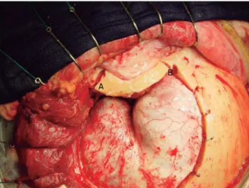
-ferior aspect of the lateral orbital wall. hen, we cut from the malar eminence to the inferior orbital issure. Another cut extends through the roof of the orbit from the level or just lateral the supraorbital foramen to the superior orbital issure, then, proceeds laterally to connect to the inferior orbital issure. Removal of the orbital roof continues with Figure 4. (A) The zygomatic osteotomy is done to the zygomatic arch anteriorly at the junction of the zygomatic arch with the temporal process of the zygomatic bone (a) and posteriorly previously to the temporomandibular joint (b). (B) Surgical view of the anterior (a) and posterior (b) zygomatic osteotomy.
bone rongeurs to the level of the planum sphenoidale me
-dially, and laterally, the lateral orbital wall is removed until the dural fold in the most lateral aspect of the superior or
-bital issure is well exposed. To aid the exposure, 4.0 nylon stitches are placed in the periorbita and after that, the orbit is retracted anteriorly.
here was not any enophtalmos among our patients (Figures 6, 7, 8 and 9).
Draping the operative ield over the bony ridge: After posi
-tioning and arrangement of rectangular cotton blocks on the free bony ridge, blue drapes are placed on the pieces of cotton, aiming to cover the supericial cranial wraps and minimize the further relection of light from the surgical microscope.
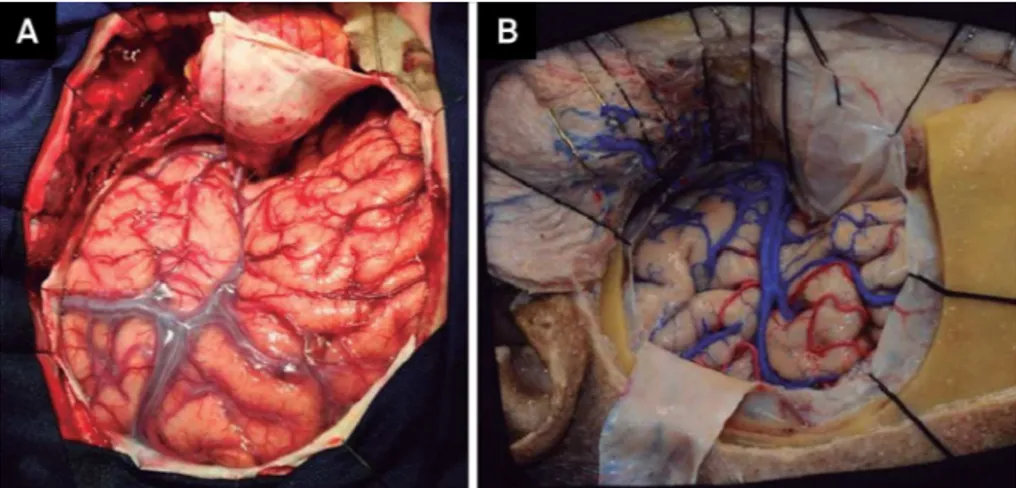
Opening the dura mater and brain exposure: he section
-ing of the dura mater should be performed in such a way that, when folded back, the external dural surface adapts itself to the bone surface without forming wrinkles or folds that might obstruct the microneurosurgical ield.
At the end of the dural opening, the shape should be of that of a large “S” so as to circumvent the temporal lobe, with the concave portion toward the rooftop free of orbit and the bot
-tom toward the edge and bot-tom of the posterior cranio-tomy. he dural incision should be initiated near the second trepanation, at the level of the most frontoparietal aspect of the dural exposure, using a scalpel blade #11, and continued in the frontal superior direction, at this point with the use of Metzenbaum scissors. It then follows toward the Sylvian is
-sure, and then upward towards the superior orbital issure and, inally, turns posteriorly, outlining the middle cranial fossa so as to fully expose the temporal lobe. he lap should be anchored with 4.0 nylon thread and pulled back in order to lift up the dural edges (Figure 10).
Opening of the Sylvian issure: he Sylvian issure is com
-posed of a supericial and a deep part. he supericial part presents a stem and three branches; the stem extends medi
-ally from the semilunar gyrus of the uncus, between the basal
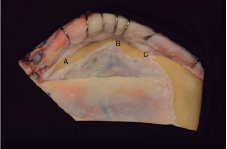
Figure 6. Orbital osteotomy from the malar eminence to the inferior orbital fissure (A) and from the level of the supraorbital foramen to the superior orbital fissure (B).
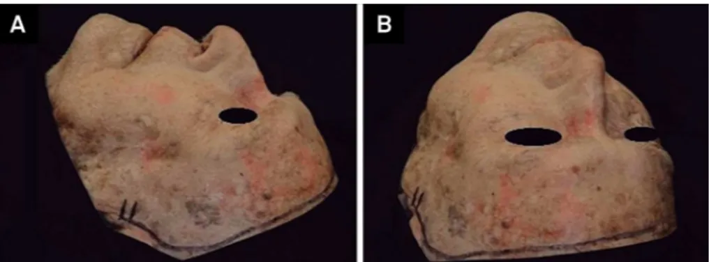
Figure 7. Three pieces of the Orbitozygomatic craniotomy: (A) zygomatic arch; (B) orbital roof; (C) Pretemporal osseous flap.
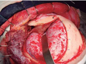
Figure 8. Cadaveric specimen showing the orbital osteotomy and the S shape dural open.
surface of the frontal lobe and the pole of the temporal lobe to the lateral end of the sphenoid ridge, where the stem di
-vides itself into anterior horizontal, anterior ascending, and posterior branches. he deep part is divided in an anterior part, the sphenoidal compartment, and a posterior part, the operculoinsular compartment. he sphenoidal compart
-ment arises in the region of the limen insulae, at the lateral margin of the anterior perforated substance. his compart
-ment is a narrow space posterior to the sphenoid ridge, be
-tween the frontal and the temporal lobes, that communi
-cates medially with the carotid cistern. he operculoinsular compartment is formed by two narrow clefts, the opercula cleft between the opposing lips of the frontoparietal and the temporal opercula and the insular cleft. he insular cleft has a superior limb located between the insula and the frontopa
-rietal opercula and an inferior limb between the insula and the temporal operculum. he opercular cleft is composed of the frontal and parietal opercula superiorly and the temporal operculum inferiorly.
When the lips of the Sylvian issure are widely separated we can see the insula. he insula connects the temporal lobe to the posterior orbital gyrus via the limen insulae. he limen insulae serves as a threshold between the carotid cistern (also called Sylvian vallecula) medially and the Sylvian issure laterally. From microsurgical and radiologic viewpoints, the insula represents the external covering of a mass comprised of the extreme, external, and internal capsules, claustrum, basal ganglia, and thalamus 19,20,21,22.
he orbitozygomatic approach proceeds with the open
-ing of the Sylvian issure and the basal cisterns. We usually open the basal cisterns before the Sylvian issure to drain the cerebrospinal luid, thus relaxing the brain, it makes the split of the Sylvian issure easier. his splitting usually begins at the level of the pars triangularis, where the space between the frontal and the temporal lobes is wider.
he basal cisterns: Orbitozygomatic craniotomy enables
the surgeon to reach the olfactory cistern, the carotid cistern,
the chiasmatic cistern, the sphenoidal compartment of the Sylvian issure, the cistern of lamina terminalis, the interpe
-duncular cistern, the ambient cistern and the crural cistern, which can be reached after the removal of the anteromedial segment of the uncus.
In order to accomplish the temporopolar approach to the interpeduncular cistern, the bridging veins draining the tem
-poral pole to the sphenoparietal sinus and cavernous sinuses may be coagulated and cut when they would be binding tem
-poral pole to middle fossa loor.
he arachnoid that binds the uncus to the oculomotor nerve and to the tentorial edge is opened, in order to achieve a nice mobility of the temporal lobe. By then, the temporal pole can be elevated superiorly through the subtemporal route and posteriorly through the temporopolar route.
he pretemporal orbitozygomatic allows the surgeon to deal with diseases arising from or extending to the anteri
-or fossa, middle fossa, sellar and parasellar regions, interpe
-duncular region, petrous apex, and upper third of the clivus. he basal exposure of the anterior and middle cranial fossa also allows the treatment of the lesions that arise from or ex
-tend to the extra dural compartment of these regions. his approach can access the sphenoidal, frontal, and ethmoidal sinuses, the components of the orbit, the cavernous sinus, the infratemporal fossa, the petrous apex, the intrapetrous internal carotid artery and the remainder of the middle cra
-nial fossa. his craniotomy allows an inferior to superior view of the microsurgical ield, very useful in aneurysms of the anterior communicating segment with posterior projec
-tion occupying the interhemispheric issure, in supra sellar lesions that project into the third ventricle and in high basi
-lar bifurcation aneurysms.
It is of singular importance a suitable head extention in or
-der to provide the best inferior to superior microsurgical view23.
he steps followed performing orbitozygomatic three pieces craniotomies have been facilitated dealing with forty-nine complex cases from 2002 to 2011.
References
1. Hakuba A, Liu S, Nishimura S: The orbitozygomatic infratemporal approach. A new surgical technique. Surg Neurol. 1986;26(3):271-6. doi:10.1016/0090-3019(86)90161-8
2. Hakuba A, Tanaka K, Suzuki T, Nishimura S. A combined orbitozygomatic infratemporal epidural and subdural approach for lesions involving the entire cavernous sinus. J Neurosurg. 1989;71(5):699-704. doi:10.3171/jns.1989.71.5.0699 3. Al-Mefty O. The cranio-orbital zygomatic approach for
intracranial lesions. Contemp Neurosurg. 1992;14(9):1-6. doi:10.1097/00029679-199214090-00001
4. Furth WR, Agur AM, Woolridge N, Cusimano MD. The orbitozygomatic approach. Neurosurgery. 2006;58(1 Suppl):ONS103-7.
doi:10.1227/01.NEU.0000197050.70397.C1
5. Zabramski JM, Kiris T, Sankhla SK, Cabiol J, Spetzler RF. Orbitozygomatic craniotomy: technical note. J Neurosurg. 1998;89(2):336-41. doi:10.3171/jns.1998.89.2.0336 6. Yasargil MG, Antic J, Laciga R, Jain KK, Hodosh RM, Smith RD.
Microsurgical pterional approach to aneurysms of the basilar bifurcation. Surg Neurol. 1976;6(2):83-91.
7. Yasargil MG, ed. Microneurosurgery. Stuttgart: Geor Thieme; 1984. Vol. 2, Basilar artery bifurcation aneurysms; p. 232-46.
8. Chaddad-Neto F, Campos Filho JM, Dória-Netto HL, Faria MH, Ribas GC, Oliveira E. The pterional craniotomy: tips and tricks. Arq Neuropsiquiatr. 2012;70(9):727-32. doi:10.1590/S0004-282X2012000900015 9. Chaddad-Neto F, Ribas GC, Oliveira E. [The pterional craniotomy
step by step]. Arq Neuropsiquiatr. 2007;65(1):101-6. Portuguese. doi:10.1590/S0004-282X2007000100021
10. Drake CG. The surgical treatment of aneurysms of basilar artery. J Neurosurg. 1968;29(4):436-46. doi:10.3171/jns.1968.29.4.0436 11. Drake CG. The treatment of aneurysms of the posterior circulation.
Clin Neurosurg. 1979;26:96-144.
12. Sano K. Temporo-polar approach to aneurysms of the basilar artery at and around the distal bifurcation: technical note. Neurol Res. 1980;2(3-4):361-7.
13. Oliveira E, Siqueira M, Tedeschi H, Peace DA. Surgical approaches for aneurysms of the basilar artery bifurcation. In: Matsushima T, editor. Cerebral aneurysms and skull base lesions. Fukuoka City: Sci Med; 1993. p. 34-42. (Matsushima T editor. Surgical anatomy for microneurosurgery; vol. 3).
14. Oliveira E, Tedeschi H, Siqueira MG, Peace DA. The pretemporal approach to the interpeduncular and petroclival regions. Acta Neurochir (Wien). 1995;136(3-4):204-11. doi:10.1007/BF01410627 15. Tedeschi H, Oliveira E, Wen HT. Pretemporal approach to basilar
bifurcation aneurysms. Tech Neurosurg. 2000;6(3):191-9. doi:10.1097/00127927-200006030-00003
16. Chaddad-Neto F, Dória-Netto HL, Campos Filho JM, Ribas ESC, Ribas GC, Oliveira E. Head positioning for anterior circulation aneurysms microsurgery. Arq Neuropsiquiatr. 2014;72(11):832-40. doi:10.1590/0004-282X20140156
17. Chaddad-Neto F, Dória-Netto HL, Campos Filho JM, Reghin-Neto M, Rothon Junor AL, Oliveira E. The far-lateral craniotomy: tips and tricks. Arq Neuropsiquiatr. 2014;72(9):699-705. doi:10.1590/0004-282X20140130
18. Chaddad-Neto F, Dória-Netto HL, Campos-Filho JM, Reghin-Neto M, Oliveira E. Pretemporal craniotomy. Arq Neuropsiquiatr. 2014;72(2):145-51. doi:10.1590/0004-282X20130202
19. Ribas GC, Ribas EC, Rodrigues CJ. The anterior sylvian point and the suprasylvian operculum. Neurosurg Focus. 2005;18(6):1-6. doi:10.3171/foc.2005.18.6.15
20. Yasargil MG. Microneurosurgery. Stuttgart: Georg Thieme; 1984. 21. Yasargil MG, Krisht AF, Türe U, Al-Mefty O, Yasargil DCH.
Microsurgery of insular gliomas: part I. Surgical anatomy of the Sylvian cistern. Contemp Neurosurg. 2002;24(11):1-8. doi:10.1097/00029679-200206010-00001