Isolated Unilateral Pulmonary Artery Agenesis. Evaluation of
Natural and Long Term Evolution after Corrective Surgery
Edmar Atik, Carla Tanamati, Luiz Kajita, Miguel Barbero-Marcial
Instituto do Coração do Hospital das Clínicas – FMUSP - São Paulo, SP - Brazil
Mailing Address: edmar Atik •
objective: Unilateral pulmonary artery agenesis is an uncommon isolated anomaly and since 1978 only 119 cases have been reported. In general the condition presents as pulmonary hypertension (PH) in children and hemoptysis in adults. Interven-tions such as pulmonary artery reconstruction and lobectomies were performed in 17% of the cases.
We analyzed four of these cases, two in natural evolution and two with late term PH regression after surgical correction.
Methods: Three 22,10 and 35 month old male children and one 20 month old female child were included in the study. The 22 month old presented right-sided heart failure (RHF) and cyanosis; the 10 month old presented RHF and the other two presented exertion fatigue. All had PH symptoms, right ventricular strain on the EKG and cardiomegaly. Cardiac catheteri-zation showed systemic pressures in the contralateral pulmonary artery, with right-sided agenesis in three of the children and left-sided agenesis in one child.
results: Surgical correction of pulmonary artery continuity was possible in the 22 month old and 10 month old using a 7mm diameter Goretex conduit between the pulmonary arteries up to the hypoplastic contralateral pulmonary hilum. There was early and late regression of the PH signs and the children remained stable during follow-up to the ages of 7 and 2.5 years, respectively. The pressure ratio between the left and right ventricles was 30 and 40%, in both cases. Pulmonary perfusion increased from 8 to 44% and from 8 to 23%, in the two cases. The same procedure was scheduled for the other patients.
conclusion: This technique has become the operation of choice for similar cases, that are rarely described in literature, even in the presence of severe PH and contralateral pulmonary artery hypoplasia.
Key words: Pulmonary artery agenesis, pulmonary hypertension, heart failure.
Unilateral pulmonary artery agenesis resulting from the lack of embryological development of either the left or right sixth aortic arch and has various clinical presentations. The most common sign during childhood is contralateral pulmonary hypertension and in adults, hemoptysis1.
In the absence of other associated heart defects the intraparenchymal pulmonary artery tree, ipsilateral to the absent right or left pulmonary artery is usually fed through the artery canal or by collateral aorta vessels, including the bronchial arteries. Nevertheless, they are insufficient to maintain an ideal pulmonary flow and the contralateral pulmonary artery usually presents in childhood as hypertension resulting from right ventricular output flow causing ipsilateral volume overload.
Early surgical indication avoids the unfavorable evolution of pulmonary hypertension to more severe and progressive degrees and prevents the development of pulmonary systemic collateral circulation, responsible for subsequent hemoptysis.
Detailed revision of the evolution and therapeutic aspects
of the various forms of the anomaly, currently recommend restoration of the pulmonary tree continuity as a first procedure and a pneumectomy or lobectomy as a second choice1.
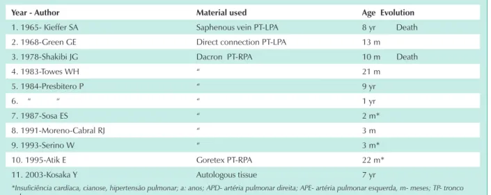
Since 1978, approximately 119 cases of this anomaly have been reported1-14. In these case reports, pulmonary artery reconstruction was performed on nine patients2,3,9-14 plus two other cases published in the 1960’s15,16. However, long term post operative evolution has not been documented.
The objective of this publication is to add three additional patients to the one patient that we previously reported2 and analyze the long term pulmonary artery
Methods
g, with an average of 9.412 g. The clinical presentation of each case, the diagnostic and therapeutic methods and post operative follow-up were reevaluated.
Pulmonary and systemic resistances were not calculated during the corrective surgery of the two cases. Since this is a retrospective study, pulmonary hypertension was evaluated using the ratio between the pulmonary arteries and aorta pressures. As a result, surgical indications were based on the combination of clinical and hemodynamic data. The fundamental clinical sign was the degree of volume overload as a result of the pathology, causing heart failure and from this cardiomegaly as seen on the chest x-ray.
Results
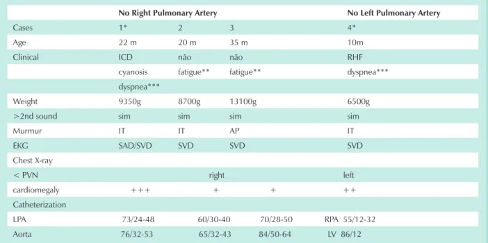
Two of the male patients and the one female patient, between the ages of 20 and 35 months, mean 25.66 months had right pulmonary artery agenesis. The other 10 month old male patient had left pulmonary artery agenesis (tab.1). The clinical presentation for the case published earlier2, was severe heart failure and cyanosis; heart failure with left pulmonary artery agenesis in one other case; and exertion fatigue for the two other cases. The physical findings corresponded to those usually found in patients with pulmonary hypertension, with an accentuated second heart sound and holosystolic murmur at the lower left sternal edge as a result of tricuspid valve insufficiency. The two patients with heart failure had systolic impulses and hepatomegaly. In the other two patients, the physical exam showed no signs of heart failure, the systolic murmur in the pulmonary cavity was minimal and the second heart sound was hyperphonetic. The electrocardiogram showed right ventricle overload in all patients. The chest
x-ray revealed a contrasting pulmonary vascular network, diminished on the artery agenesis side and increased in the contralateral region, as well as cardiomegaly caused by the enlarged right cavities in the two heart failure patients. The echocardiogram was also conclusive for unilateral agenesis of one of the pulmonary arteries and the hemodynamic study highlighted the presence of pulmonary artery hypertension in all patients. The angiocardiography showed that the corresponding pulmonary arteries were missing but a retrograde injection in the pulmonary veins enabled visualization of the hypoplastic ipsilateral intraparenchymal pulmonary artery tree (fig.1). Two patients were submitted to surgery for the placement of a 7mm diameter Goretex conduit that in one patient was implanted between the pulmonary trunk and the right pulmonary artery (fig.1) and in the other between the pulmonary trunk and the left pulmonary artery. The heart failure symptoms were resolved in both patients with disappearance of the murmur caused by the tricuspid insufficiency; the electrocardiography signs were regularized and the chest x-ray showed cardiomegaly regression (fig.2). The post operative hemodynamic study showed pulmonary hypertension regression in the first patient with pressures of 40/10 and an average of 20 mmHg (62 months after) and in the second patient pressures were reduced to 45/12 mmHg (27 months after) with a reduction in the pressures between the left pulmonary artery and left ventricle from 0.64 to 0.44. The pulmonary scintigraphy confirmed increased right pulmonary perfusion from 9% before the surgery to 44% after the surgery in the first case and increased left pulmonary perfusion in the second case from 8 to 23%, during the same evolution timeframe. The early and late term
post-no right Pulmonary Artery no left Pulmonary Artery
Cases 1* 2 3 4*
Age 22 m 20 m 35 m 10m
Clinical ICD não não RHF
cyanosis fatigue** fatigue** dyspnea*** dyspnea***
Weight 9350g 8700g 13100g 6500g
>2nd sound sim sim sim sim
Murmur IT IT AP IT
EKG SAD/SVD SVD SVD SVD
Chest X-ray
< PVN right left cardiomegaly +++ + + ++ Catheterization
LPA 73/24-48 60/30-40 70/28-50 RPA 55/12-32 Aorta 76/32-53 65/32-43 84/50-64 LV 86/12
PC- pulmonary cavity; RPA- right pulmonary artery; LPA- left pulmonary artery; g- grams; RHF- right-sided heart failure; TI- tricuspid insufficiency;
m-months; PVN- pulmonary vascular network; LV- left ventricle; RAO- right atrial overload; RVO- right ventricular overload; *: patients operated on; **: exertion fatigue; ***: dyspnea at rest.
Post- operative
AD (RA): 7 VD (LV): 86/15 APE (LPA): 73/24-48 APD (RPA): 68/9-32
AE (L A): 6 VE (LV): 88/12 Ao: 76/32-53 APE (LPA): 69/7-31
Fig. 1 - Preoperative angiograph of the 22 month old patient. View “a” shows the continuity of the left pulmonary artery with the pulmonary trunk and the absence of right pulmonary artery filling. View “b” shows opacification after retrograde intravenous injection in the pulmonary vein. In view “c” the right ventricle is dilated and hypercontractile. View “d” shows the posterior displacement of the left ventricle. Views “e” and “f” show the continuity of the pulmonary tree immediately following corrective surgery using a Goretex conduit implant between the pulmonary trunk and right pulmonary hilum (arrow). The pressures recorded are right cavity systemic pressures with reductions immediately following surgery. RA- right atrium; LA- left atrium; Ao- aorta; RPA- right pulmonary artery; LPA- left pulmonary artery; RV- right ventricle; LV- left ventricle.
a b
c d
e
f
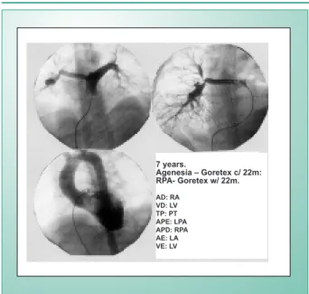
operative angiographies emphasized the pulmonary artery tree restoration as shown for the first case (fig. 1 and 3). After an average long term post operative evolution of 50 months, both patients were functional class I without any residual cardiopathy symptoms. The other two patients are waiting for the same type of corrective surgery. Their operations have not been confirmed largely due to their recent diagnoses. In addition, they have few symptoms and the natural evolution of pulmonary hypertension is being followed as demonstrated in their hemodynamic studies.
Discussion
From the 108 cases reported of this anomality between 1978 and 2000 and compiled by Ten Harkel and associates1, there was a clear prevalence of right pulmonary artery agenesis which was present in 67 patients (63%). In this material, signs of pulmonary hypertension were confirmed in 44% (25/57 patients), dyspnea in 40% (36/92 patients), pulmonary infections, chest pain and pleural effusion in 37% (35/94 patients) and hemoptysis in 20% (18/92 patients). In the most unusual presentation, 14 (12.9%) of the 108 patients were asymptomatic.
Since 2000, 11 additional cases of this anomality have been reported3-8. Only one of these documented cases underwent corrective surgery for a connection between the pulmonary trunk and right pulmonary artery, which hemodynamically restored the pulmonary artery tree3. The
connective material used in this seven year old patient was autologous pericardium.
This procedure, using other connective materials, had only been conducted in eight previous cases2,9-14. Pneumectomies or lobectomies to control hemoptysis were performed on nine other patients from the 108 cases reported earlier, for a total of 17 (7%) therapeutic procedures for this anomality between 1978 and 20001.
During this timeframe, three of the nine patients submitted to corrective surgery for pulmonary artery vascularization presented heart failure and associated cyanosis2,13,14, including one of our previously published case2. The other six3,9-12, showed signs of pulmonary hypertension and heart failure but no right-to-left shunt.
Prior to 1978, two other patients underwent surgery. The first was reported in 1965 by Kieffer and associates15 and circulation was restored by connecting the pulmonary trunk to the left pulmonary artery using a saphenous vein in an eight year old child. The second case in 1968, reported by Green and associates16 directly connected the pulmonary trunk to the left pulmonary artery in a thirteen month old baby (tab 2).
It is emphasized that, generally speaking, long term evolution details for this procedure after artery connection and consequent restoration of the pulmonary artery tree have not been published.
material, the favorable long term evolution results in order to distinguish this procedure as the best option for this rare pathology. Long term follow-up of our two patients submitted to the procedure revealed resolution of the heart failure
condition and regression of the pulmonary hypertension. In the most remarkable case associated with cyanosis and a left atrium foramen ovale shunt, the procedure regularized the cardiac cavity, eliminated the pulmonary vascular network
Preop. - 22 m Postop. - 7 anos
Pulmonary Scintigraphy
Preop Postop
RPA 8% 44%
LPA 8% 23%
Fig. 2 - The chest x-ray film on the left-hand side taken during complementary preoperative evolution tests of the 22 month old patient, emphasize the significant
cardiomegaly and enlargement of the left pulmonary vascular network that were regularized as shown on the post operative chest x-ray film taken when the patient was 7 years old. The electrocardiogram clearly demonstrates the regression of the right ventricular overload until regularization of the electrical potentials at late post operative follow-up.
1 year and 10 months
1 year and 11 months (immediate period after connection with LPA)
7 years
year - Author Material used Age evolution
1. 1965- Kieffer SA Saphenous vein PT-LPA 8 yr Death
2. 1968-Green GE Direct connection PT-LPA 13 m
3. 1978-Shakibi JG Dacron PT-RPA 10 m Death
4. 1983-Towes WH “ 21 m
5. 1984-Presbitero P “ 9 yr
6. “ “ “ 1 yr
7. 1987-Sosa ES “ 2 m*
8. 1991-Moreno-Cabral RJ “ 3 m
9. 1993-Serino W “ 3 m*
10. 1995-Atik E Goretex PT-RPA 22 m*
11. 2003-Kosaka Y Autologous tissue 7 yr
*Insuficiência cardíaca, cianose, hipertensão pulmonar; a: anos; APD- artéria pulmonar direita; APE- artéria pulmonar esquerda, m- meses; TP- tronco
pulmonar.
contrast as shown on the chest x-ray, corrected the right ventricle overload, stabilized the electrical potentials and consequently increased pulmonary flow which was measured by intravenous injection with technetium (fig.2).
The prolonged post operative timeframe of this satisfactory evolution is sufficient to indicate this therapeutic procedure
for these patients, particularly in the early stages of life when it is hoped that complete regression of the pulmonary vascular alterations will occur.
The pulmonary artery reconstruction procedures described in literature2,3,9-16, were performed on patients between the ages of two months and nine years with an average age of 34 months. Five were younger than 1 year old9-11,13,14, with an average of 6 months, three were between one and two years old2,12,16, with an average age of 18.6 months, and three were 7, 9 and 8 year old children3,10,15. The overall mortality was 2 patients9,15 (18.1%) from the 11 who underwent the surgery.
It is hoped that this procedure will be standardized and that its viability will not be questioned especially when performed on young children.
In relation to our two natural evolution patients that have not yet undergone surgery, the lack of symptoms such as exertion fatigue did not precipitate the immediate surgical indication, but certainly would be beneficial as seen in other similar cases reported in literature, even with children older than seven3,10,15.
In this context, it is emphasized that the surgical indication procedure in general should prevail, but obviously in the case of a clinical picture of cardiac volume overload, the presence of excessive pulmonary flow should indicate pressure overload, which is predominant in irreversible pulmonary hypertension conditions.
Potential conflict of interest
No potential conflict of interest relevant to this article was reported.
References
1. Ten Harkel AD, Blom NA, Ottenkamp J. Isolated unilateral absence of a pulmonary artery: a case report and review of the literature. Chest 2002;122:1471-7.
2. Atik E, Barbero-Marcial M, Kajita L, et al. Agenesis of the right pulmonary artery with severe pulmonary hypertension, attenuated by surgical correction. Arq Bras Cardiol 1995; 64:133-6.
3. Kosaka Y, Kurosawa H, Hoshino S, Shinoka T, Isomatsu Y, Tsuji Y. Surgery for unilateral absence of pulmonary artery using autologous tissue. Ann Thorac Surg 2003;76:1281-3.
4. Mahnken AH, Wildberger JE, Spuntrup E, Hubner D. Unilateral absence of the pulmonary artery associated with coronary to bronchial artery anastomosis. J Thorac Imaging 2000;15:187-90.
5. Maeda S, Suzuki S, Moriya T, et al. Isolated unilateral absence of a pulmonary artery: influence of systemic circulation on alveolar capillary vessels. Pathol Int 2001;51-649-53.
6. Kadir IS, Thekudan J, Dheodar A, Jones MT, Carroll KB. Congenital unilateral pulmonary artery agenesis and aspergilloma. Ann Thorac Surg 2002;74: 2169-71.
7. Apostolopoulou SC, Kelekis NL, Brountzos EN, Rammos S, Kelekis DA. Absent pulmonary artery in one adult and five pediatric patients: imaging, embriology, and therapeutic implications. Am J Roentgenol 2002; 179: 1253-60.
8. Farghly E, Bousamra M 2nd. Hemoptysis resulting from unilateral pulmonary artery agenesis. Ann Thorac Surg 2002;74:255-7.
9. Shakibi JG, Rastan H, Nazarian I, Paydar M, Aryanpour I, Siassi B. Isolated unilateral absence of the pulmonary artery: review of the world literature and guidelines for surgical repair. Jpn Heart J 1978;19: 439-51.
10. Presbitero P, Bull C, Haworth SG, de Leval MR. Absent or occult pulmonary artery. Br Heart J 1984;52:178-185.
11. Moreno-Cabral RJ, McNamara JJ, Reddy VJ, Caldwell P. Unilateral absent pulmonary artery: surgical repair with a new technique. Thorac Cardiovasc Surg 1991;102:463-5.
12. Towes WH, Pappas G. Surgical management of absent right pulmonary artery with associated pulmonary hypertension. Chest 1983;84:497-9.
13. Serino W, Argento G, Fiorilli R, Di Benedetto G, Velitti F. The isolated agenesis of a branch of the pulmonary artery: a case report; its diagnosis and therapy. Cardiologia 1993;38:531-4.
14. Sosa ES, Ba M, Kratz C, et al. Congenital unilateral absence of a pulmonary artery. Chir Pediatr 1987;28:204-8.
15. Kieffer SA, Amplatz K, Anderson RC, lillehei CW. Proximal interruption of a pulmonary artery. Am J Roentgenol Radium Ther Nucl Med 1965; 95:592-7.
16. Green GE, Reppert EH, Cohlan SQ, Spencer FC. Surgical correction of absence of proximal segment of left pulmonary artery. Circulation 1968;37:62-9.
7 years.
Agenesia – Goretex c/ 22m: RPA- Goretex w/ 22m.
AD: RA VD: LV TP: PT APE: LPA APD: RPA AE: LA VE: LV
Fig. 3 - Post operative angiography of the 22 month old case when the