Arq Bras Oftalmol. 2008;71(2):262-4
Hematoma subperiósteo e compressão orbitária após trauma frontal leve na
anemia falciforme: relato de caso
Trabalho realizado no Departamento de Oftalmologia, Otorrinolaringologia, e Cirurgia de Cabeça e Pescoço da Faculdade de Medicina de Ribeirão Preto - Universi-dade de São Paulo - USP - Ribeirão Preto (SP) - Brazil. 1From Departamento de Oftalmologia, Otorrinolaringo-logia, e Cirurgia de Cabeça e Pescoço da Faculdade de Medicina de Ribeirão Preto - Universidade de São Paulo - USP - Ribeirão Preto (SP) - Brazil.
2From Departamento de Oftalmologia, Otorrinolaringo-logia, e Cirurgia de Cabeça e Pescoço da Faculdade de Medicina da USP - Ribeirão Preto (SP) - Brazil. 3From Departamento de Oftalmologia,
Otorrinolaringo-logia, e Cirurgia de Cabeça e Pescoço da Faculdade de Medicina da USP - Ribeirão Preto (SP) - Brazil. 4From Departamento de Oftalmologia,
Otorrinolaringo-logia, e Cirurgia de Cabeça e Pescoço da Faculdade de Medicina da USP - Ribeirão Preto (SP) - Brazil. Endereço para correspondência: Antonio Augusto Velasco e Cruz. Departamento de Oftalmo, Otorrino e Cabeça e Pescoço - Hospital das Clínicas de Ribeirão Preto - Campus. Av. Bandeirantes, 3.900 - Ribeirão Preto (SP) CEP 14049-900
E-mail: [email protected] Recebido para publicação em 16.05.2007 Última versão recebida em 07.08.2007 Aprovação em 27.08.2007 Fernando Procianoy1
Mauro Brandão Filho2
Antonio Augusto Velasco e Cruz3
Victor Marques Alencar4
Subperiosteal hematoma and orbital compression
syndrome following minor frontal trauma in sickle
cell anemia: case report
Keywords: Hematoma/etiology; Craniocerebral trauma/complications; Orbit/surgery; Anemia, sickle-cell; Optic nerve injuries; Frontal bone; Case reports [Publication type]
We report the case of an 11-year-old girl with sickle cell disease who presented to the emergency room after being hit by a mud pie in the left frontal region. Examination evidenced left eye proptosis, eyelid swelling, reduced visual acuity and afferent pupillary defect, without any in-flammatory signs such as fever, hyperemia or tenderness. Computed tomography of the orbits showed a large superomedial subperiosteal hematoma in the left orbit. The patient was treated with canthotomy, cantholysis and surgical draining of the hematoma. Two days after drainage she persisted with a subperiosteal hematoma and low visual acuity. A wide exploration of the orbital roof through a lid crease approach disclosed a thickened superior orbital rim with multiple bone defects along the roof and with continuous bleeding. Hemostasis was accomplished with bone wax. Orbital compression was resolved and the patient recovered her previous normal visual acuity.
ABSTRACT
RELATOS DE CASOS
INTRODUCTION
Sickle cell disease (SCD) is a generic term used to designate a group of inherited hemolytic disorders caused by a mutation in the gene that
enco-des hemoglobin(1). This point mutation (GAG>GTG), which originated
50,000 years ago in Africa, provokes an amino acid substitution (valine for
glutamic acid) at position 6 of the β chain of the globin subunit of
hemo-globin. As a result, instead of the normal hemoglobin of adulthood
(hemo-globin A), an abnormal molecule (hemo(hemo-globin S) is formed(2). This molecule
tends to polymerize on deoxygenation deforming the red blood cells into
the characteristic sickle shape(1-2). The disease follows a simple recessive
Mendelian pattern and thus is acquired only when the S gene is inherited from both parents (homozygous SS disease). Since there are a variety of abnormal hemoglobin mutants, other genotypes such as S/C, S/D, S/B+
tha-lassemia also induce sickle cell disease(1-2). However, the designation
si-ckle-cell anemia is restricted to the SS form(1).
The pathophysiology of all clinical manifestations of SCD is linked to the less pliable and stiff red blood cells which block blood flow
(vaso-occlu-sion) and are prematurely destroyed (hemolysis)(1-2). Skeletal changes are
syndro-Arq Bras Oftalmol. 2008;71(2):262-4
263 Subperiosteal hematoma and orbital compression syndrome following minor frontal trauma in sickle cell anemia: case report
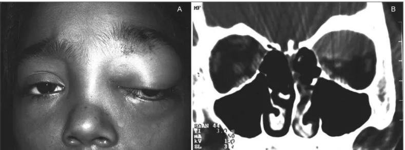
Figure 1 - A: Non-axial left eye proptosis and upper eyelid edema immediately after frontal trauma. B: Coronal computed tomography scan immediately after trauma. Notice in this soft-tissue window a large subperiosteal hematoma and an enlarged left frontal bone.
me has been reported many times(3). These cases have been
typically associated with inflammatory signs mimicking orbital
cellulitis(3). The occurrence of trauma-related subperiosteal
hematoma without inflammatory signs is rare.
CASE REPORT
An 11-year-old girl with SCD presented to the emergency room with visual loss after being hit in the left frontal region by a mud pie. On examination, the left eye was grossly prop-totic with inferolateral dystopia, important eyelid swelling and afferent pupillary defect (Figure 1A). Visual acuity was 20/20 in the right eye and counting fingers in the left eye. Slit lamp examination was normal in both eyes. Computed tomography of the orbits showed a superomedial subperiosteal hematoma (Figure 1B). The patient underwent an urgent canthotomy, can-tholysis and drainage of the hematoma through a lid crease approach. On the first postoperative day proptosis was re-duced, the pupillary reflexes were normal and the visual acuity improved to 20/40. However, as the globe displacement per-sisted the patient was transferred to the Oculoplastic Clinic of the School of Medicine of Ribeirão Preto. A second CT scan of the orbits showed an important subperiosteal hemorrhage. Hy-pertrophy of the diploic spaces of the calvarium bones was evi-dent, especially in the left frontal bone (Figure 2). New surgical exploration was performed through the same eyelid crease approach and the periosteum was widely opened at the superior orbital rim. Exposure allowed the visualization of multiple bone erosions on the orbit roof with persistent bleeding (Figure 3). Hemostasis was accomplished only with the use of bone wax. The patient’s evolution was excellent with no recurrence of bleeding. On the fourth postoperative day the visual acuity had returned to 20/20 in the left eye. There were no more episodes of bleeding or of orbital disease in a 1-year follow-up.
DISCUSSION
Orbital compression syndrome has already been described in patients with SCD 3, including a first report in the Brazilian
literature(4). However, the usual presentation is an acute event
dominated by inflammatory signs resulting from bone infarc-tion. In the present case, after a minor trauma the patient presented with an important subperiosteal hematoma without signs of inflammation. Surgical exploration revealed that the frontal bone was abnormally thickened with multiple holes on the orbital rim and along the roof (Figure 3). We believe that this abnormality was caused by the bone marrow hyperplasia found at several skeletal sites including the skull. In normal adults bone marrow is found mainly in the axial skeleton. However, in children with SCD the marrow is expanded into all of the bones. In the skull the typical finding is as an expansion of the diploic space with thinning of both outer and inner tables. This process is an attempt of the organism to increase erythrogenesis and thus represents a response to chronic anemia. Bone marrow hyper-plasia, which is usually more severe in sickle cell thalassemia, can be demonstrated by a variety of imaging techniques including scans with radionuclides, magnetic resonance imaging,
compu-ted tomography, and plain films(5). In our case bone marrow
hyperplasia of the roof was the underlying condition that caused bleeding after the minor frontal trauma. This event is similar to hemarthrosis, an uncommon complication occurring in sickle cell
anemia(5). Orbital surgeons should be aware of the fact that
orbital bleeding without signs of infarction is due to an intrinsic bone weakening that must be specifically addressed.
RESUMO
264Subperiosteal hematoma and orbital compression syndrome following minor frontal trauma in sickle cell anemia: case report
Arq Bras Oftalmol. 2008;71(2):262-4
Figure 2 - Computed tomography scan of the orbits (bone window) two days after the first drainage. Recurrent hematoma in the left orbit. Bone marrow hyperplasia is evident in both coronal (A) and axial (B) scans.
Figure 3 - View of the superior orbital rim during subperiosteal hematoma exploration. The frontal bone is thickened with multiple
holes leading to profuse bleeding.
de barro na região frontal esquerda. Apresentava ao exame proptose do olho esquerdo, edema palpebral, diminuição da acui-dade visual e defeito pupilar aferente, sem quaisquer sinais inflama-tórios como febre, hiperemia ou aumento de sensibilidade. A tomografia computadorizada de órbitas demonstrou um extenso hematoma subperiósteo superomedial na órbita esquerda. A
pa-ciente foi tratada com cantotomia, cantólise e drenagem cirúrgica do hematoma. Dois dias após a drenagem, ela permaneceu com um hematoma subperiósteo e a acuidade visual diminuída. Uma ampla exploração através de incisão no sulco palpebral superior revelou um rebordo orbitário superior espessado, e múltiplos defeitos ós-seos ao longo do teto da órbita com sangramento persistente. Foi realizada hemostasia com cera óssea. A compressão orbitária foi resolvida, e a paciente recuperou a acuidade visual normal prévia.
Descritores: Hematoma/etiologia; Trauma crânio-cerebral/com-plicações; Órbita/cirurgia; Anemia falciforme; Traumatismos do nervo óptico; Osso frontal; Relatos de casos [Tipo de publicação]
REFERENCES
1. Serjeant GR. Sickle-cell disease. Lancet. 1997; 350(9079):725-30. Comment in: Lancet. 1997;350(9092):1710.
2. Dover GJ, Platt OS. Sickle cell disease. In: Nathan DG, Oski FA, editors. Hematology of infancy and childhood. Philadelphia: Saunders; 2003. p.790-841. 3. Ganesh A, William RR, Mitra S, Yanamadala S, Hussein SS, Al-Kindi S, et al. Orbital involvement in sickle cell disease: a report of five cases and review literature. Eye. 2001;15(Pt 6):774-80.
4. Marback EF, Marback PMF, Marback RL, Sampaio CM, Sé DCS. Síndrome de compressão orbitária relacionada à anemia falciforme. Arq Bras Oftalmol. 1999;62(5):631-4.