Instituto Dante Pazzanese de Cardiologia
Mailing address: Auristela Isabel de Oliveira Ramos - Av. Miruna, 327/44 Cep 04084-001 - São Paulo, SP, Brazil - E-mail: [email protected] English version by Stela Maris C. e Gandour
Arq Bras Cardiol, volume 81 (nº 4), 393-8, 2003
Auristela I. O. Ramos, Rui F. Ramos, Dorival J. D. Togna, Antoninho S. Arnoni, Rodolfo Staico, Mercedes M. Galo, Zilda M. Meneghelo
São Paulo, SP - Brazil
Fibrinolytic Therapy for Thrombosis in Cardiac Valvular
Prosthesis Short and Long Term Results
Thrombosis in a cardiac valvular prosthesis is a rare, but severe complication. Its incidence ranges from 0.5% to 6% per patient per year in the aortic and mitral positions, de-pending on the degree of anticoagulation and the prosthetic design 1. In 1971, Luluaga et al 2 reported for the first time the
successful use of fibrinolytic agents for the treatment of thrombosis in a Starr-Edwards prosthesis in the tricuspid position. From that time onwards, several cases have been reported 3-6. The objective of this study was to analyze the
use of the fibrinolytic agent, streptokinase, in the treatment of prosthetic thromboses.
Methods
This study prospectively analyzed patients with valvu-lar prosthetic thrombosis diagnosed by clinical history and examination, transesophageal Doppler-echocardiography, and cinefluoroscopy.
The study comprised patients with diagnostic confir-mation of prosthetic thrombosis, who were hemodynami-cally stable and considered of high surgical risk or with a contraindication for prosthesis replacement surgery. Patients with inadequate anticoagulation control, either due to lack of adherence to treatment, or lack of conditions to undergo periodical controls, and those with large thrombi in the atrial cavity were excluded from the study.
The diagnostic hypothesis of prosthetic thrombosis was raised when the patient reported worsening of his func-tional class, appearance of signs of low cardiac output, a history suggestive of transient embolism, or when the pa-tient noticed a change in the intensity of the prosthetic click. The auscultatory findings of muffling of the metallic click, as well as the appearance or intensification of preexisting murmurs, contributed to the diagnostic suspicion. The fluo-roscopy performed in the hemodynamics laboratory served as triage for all patients suspected of having prosthetic obs-truction. Through fluoroscopy, the opening angle of the prosthesis, formed between the disk and the prosthetic ring, and the mobility of the disks were analyzed. The analysis of
Objective - To assess the short- and long-term results of the use of streptokinase (SK) for the treatment of throm-boses in cardiac valvular prostheses.
Methods - Seventeen patients with cardiac prosthetic thrombosis diagnosed by clinical, echocardiographic, and radioscopic findings underwent fibrinolytic treatment with a streptokinase bolus of 250,000 U followed by 100.000 U/hour. Short- and long-term results were asses-sed by radioscopy and echocardiography.
Results - Of the 17 patients, 12 had mechanical dou-ble-disk prostheses (4 aortic, 6 mitral, 2 tricuspid), 4 had single-disk prostheses (2 aortic, 1 mitral, and 1 tricuspid), and 1 had a tricuspid bioprosthesis. The success rate was 64.8%, the partial success rate was 17.6%, and the nonsuc-cess rate was 17.6%. All patients with a double-disk pros-thesis responded, completely or partially, to the treatment. None of the patients with a single-disk prosthesis had com-plete resolution of the thrombosis. The time of streptokinase infusion ranged from 6 to 80 hours (mean of 56 h). The mor-tality rate due to the use of streptokinase was 5.8% and was secondary to cerebral bleeding. During streptokinase infusion, 3 (17.6%) embolic episodes occurred as follows: 1 cerebral, 1 peripheral, and 1 coronary. The rethrombosis index was 33% in a mean follow-up of 42 months.
Conclusion - The use of fibrinolytic agents was effec-tive and relaeffec-tively safe in patients with primary thrombosis of a double-disk prosthesis. A fatal hemorrhagic cation occurred in 1 (5.8%) patient, and embolic compli-cations occurred in 3 (17.6%) patients. In a mean 42-month follow-up, 67% of the patients were free from re-thrombosis.
the disks requires an incidence in which the profile of the disks is filmed, and in which the disks are perpendicular to the prosthetic ring (figs. 1 and 2). Fluoroscopy was perfor-med prior to beginning streptokinase infusion and was re-peated every 24 hours to assess response to treatment.
Transesophageal Doppler-echocardiography comple-mented the clinical and fluoroscopic diagnosis. Echocardio-graphy made possible the analysis of the mobility of the disks, the visualization of possible thrombi and their charac-teristics, such as size, location, and mobility, in addition to the calculation of transprosthetic gradients, valvular area, ventricular function, and the diameters of the cardiac cavi-ties. The echocardiographies were performed before and after streptokinase infusion.
The diagnosis was established by associating the clinical findings and the echocardiographic and fluoroscopic findings. Streptokinase was the fibrinolytic agent used in all pa-tients. It was intravenously administered in a bolus of 250,000 U for 30 minutes, followed by the infusion of 100,000 U/hour, in the coronary unit. Streptokinase infusion was suspended when the mobility of the prosthetic disks was normalized on fluoroscopy, in case of hemorrhagic compli-cation, or when no improvement was observed in the mobi-lity of the disks 72 hours after drug infusion. After suspen-sion of the fibrinolytic agent, intravenous heparin was ini-tiated, as soon as the partial thromboplastin time (PTT) was shorter than twice the control PTT. Oral anticoagulation with phenprocoumon was reinitiated 24 hours after the in-terruption of streptokinase in the patients who responded to treatment. They were discharged from the hospital recei-ving oral anticoagulation with the dose adjusted according to the International Normalized Ratio (INR) between 2.5 and 3.5 in association with 100 mg of aspirin per day.
Late follow-up was performed on an outpatient care basis and with fluoroscopic and echocardiographic control every 6 months or when symptoms suggestive of thrombo-sis recurrence appeared.
Successful treatment was defined as an improvement in clinical findings in association with normalization of the mobility of the disks and a reduction in the transprosthetic gradient. Partial success was defined as the improvement in clinical findings in association with a reduction in the trans-prosthetic gradient, but, on fluoroscopy, some reduction in the mobility of the disks still persisted. Nonsuccess was de-fined as the lack of resolution of the thrombosis, or when the patient had fatal complications.
Results
From January 1993 to July 2002, 17 inconsecutive pa-tients, who met the inclusion criteria, received streptokinase for the treatment of thrombosis in the cardiac valvular pros-theses. The mean age was 41 (12 to 60) years. Twelve were females. Twelve had mechanical double-disc prostheses (4 aortic, 6 mitral, 2 tricuspid), 4 had single-disk prostheses (2 aortic, 1 mitral, 1 tricuspid), and 1 had a porcine valvular prosthesis. Of these 12 patients, 8 had already undergone 2 or more previous valvular replacements. The INR was adequate, ie, between 2.5 and 3.5, in only 6 (35%) patients. Eleven (65%) had inadequate anticoagulation with INR < 2, due to the incorrect use of the medication or suspension of the anticoa-gulant agent for surgery. The patient with the porcine pros-thesis was not receiving coumarin anticoagulants.
On physical examination, signs of congestive heart fai-lure, appearance or intensification of murmurs of stenosis, and muffling of the metallic click were found in most patients. The symptom most frequently reported by the pa-tients was worsening of their physical capacity with the ap-pearance of dyspnea associated with the perception of muf-fling of the metallic click of the prosthesis. One patient repor-ted precordial pain with characteristics similar to those of angina, and another patient had an episode suggestive of transient cerebral ischemia with short-duration dyslalia. The duration of the symptoms related to a prosthetic throm-bosis ranged from 3 to 90 days.
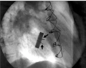
Fig. 1 - Radioscopy of the metallic double-disk prosthesis in the mitral position, prior to fibrinolysis. The upper arrow shows the small opening of the disk and the lower arrow shows the still disk.
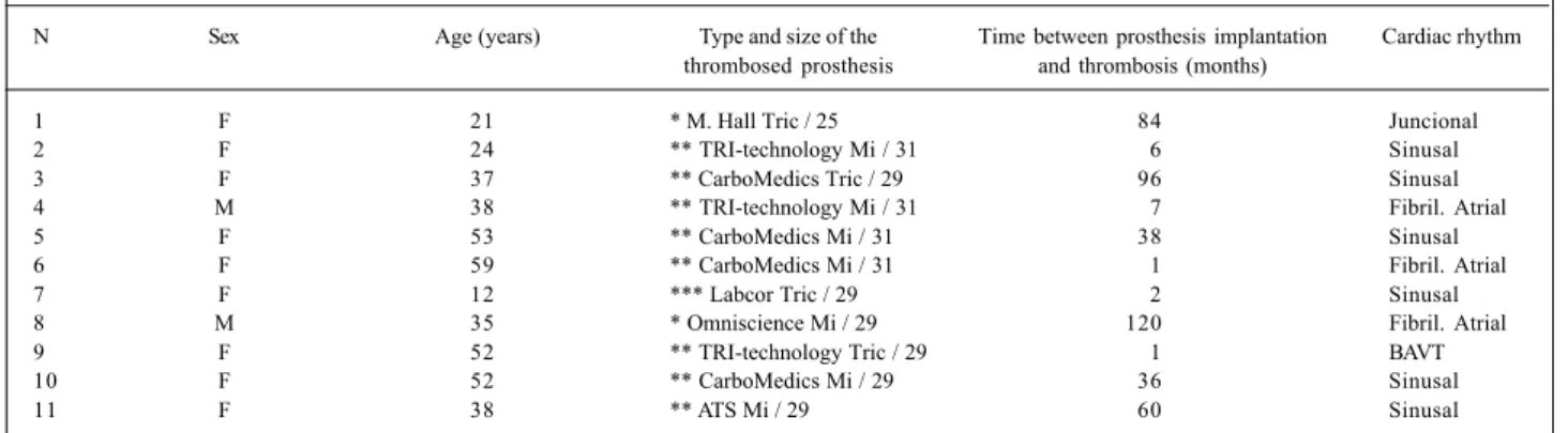
The demographic characteristics, the model of the prosthesis, the time elapsed between prosthesis implanta-tion and thrombosis, and the cardiac rhythm of patients with a prosthesis in the aortic and atrioventricular position are shown in tables I and II.
Of the 6 patients with an aortic prosthesis, 4 had double-disk prostheses and 2 had single-disk prostheses. Most patients had sinus rhythm. The time elapsed between prosthesis implantation and thrombosis ranged from 8 to 136 (mean of 72) months.
Of the 11 patients with atrioventricular prostheses (3 tricuspid and 8 mitral), 2 had single-disk prostheses, 8 had double-disk prostheses, and 1 had a bioprosthesis. Three patients had atrial fibrillation, 6 had sinus rhythm, 1 had junctional rhythm, and 1 had pacemaker rhythm. The interval between the implantation of the valvular prosthesis and the diagnosis of thrombosis ranged from 1 to 120 (mean of 41) months. Two patients were still at the hospital after prosthe-sis implantation when thromboprosthe-sis occurred. One of them had congenital heart disease, a very dilated right atrium with a tricuspid bioprosthesis complicated with respiratory in-fection and heart failure. The other patient had mitral and tri-cuspid metallic prostheses, which evolved with total atrio-ventricular block, which required heparin to replace phen-procoumon for definitive pacemaker implantation. Both pa-tients were considered at high risk for reoperation.
In 11 of the 17 patients, the mobility of the disks was shown on fluoroscopy, coinciding with the drop in
trans-prosthetic gradients, corresponding to a success rate of 64.8%. Despite the drop in gradients, the disks maintained some degree of difficulty in opening in 3 patients, corres-ponding to a partial success rate of 17.6%. The mean of the maximum aortic gradients dropped from 67 mmHg, prior to streptokinase, to 27 mmHg, after streptokinase; the mean of the mean atrioventricular gradients dropped from 14.5 mmHg, prior to streptokinase, to 4.9 mmHg, after streptoki-nase. The nonsuccess rate was 17.6%.
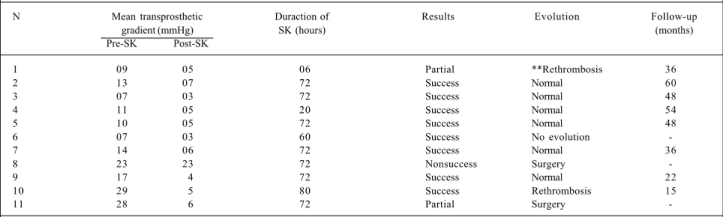
The duration of streptokinase infusion ranged from 6 to 80 (mean of 56) hours, being 34 hours in the patients who partially responded to the fibrinolytic agent, 44 hours in those who achieved no success, and 66 hours in those with resolution of the thrombosis (Tables III and IV).
Of the 4 patients with a single-disk prosthesis, only 1 (25%) patient partially benefited from the fibrinolytic treat-ment. One patient died due to an intracranial hemorrhage, and the other 2 were referred for prosthesis replacement, but with no diagnostic confirmation by the surgeon, who repor-ted the presence of a tissue formation suggestive of pan-nus surrounding the prosthetic ring.
Of the 12 patients with a double-disk prosthesis, com-plete resolution of the thrombosis occurred in 10 (83%). Of the 2 patients with partial success, 1 was clinically mana-ged, and the other was referred for surgical treatment, during which the prosthetic thrombosis was confirmed.
The mean time interval between prosthetic implanta-tion and thrombosis was longer in the single-disk prosthe-ses than in the double-disk prostheprosthe-ses (103 vs 33 months).
Table I – Characteristics of the patients with a prosthesis in the aortic position
N Sex Age (years) Type and size of the Time between prosthesis implantation Cardiac rhythm thrombosed prosthesis and thrombosis (months)
1 F 60 ** CarboMedics Ao / 25 8 Sinus
2 M 52 ** CarboMedics Ao / 25 108 Atrial flutter
3 F 52 * Omniscience Ao / 21 136 Sinus
4 M 40 * Omniscience Ao /25 72 Sinus
5 F 42 ** St Jude Ao 36 Sinus
6 M 32 ** St Jude Ao 48 Sinus
F = female; M = male; * = single-disk; ** = double-disk; Ao = aortic.
Table II - Characteristics of the patients with prosthesis in the mitral and tricuspid positions
N Sex Age (years) Type and size of the Time between prosthesis implantation Cardiac rhythm thrombosed prosthesis and thrombosis (months)
1 F 21 * M. Hall Tric / 25 84 Juncional
2 F 24 ** TRI-technology Mi / 31 6 Sinusal
3 F 37 ** CarboMedics Tric / 29 96 Sinusal
4 M 38 ** TRI-technology Mi / 31 7 Fibril. Atrial
5 F 53 ** CarboMedics Mi / 31 38 Sinusal
6 F 59 ** CarboMedics Mi / 31 1 Fibril. Atrial
7 F 12 *** Labcor Tric / 29 2 Sinusal
8 M 35 * Omniscience Mi / 29 120 Fibril. Atrial
9 F 52 ** TRI-technology Tric / 29 1 BAVT
10 F 52 ** CarboMedics Mi / 29 36 Sinusal
11 F 38 ** ATS Mi / 29 60 Sinusal
The most common hemorrhagic complication was hema-toma in the venous puncture site. One (5.8%) fatal cranial bleed occurred, as already cited, in a patient with a single-disk prosthesis in the 36th hour of streptokinase infusion.
Three (17.6%) embolic phenomena occurred. One patient had peripheral embolism to the left radial artery, which resolved with maintenance of streptokinase. A se-cond patient had an ischemic stroke; streptokinase infusion was interrupted, bleeding was ruled out on cranial tomogra-phy, and, after 2 weeks, the patient was referred for surgery. The third patient had intense precordial pain with elevation of the ST segment in the anterior wall; he was referred to the hemodynamics laboratory. Embolism to the anterior des-cending artery was diagnosed and treated with angioplasty and stent implantation. Two hours later, the patient had a new episode of pain and elevation of the ST segment in the inferior wall, returning to the hemodynamics laboratory, where embolism to the right coronary artery was diagnosed and treated with angioplasty and stent implantation.
Of the 13 patients being discharged from the hospital, 1 did not return for follow-up and the other 12 underwent a mean follow-up of 42 months. Four patients (33%) had clini-cal, fluoroscopic, and echocardiographic rethrombosis within a mean interval of 16 months. Of these 4 patients, 2 had single-disk prostheses with residual difficulty in disk mobility; the other 2 had double-disk prostheses and suc-cessfully responded to the initial treatment. One patient with a mitral double-disk prosthesis refused to undergo the
surgical treatment, and as the patient had already received streptokinase within the 12 preceding months, the following medication scheme was prescribed: 10,000 U of subcuta-neous heparin, twice a day, 200 mg of aspirin, and an oral an-ticoagulant agent for 3 months. The patient evolved with normalization of the mobility of the disks and a drop in the transprosthetic gradients, which remained unaltered after 7 months. For the other 3 patients, a new fibrinolytic treat-ment was indicated: 1 patient with an aortic double-disk prosthesis successfully responded, but, 12 months later, a new thrombosis occurred in the prosthesis, and the patient was referred for valvular replacement, during which the diagnosis was confirmed. The other patient with an aortic double-disk prosthesis had an episode of cerebral embo-lism, and, after stabilization, was referred for surgery, during which thrombosis was confirmed. The patient with a tri-cuspid single-disk prosthesis successfully responded to streptokinase infusion, and, on the control performed after 17 months of follow-up, the prosthesis was functioning normally. The patient with the bioprosthesis is asymptoma-tic and the transprostheasymptoma-tic gradient on echocardiography was low on the 36-month follow-up.
Discussion
The diagnosis of prosthetic thrombosis should always be remembered in patients with clinical findings of embolism
Table III - Aortic prosthesis. Short and long-term results
N Maximum transprosthetic Duration of SK Results Evolution Follow-up
gradient (mmHg) (hours) (months)
Pre-SK Post-SK
1 68 24 24 Partial ** Rethrombosis 39
2 33 17 72 Success Normal 51
3 110 60 36 * Nonsuccess -
-4 64 64 24 Nonsuccess Surgery
-5 102 42 72 Success **Rethrombosis 11
6 80 30 6 Success Normal 6
SK = streptokinase; * = death; ** = fibrinolytic.
Table IV - Mitral and tricuspid prosthesis. Short and long-term results
N Mean transprosthetic Duraction of Results Evolution Follow-up
gradient (mmHg) SK (hours) (months)
Pre-SK Post-SK
1 09 05 06 Partial **Rethrombosis 36
2 13 07 72 Success Normal 60
3 07 03 72 Success Normal 48
4 11 05 20 Success Normal 54
5 10 05 72 Success Normal 48
6 07 03 60 Success No evolution
-7 14 06 72 Success Normal 36
8 23 23 72 Nonsuccess Surgery
-9 17 4 72 Success Normal 22
10 29 5 80 Success Rethrombosis 15
11 28 6 72 Partial Surgery
or heart failure of recent onset, and symptoms of low cardiac output, mainly in patients with inadequate anticoagulation.
The isolated calculation of transprosthetic gradients is not sufficient to establish the diagnosis of prosthetic throm-bosis, because these gradients may be elevated due to chan-ges in flow, especially in the single-disk prostheses and those with small diameters. Therefore, a serial echocardiographic assessment is fundamental for the patients, to allow a longi-tudinal comparison of the gradients and the differential diagnosis between acute dysfunction due to thrombosis and that due to prosthesis-patient mismatch. In these cases, the clinical scenario is not acute, the prosthesis being usually implanted in the patient while still young. However, the diag-nosis of thrombosis may be confirmed if images of the throm-bus or anomalies in the mobility of the disks are visualized on transesophageal echocardiography or fluoroscopy. Fluoros-copy has helped in diagnosing prosthetic obstruction due to thrombosis, mainly in double-disk prostheses, in which the 2 disks may be compared in regard to mobility and opening angles.
The distinction between pannus and thrombus is complex, because both may present as masses and impede opening and closing of 1 or 2 disks. The old prostheses and the chronicity of the clinical findings were the major clinical characteristics found in a series of 12 patients with pannus reported by Sanchez et al 7.
Thrombosis of the prosthesis is a permanent risk for car-diac valvular prostheses, despite the anticoagulant therapy and improvement in their performance. The surgery for pros-thesis replacement has been the traditional treatment, but the surgical risk ranges from 0 to 69%, according to some se-ries8,11,12, causing the search for other therapeutic possibilities.
Fibrinolytic therapy has been described as an alterna-tive to surgical treatment, being considered the first-line treat-ment in prosthetic thrombosis in the tricuspid position 8,9.
The use of a fibrinolytic agent for treating thrombosis in val-vular prostheses in the mitral and aortic positions remains controversial. Some authors have indicated fibrinolytic the-rapy only for critically ill patients, in NYHA functional class III or IV, in whom the surgical intervention is of high risk, or in patients with contraindications 8-12. The controversy in
re-gard to the use of fibrinolytic therapy in patients in functional class I or II is based on the low surgical risk observed in this group of patients as compared with the thromboembolic risk caused by fibrinolysis, which ranges from 12 to 17% 10-13. On
the other hand, some authors have indicated fibrinolysis as the first line of therapy in patients with St. Jude prostheses with a low risk of permanent complications and an excellent chance of success 3. Among us, Campagnucci et al 5 reported
the results of fibrinolytic therapy for treating prosthetic thrombosis in 8 patients with a success rate of 100% and fatal complication rate of zero. More recently, Baptista Filho et al 6
reported the successful use of streptokinase in a 75-year-old female patient with thrombosis in a metallic prosthesis in the
mitral position on the 45th postoperative day of myocardial revascularization.
In our study, we selected the stable patients or those with elevated surgical risk due to antecedents of 1 or more previous surgeries, or patients, who, due to some reason, had their anticoagulation temporarily suspended or poorly controlled, ie, who had a transient reason for having a pros-thetic thrombosis.
The subcutaneous use of heparin in association with an anticoagulant and an antiplatelet agent has also been in-dicated for patients with partial thrombosis of the prosthesis who are mildly symptomatic 8. In this series, 1 patient with
rethrombosis successfully responded to that therapeutic strategy.
One patient died and the mortality rate (5.8%) was ac-ceptable considering the severity of prosthetic thrombosis. The risk of embolism was greater than that of bleeding (15% vs 5.8%). Peripheral embolism was resolved with mainte-nance of the fibrinolytic treatment in agreement with the lite-rature 14. The patient who had a stroke had his streptokinase
suspended, underwent cranial tomography to rule out bleeding, and was referred for surgery 2 weeks later. The third patient complicated with 2 coronary embolisms, which were treated with a percutaneous intervention. A hemorrha-gic complication was fatal in 1 patient.
Although the immediate postoperative period may be considered a relative contraindication to fibrinolytic thera-py, 2 patients with prostheses in the tricuspid position re-ceived streptokinase while at the hospital, approximately 30 days after valvular replacement, and developed no hemor-rhagic complications 8,15.
In patients with single-disk prostheses, the success rate was zero, the nonsuccess rate was 75%, and partial success was obtained only in 1 patient, who evolved with rethrombosis of the prosthesis in the 19th month of evolu-tion. This may have been due to the advanced age of the prostheses (mean time of implantation of 103 months) and to their own design. These factors propitiate the formation of pannus and allow the fibrinolytic therapy to partially solve the prosthetic obstruction or not alter it at all.
On the other hand, the nonsuccess rate was zero in the double-disk prostheses with a shorter interval of implanta-tion, thrombosis being the primary cause of obstruction.
Based on the mean time of streptokinase infusion, the nonsuccess rate was also observed to be greater in those re-ceiving streptokinase for a shorter period.
1. Edmunds LH. Thromboembolic complications of current cardiac valvular prostheses. Ann Thorac Surg 1982;34:96-106.
2. Luluaga IT, Carrera D, D’Oliveira J, et al. Successful thrombolytic therapy after acute tricuspide valve obstruction. Lancet 1971;1:1067-8.
3. Silber H, Khan SS, Matloff JM, Chaux A, DeRobertis M, Gray R. The St. Jude valve: thrombolysis as the first line of therapy for cardiac valve thrombosis. Circulation 1993;87:30-7.
4. Vasan RS, Kaul U, Sanghvi S, et al. Thrombolytic therapy for prosthetic valve: a study based on serial Doppler-echocardiographic evaluation. Am Heart J 1992;123:1575-80.
5. Campagnucci VP, Sukuzi HY, Franken RA, Rivetti LA. Terapêutica trombolítica nas tromboses de próteses mecânicas. Arq Bras Cardiol 1994, 63:35-8. 6. Baptista Filho MLA, Succi JE, Galantier M, et al. Terapêutica trombolítica em
trom-bose de prótese mitral e átrio esquerdo. Rev Bras Cardiol Invas 2001;9:34-8. 7. Sänchez Ramos JG, Diaz J, Moreno G, Navarrete A, Prades I, Martin P. Predictores
clínicos de trombosis em prótesis valvulares mecânicas. Rev Soc Andal Card 1998;16:45
References
8. Lengyel M, Fuster V, Keltai M, et al. Guidelines for management of left-sided prosthetic valve thrombosis: a role for thrombolytic therapy. J Am Coll Cardiol 1997;15:1521-6
9. Peterfly A, Henke A, Savidge GF, Landon C, Bjork VO. Late thrombotic malfunc-tion of the Bjork-Shiley tilting disc valve in the tricuspide posimalfunc-tion. Scand J Thorac Cardiovasc Surg 1980;14:33-8.
11. Birdi I, Angelini GD, Bryan AJ. Thrombolytic therapy for left-side prosthetic valve thrombosis. J Heart Valve Dis 1995;4:154-9.
12. Roudat R, Labbe T, Lorient Roudaut MF, et al. Mechanical cardiac valve throm-bosis: is fibrinolysis justified? Circulation 1992:86 (suppl II) II-8-15. 13. Witchitz S, Veyrat C, Moisson P, Scheinman N, Rozenstajn L. Fibrinolytic
treat-ment of thrombus on prosthetic heart valves. Br Heart J 1980;44:545-54. 14. Kurzrock S, Singh AK, Most AS, Williams DO. Thrombolytic therapy for
pros-thetic cardiac valve thrombosis. J Am Coll Cardiol 1987;9:592-8.
15. Asante Korang A, Sreeram N, McKay R, Arnold R. Thrombolysis with tissue ty-pe plasminogen activator following cardiac surgery in children. Int J Cardiol 1992;35:317-22.
Editor da Seção de Fotografias Artísticas: Cícero Piva de Albuquerque