Instituto de Cardiologia do Rio Grande do Sul/Fundação Universitária de Cardiologia Mailing address: Stelamaris Luchese - Unidade de Pesquisa do IC/FUC Av. Princesa Isabel, 395 - 90620-001 - Porto Alegre, RS – Brazil E-mail: [email protected]
Received - 7/30/02 Accepted - 1/14/03
Arq Bras Cardiol, volume 81 (nº 4), 405-10, 2003 Stelamaris Luchese, João L. Mânica, Paulo Zielinsky
Porto Alegre, RS - Brazil
Intrauterine Ductus Arteriosus Constriction. Analysis
of a Historic Cohort of 20 Cases
Premature constriction of the ductus arteriosus, in the absence of triggering factors, is considered a rare alteration, and experience with this defect is poor, both during preg-nancy and in the neonatal period. Studies have demonstra-ted evidence of important repercussions, leading to heart fai-lure and hydrops and may result in fetal or neonatal death in long-term cases 1-3. The delay in diagnosis can lead to a
per-sistent pulmonary hypertension in newborns, sometimes not responsive to available therapeutic interventions 4,5.
Fortu-nately, early diagnosis enables therapeutic intervention with improvement in the prognosis. If associated with the use of prostaglandin inhibitors and if the causal agent is removed in the beginning of the clinical picture, total recovery of the al-terations may occur, without evidence of neonate complica-tions 6,7. However, constriction of the ductus arteriosus may
be related to severe alterations when the diagnosis is delayed, fetal monitoring with echocardiography being of utmost im-portance 8.
Methods
We reviewed the examinations and charts of the pregnant women undergoing fetal echocardiography at the Fetal Cardio-logy Unit (FCU) of the Instituto de Cardiologia do Rio Grande do Sul, from August 1997 to August 2001, in a population of 7000 pregnant women. To obtain the echocardiograms, we used ATL, (Advanced Technological Laboratories), model ULTRAMARK 9 Digital Plus Acuson model XP-10, and Acuson Aspen, with convex transducers 7 or 5 MHz and/or a sectorial phased array of 3.5 or 5 MHz. All examinations were performed by ICU pediatric cardiologists with experience in fetal echocardiography. Constriction of the ductus was determined by the presence of turbulent flow in the ductus arte-riosus, associated with systolic velocity greater than 1.4 m/s and diastolic velocity greater than 0.3 m/s, in association with a pulsatility index smaller than 1.9 9, which was calculated by the
systolic velocity minus the diastolic velocity, divided by the mean velocity. In patients with total occlusion of the ductus ar-teriosus, the absence of ductal flow was analyzed.
Twenty fetuses fulfilled the diagnostic criteria and
for-Objective – To describe the relative incidence, presentation, and evolvement of fetuses with early ductus constriction.
Methods – Twenty fetal echocardiograms indicating ductus constriction were reviewed in a population of 7000 pregnants.
Results – The cases were divided into group A (rela-ted to maternal use of cyclooxygenase inhibitors [n=7] and group B (idiopathics [n=13]). Mean gestational age was 32.5±3.1 (27-38) weeks and maternal age was 28.2±8.5 (17-42) years. Mean systolic velocity in the ductus was 2.22±0.34 (1.66-2.81) m/s, diastolic velocity 0.79±0.28 (0.45-1.5) m/s, and pulsatility index 1.33±0.36 (0.52-1.83). Two cases of ductal occlusion were noted. In 65% of the cases, an increase occurred in the right cavi-ties; in 90% of the cases, tricuspid or pulmonary regurgi-tation, or both, occurred, with functional pulmonary atre-sia in 1 case. Diastolic velocity was greater in group A (1.13±0.33) than in group B (0.68±0.15) (P=0.008). The other data were similar in the 2 groups. The evolvement was not favorable in 4 patients from group B, including 1 death and 2 cases of persistent pulmonary hypertension.
Conclusion – The high incidence of idiopathic cons-triction of the ductus arteriosus suggests that its diagnosis is underestimated and that many cases of persistence of fetal circulation in newborns may be related to constric-tion of the ductus arteriosus not diagnosed during intrau-terine life. Group B had a lower severity but a risk of an unfavorable evolvement, suggesting a distinct alteration.
med the study group. The cases detected were followed up with fetal or neonatal echocardiography, and charts were re-viewed. We did not include fetuses whose follow-up was not performed or those who had structural heart defects. The ca-ses were divided into group A, with 7 fetuca-ses in which the cause was secondary to maternal use of prostaglandin inhibi-tors, and group B, with 13 fetuses in which the cause was not determined and was therefore considered idiopathic. Gesta-tional age was determined based on the last menstrual period. Maternal age and obstetric history including the number of pregnancies and labors were obtained from chart data. He-modynamic involvement was considered mild in the presen-ce of mild tricuspid regurgitation and/or mild pulmonary re-gurgitation; moderate in the presence of tricuspid or pulmo-nary regurgitation associated with right cavity enlargement; severe in the presence of severe tricuspid and/or pulmonary regurgitation, pulmonary functional atresia, cavity enlarge-ment with right ventricular hypertrophy and/or contractile dysfunction, or total occlusion of the ductus arteriosus, fetal hydrops, and, alternatively, in the presence of a pulsatility index lower than 1 associated with any hemodynamic reper-cussions. To evaluate hemodynamic involvement between the groups, the severity score was formulated, considering 0 the absence of hemodynamic alterations, 1 the mild ment, 2 the moderate involvement, and 3 the severe involve-ment. Evolvement was considered favorable when regres-sion of the alterations occurred and in the absence of any as-sociated complications.
In the statistical analysis, numerical data are presented as mean ± standard deviation (SD), median and interquartile range (25th percentile [P25], 75th percentile [75]). For com-parison of the variables, gestational age and maternal age, systolic velocity, diastolic velocity and pulsatility index, whose distributions were symmetric (normal) between the groups, the Student t test was used. For the analysis of the severity score variable, the Mann-Whitney U test was used, and for the evolvement variable, Fisher’s exact test was used, and odds ratio and 95% confidence interval (CI 95%) were calculated. In the statistical analysis, we accepted P<0.05 as statistically significant.
Results
The group studied comprised 20 fetuses with diagno-ses of premature constriction of the ductus arteriosus, and we performed 47 fetal echocardiograms. The same examiner with expertise in fetal cardiology diagnosed 85% (17) of the cases. Gestational age ranged from 27 to 38 weeks, with a mean age of 33±2.9 weeks and a median of 28 weeks. Mater-nal age ranged from 17 to 42 years, with a mean age of 27± 8.4 years and a median of 28 years. Concerning obstetric history, the median number of pregnancies was 2 and the median number of labors was 1. The maximum number of pregnancies reported was 7 for 6 labors.
Comparing groups A and B, we did not observe sig-nificant differences regarding gestational age and maternal age (P=0.19 and P= 0.58, respectively) (tab. I).
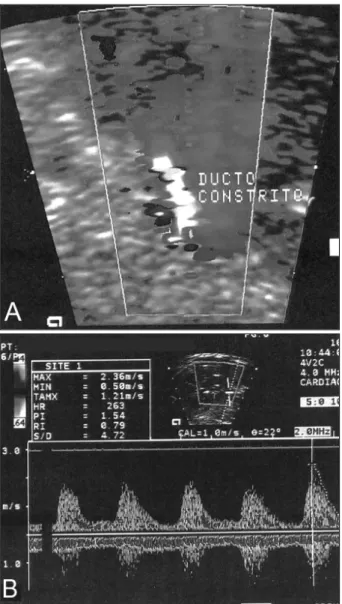
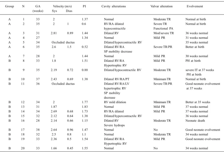
The study of velocity flow from the patent ductus ar-teriosus demonstrated systolic velocity peak, ranging from 1.66 to 2.81 m/s (mean = 2.22±0.34m/s), and diastolic velocity ranging from 0.45 to 1.5 m (mean = 0.79± 0.28m/s). Mean pulsatility index was 1.33±0.36, ranging from 0.52 to 1.83 (fig. 1A, B). In table II, we describe the total and comparative values of systolic and diastolic velocities and pulsatility index between the patients whose cause was related to the use of cyclooxygenase-inhibiting drugs (group A) and the patients whose cause was not deter-mined (group B). In 2 fetuses, we verified total occlusion of the ductus arteriosus (tab. II).
Regarding cause, only 7 of the 20 cases were related to the use of cyclooxygenase-inhibiting drugs (group A), cor-responding to 35% of the cases. Associated drugs include indomethacin in 3 pregnant women, aspirin in 2 women, and sodium diclofenac in 2 women. In the other 13 fetuses (group B) no maternal history existed of drugs capable of interfering with the prostaglandin route. Table III demons-trates maternal and gestational age, the number of pregnan-cies and labors, the indication for examination and the medi-cations used.
In table IV, we list all the patients from the sample, and we describe the echocardiographic data, including evalua-tion of the ductus flow in the Doppler examinaevalua-tion, hemody-namic repercussions (cardiac cavity involvement, valvular alterations, signs of cardiac failure), and the evolvement of the patients. Regarding hemodynamic repercussions, apart from 2 fetuses (10%), the other patients experienced tricus-pid and/or pulmonary regurgitation, which varied from mild to severe, with functional pulmonary atresia in 1 fetus. Thir-teen fetuses (65%) had an increase in the right atrium or right ventricle, which was important in most cases. Hypertrophy, or right ventricle contractile dysfunction, or both, occurred in 7 cases (35%). One of the fetuses had signs of decompen-sated heart failure with severe hydrops. In 2 fetuses, one in each group, with important hemodynamic repercussions, the septum primum was displaced to the left, with a decrease in its mobility, and in one of these fetuses we also observed pulsatile umbilical venous flow.
All the 7 cases with a history of the use of prostaglandin drug inhibitors evolved favorably after suspension of the medications. Five patients underwent control fetal echo-cardiographic examination, performed 4 days and 7 weeks after suspension of the medication, which demonstrated normaliza-tion of the altered findings. Two fetuses were followed-up after birth and both evolved without complications.
Table I – Gestational and maternal age
Age years Group A (n=7) Group B (n=13) P Mean (SD) Median Mean (SD) Median
Gestational 31.9 ± 3.3 33 32.9 ± 3.1 33 0.19 Maternal 29 ± 8.9 28 27.8 ± 8.6 28 0.58
In the 13 cases where the cause was not determined, the evolvement was the following: in 9 cases (69.2%), the echocardiogram demonstrated normalization of flows, 6 du-ring fetal life and 3 dudu-ring the neonatal period. One fetus in this group had ductus occlusion; the diagnosis was confirmed just after birth, the right cavity enlargement re-gressed, and the follow-up was satisfactory. In one case, where persistence of alterations in the fetal echocardiogram was observed, the evolvement was favorable after birth, no further abnormalities were detected, and the patient
evolved without complications. Two patients, with worse-ning in the control fetal echocardiogram, had persistent pul-monary hypertension as newborns, requiring mechanical ventilation and prolonged hospitalization; afterwards, these alterations regressed and the patients evolved wi-thout apparent sequelae. One patient with clinical features of severe hydrops evolved in preterm delivery nonrespon-sive to inhibition of premature labor, and the newborn, extre-mely premature, died within the first hours of birth. In 4 pa-tients with unfavorable results, 1 death occurred before the institution of adequate therapeutic measures. In another fetus, with worsening in contractile dysfunction in the con-trol examination performed in the 37th week of pregnancy, interruption of pregnancy was recommended. In the other 2 fetuses, we opted for expecting conduct, monitoring with fetal echocardiography (tab. IV).
Table V demonstrates the evolvement of patients in both groups, classifying it as favorable or unfavorable. Table VI presents a severity score for each of the groups.
Discussion
Doppler echocardiography evaluation is sensitive for diagnosing intrauterine constriction of the ductus arterio-sus. To avoid confusion in the diagnosis because of an in-crease in ventricular output, only fetuses with a pulsatility index lower than 1.9 were included in the study 9. In one of
the fetuses admitted with a diagnosis of aortic coarctation, the examination was repeated in the neonate period, this diagnosis then being eliminated.
Ductal constriction usually occurs after the use of cy-clooxygenase-inhibiting drugs and is reversible after dis-continuation of the medication, especially when diagnosed early 10. With the increase in gestational age, the ductus
be-comes more sensitive to constricting factors 8,11,
demons-trating an increased incidence of ductus constriction after the 31st gestational week 12, being rare before the 27th
week. According to these data, we observed a mean gesta-tional age of 33 weeks and a minimum of 27 weeks.
Ductal constriction is rare, when not related to the use of prostaglandin inhibitors 13-15. The high number of
idio-pathic cases raises some questions, among them the possi-bility that this diagnosis is already underestimated. The per-formance of fetal echocardiography in a referral service, conducted by an experienced examiner, may have reduced the false-negative results. We observed a high number of cases coming from the private sector, which are usually
mo-Table II – Flow in ductus arteriosus
Group A Group B Total P
Mean (SD) Mean (SD) Mean (SD)
Systolic velocity (m/s) 2.28 ± 0.39 2.20 ± 0.33 2.22 ± 0.34 0.68
Diastolic velocity (m/s) 1.13 ± 0.33 0.67 ± 0.17 0.78 ± 0.29 0.008
Pulsatility index 1.12 ± 0.43 1.43 ± 0.28 1.34 ± 0.36 0.08
Student t test.
Table III- Clinical characteristics of cases
Group N GA Mother Gestation/ Indication Medications
(weeks) (years) labor
A 1 33 42 IV/III Maternal heart disease AAS, amiodarone, digital
A 2 35 39 II/I DMG, polyhydramnios cardiomegaly Sodium diclofenac
A 3 31 24 II/I Hypertension, polyhydramnios Indomethacin
A 4 27 28 III/II Suicide attempt Indomethacin
A 5 34 30 VII/VI GDM, polyhydramnios Indomethacin
A 6 35 23 II/I Fetal arrhythmia Sodium diclofenac
A 7 28 17 I/0 Diabetes mellitus AAS, Levonorgestrel + Ethynyl estradiol
B 8 33 19 I/0 Oligohydramnios/IURG No
B 9 27 32 II/I Gemellary/hydrocephali No
B 10 37 17 I/0 Clinical appointment (day F) No
B 11 36 35 II/I Suspect of AOCO No
B 12 34 21 I/0 Clinical appointment No
B 13 31 32 V/III GDM No
B 14 34 36 III/I Clinical appointment No
B 15 32 25 II/0 Clinical appointment Bromhexine Chlorinate
B 16 28 41 III/II Severe hydrops No
B 17 38 17 I/0 Right kidney hypoplasia No
B 18 32 20 II/I Single umbilical artery Isometheptene
B 19 33 28 II/I Fetal arrhythmia No
B 20 33 39 IV/I Clinical appointment Propolis
AOCO- aortic coarctation; GA- gestational age; GDM- gestational diabetes mellitus; IUGR- intrauterine retard growth.
Table IV – Doppler-echocardiography and evolvement data
Group N GA Velocity (m/s) PI Cavity alterations Valvar alteration Evolvement (weeks) Sys Dias
A 1 33 2 1.37 Normal Moderate TR Normal at birth
A 2 35 2 1 0.6 RV/RA dilated Severe TR Normal at birth
hypocontractile Functional PA
A 3 31 2.81 0.89 1.44 Dilated RV Mod/severe TR 36 weeks normal
A 4 27 1.34 Normal Mild PR 31 weeks normal
A 5 34 Occluded ductus Dilated/hypercontractile RV 35 weeks normal
A 6 35 2.6 1.5 0.52 Dilated RV/RA Severe TR/PR Better at birth
SP mobility decrease
A 7 28 2 1.44 Normal Mild PR 30 weeks normal
B 8 33 1.8 1.51 Dilated RV/RA Mild PR PH at birth
Hypertrophic RV
B 9 35 2.19 0.72 0.88 Dilated/hypocontractile RV Moderate TR severe IT at 37 weeks PH at birth
B 10 37 2.43 0.69 1.38 Dilated RV/RA/PT Minimum TR Normal at birth
B 11 36 Occluded ductus Dilated RV/RA/LV Severe TR/PR Good neonate evolvement
hypertrophic RV at 37 weeks
SP mobility decrease
B 12 34 2 1.77 RV mild dilation Minimum TR Better at 35 weeks
B 13 31 1.87 1.83 Normal Mild PR 37 weeks normal
B 14 34 2.69 0.68 1.73 RV/RA dilated Mild PR 37 weeks normal
B 15 32 2.12 0.64 1.38 Dilated/hypercontractile RV 36 weeks normal
B 16 28 2.14 0.66 1.15 Dilated RV Moderate TR Neonate death
Severe hydrops
B 17 38 2.64 0.96 1.47 Normal No Good neonate evolvement
B 18 32 2.5 0.8 1.1 Normal Moderate TR 34 weeks normal
B 19 33 2.36 0.5 1.54 Dilated RV/RA Mild PR Good neonate evolvement
Hypertrophic RV
B 20 33 1.66 0.45 1.55 Normal No 34 weeks normal
Table V - Evolvement
Favorable Unfavorable
Group A 7 0
Group B 9 4
Fisher’s exact test = 0.25; odds ratio = 7.1 (IC 95% 0.33 – 153.77).
Table VI – Hemodynamic involvement
Severity score
Mean (SD) Median (P25 – P75)
Group A 2.14 ± 0.9 2 (1.0 – 3.0) Group B 2.08 ± 1.12 2 (1.8 – 3.0)
Mann-Whitney test; p=10.
nitored with obstetric echocardiography, increasing the possibility of finding abnormalities. The fact that all preg-nant women sent for clinical appointments were from group B (undetermined causes) reinforces the importance of fetal echocardiography.
A study evaluating the presence of antiinflammatory medication in the meconium 16 demonstrated a positive
as-sociation with pulmonary hypertension in newborns. There was a weak agreement with maternal history, probably be-cause of free access to combined medications 16. It is
ques-tionable whether a substance with antiinflammatory action used as tea or natural medication may be deleterious. For this investigation, a directed questionnaire may be required to meet the Bradford-Hill casualty criteria. The association with drugs that enhance the sensitivity of the ductus arte-riosus, such as retinoic acid 17,18, should be investigated. We
reported the use of other medications in both groups, however, with unknown effects on prostaglandin inhi-bition. We did not observe the association of corticoid with indomethacin, that are synergic factors 20.
Regarding echocardiographic data, diastolic velocity was significantly greater in the group with undetermined causes (group B). As 65% of the patients (13/20) had right ventricular dilation or contractile dysfunction, or both, the evaluation of systolic velocity may be hindered, just as the pulsatility index is, because its assessment requires systolic velocity. Diastolic velocity may be a more reliable index, suggesting that the group related to the use of drugs is more affected, thus having a more favorable prognosis. Most patients (90%) had functional involvement, 75% (15/ 20) were moderate to severe. We did not observe differen-ces in the severity between groups, and one case occurred in each group of total occlusion of the ductus arteriosus and decrease in the mobility of the septum primum .
According to data from the literature, sodium diclo-fenac and indomethacin are more powerful inhibitors of cy-clooxygenase than aspirin is 21. In this study, all cases
re-lated to the use of prostaglandin inhibitors (group A) evol-ved favorably. It is expected that cardiac function
normali-zes within 24 hours, or a few days after medication is sus-pended 10. We observed normalization in control
examina-tions 4 days to 7 weeks after drug suspension.
In the 13 cases where the use of prostaglandin inhibi-tors was not identified (group B), 9 fetuses (69.3%) impro-ved in clinical condition, 4 fetuses (30.7%) showed worse-ning on the fetal echocardiogram, 2 fetuses had persistence of fetal circulation after birth, and 1 fetus died. Responsible factors for unfavorable evolvement may be related to intrin-sic alterations in the ductus arteriosus, interfering with the prostaglandin or nitric oxide route, or even enhancing sen-sitivity to extrinsic constrictor factors 22-24. Another
possi-bility is the potential delay in diagnosis and the impossibi-lity of withdrawing the causal agent. With the available data, we may estimate a -7.1-times greater relative risk for an unfa-vorable evolvement in patients with undetermined causes (group B), although it has not reached statistical significan-ce (CI 95% >0.33 -153.77).
After ductal mechanical occlusion 25 or secondary to
antiinflammatory medication 26,27, the increase in muscular
pulmonary artery and pulmonary hypertension was de-monstrated 3,12,28,29. The patients from this study that
evol-ved with persistence of fetal circulation after birth needed prolonged neonatal intensive care unit hospitalization but had a good response to treatment and satisfactory evolve-ment. A prior intrauterine diagnosis may have contributed favorably. It is estimated that pulmonary hypertension in newborns is idiopathic in 23% of cases 30. It may be
sugges-ted that these cases are relasugges-ted to undocumensugges-ted ductal constriction in fetal life, because of the number of idiopathic cases observed in this study, 15% of which (2/13) had pul-monary hypertension. None of these cases was related to maternal diabetes, a condition that may be associated 30.
Echocardiography is indicated in nonimmune fetal hydrops, aiming at discarding ductus arteriosus constric-tion 3,31,32. The fetus with cavity enlargement and severe
hy-drops evolved with premature labor and died. In this case, we cannot infer the duration of ductal occlusion because of the precocious death.
In previous studies 33,34, we observed that the use of
nonhormonal antiinflammatory medications during preg-nancy was related to the increase in ductus arteriosus pa-tency in newborns. None of our patients had this compli-cation; however, examining this was not the purpose of this study.
Currently, measures aiming at relaxation of the ductus arteriosus, in fetal life, are not available. Prostaglandin use in human fetuses for this purpose was not described. Experi-mental studies 35 demonstrate positive effects with the use of
endothelin antagonists, decreasing the consequences of ductal constriction. The possible role of nitric oxide in dilating the fetal ductus arteriosus has already been suggested 23.
Further studies are necessary aimed at specific treatment of premature constriction of the ductus arteriosus.
echo-cardiographic data demonstrated lower severity in the group with idiopathic constriction than in the group with ductal constriction secondary to maternal use of nonsteroi-dal antiinflammatory medication, but with a risk of
unfavo-References
1. Hofstadler G, Tulzer G, Altmann R, Schmitt K, Danford D, Huhta JC. Sponta-neous closure of the human fetal ductus arteriosus: a cause of fetal congestive heart failure. Am J Obstet Gynecol 1996;174:879-83.
2 Mielke G, Steil E, Breuer J, Goelz R. Circulatory changes following intrauterine closure of the ductus arteriosus in the human fetus and newborn. Prenat Diagn 1998;18:139-45.
3. Downing GJ, Thibeault DW. Pulmonary vasculature changes associated with idiopathic closure of the ductus arteriosus and hydrops fetalis. Pediatr Cardiol 1994;15:71-5.
4. Zenker M, Klinge J, Krüger C, Singer H, Scharf J. Severe pulmonary hypertension in a neonate caused by premature closure of the ductus arteriosus following maternal treatment with diclofenac: a case report. J Perinat Med 1998;26,231-4. 5. Niebyl JR, Witter FR. Neonatal outcome after indomethacin treatment for preterm
labor. Am J Obstet Gynecol 1986;155:747-9.
6. Moise KJ Jr. Effect of advancing gestational age on the frequency of fetal ductal constriction in association with maternal indomethacin use. Am J Obstet Gynecol 1993;168:1350-3.
7. Bivins HA, Jr Newman RB, Fyfe DA, Campbell BA, Stramm SL. Randomized trial of oral indomethacin and terbutaline sulfate for the long-term suppression of preterm labor. Am J Obstet Gynec 1993;169:1065-70.
8. Vermillion ST, Scardo JA, Lashus AG, Wiles HB. The effect of indomethacin tocolysis on fetal ductus arteriosus constriction with advancing gestational age. Am J Obstet Gynecol 1997;177:256-61.
9. Tulzer G, Gudmundsson S, Sharkey AM, Wood DC, Cohen AW, Huhta JC. Doppler echocardiography of fetal ductus arteriosus constriction versus increased right ventricular output. J Am Coll Cardiol 1991;18:532-6. 10. Moise KJ Jr, Huhta JC, Sharif DS et al. Indomethacin in the treatment of premature
labor. Effects on the fetal ductus arteriosus. N Engl J Med 1988;319:327-31. 11. Tynan M. The ductus arteriosus and its closure. N Engl J Med 1993;18: 1570-2. 12. Turner GR, Levin DL. Prostaglandin synthesis inhibition in persistent
pulmonary hypertension of the newborn. Clin Perinatol 1984;11:581-9. 13. Harlass FE, Duff P, Brady K, Read J. Hydrops fetalis and premature closure of the
ductus arteriosus: a review. Obstet Gynecol Survey 1989;44:541-3. 14. Mielke G, Steil E, Gonser M. Prenatal diagnosis of idiopathic stenosis of the
ductus arteriosus associated with fetal atrial flutter. Fetal Diagn Ther 1997;12:46-9.
15. Yaman C, Arzt W, Tulzer G, Tews G. Spontaneous constriction of the fetal ductus arteriosus. Z Geburtshilfe Neonatol 1999;203:44-6.
16. Alano MA, Ngougmna E, Ostrea Jr EM, Konduri GG. Analysis of nonsteroidal antiinflammatory drugs in meconium and its relation to persistent pulmonary hypertension of the newborn. Pediatrics 2001;107:519-23.
17. Momma K, Toyono M, Miyagawa-Tomita S. Accelerated maturation of fetal ductus ar-teriosus by maternally administered vitamin A in rats. Pediatr Res 1998;43:629-32. 18. Wu G-R, Jing S, Momma K, Nakanishi T. The effect of vitamin A on contraction of
the ductus arteriosus in fetal rat. Pediatric Research 2001;49:747-54. 19. Brezinka CH, Gittenberger-de Groot AC, Wladimiroff JW. The fetal ductus
arteriosus, a review. Zentralbl Gynakol 1993;115:423-32.
20. Levy R, Matitiau A, Ben Arie A, Milman D, Or Y, Hagay Z. Indomethacin and corticosteroids: an additive constrictive effect on the fetal ductus arteriosus. Am J Perinatol 1999;16:379-83.
21. Momma K, Hagiwara H, Konishi T. Constriction of fetal ductus arteriosus by non-steroidal anti-inflamatory drugs: study of additional 34 drugs. Prostaglan-dins 1984;28:527-36.
22. Heymann MA, Rudolph AM. Control of the ductus arteriosus. Physiol Rev. 1975;55:62-78.
23. Momma K, Toyono M. The role of nitric oxide in dilating the fetal ductus arteriosus in rats. Pediatr Res 1999;46:311-5.
24. Coceani F, Liu YA, Seidlitz E, Kuwaki T, Ackerley C, Yanagisawa M. Deletion of the endothelin-A-receptor suppresses oxygen-induced constriction but not postnatal closure of the ductus arteriosus. J Cardiovasc Pharmacol 2000; 36(suppl1):S75-7.
25. Wild LM, Nickerson PA, Morin FC. Ligating the ductus arteriosus before birth remodels the pulmonary vasculature bed of the lamb. Pediatr Res 1989;25:251-7. 26. Levin D, Fixler DE, Morris FC, Tyson J. Morphologic analysis of the pulmonary vascular bed in infants exposed in utero to prostaglandin synthetase inhibitors. J Pediatr 1978;92:478-83.
27. Murphy JD, Rabinovich M, Goldstein JD, Reid LM. The structural basis of persis-tent pulmonary hypertension of the newborn infant. J Pediatr 1981;98:962-7. 28. Levin DL, Hyman AI, Heymann MA, Rudolph AM. Fetal hypertension and the
development of increased pulmonary vascular smooth muscle: A possible me-chanism for persistent pulmonary hypertension of the newborn infant. J Pedia-trics 1978;92:265-9.
29. Manchester D, Margolis HS, Sheldon RE. Possible association between maternal indomethacin therapy and primary pulmonary hypertension of the newborn. Am J Obstet Gynecol 1976;126:467-9.
30. Van Marter LJ, Leviton A, Allred EN et al. Persistent pulmonary hypertension of the newborn and smoking and aspirin and nonsteroidal antiinflammatory drug consumption during pregnancy. Pediatrics 1996;97:658-63.
31. Momma K, Nishihara S, Ota Y. Constriction of the fetal ductus arteriosus by glucocorticoid hormones. Pediatr Res 1981;15:19-21.
32. Räsänen J, Debbs RH, Wood DC, Weiner S, Weil SR, Huhta JC. Human fetal right ventricular ejection force under abnormal loading conditions during the second half of pregnancy. Ultrasound Obstet Gynecol 1997;10:325-32.
33. Norton ME, Merrill J, Cooper BAB, Kuller JA, Clyman RI. Neonatal complica-tions after the administration of indomethacin for preterm labor. N Engl J Med 1993;329:1602-7.
34. Hammerman C, Glaser J, Kaplan M, Schimmel MS, Ferber B, Eidelman AI. Indome-thacin tocolysis increases postnatal patent ductus arteriosus severity. Pedia-trics 1998;102:56.
35. Takizawa T, Horikoshi E, Shen MH, et al. Effects of TAK-044, a nonselective en-dothelin receptor antagonist, on the spontaneous and indomethacin- or methy-lene blue-induced constriction of the ductus arteriosus rats. J Vet Med Sci 2000;62:505-9.