Faculdade de Medicina de Botucatu, UNESP
Mailing address: Henrique Barbosa Ribeiro - Faculdade de Medicina de Botucatu, UNESP - Depto de Clínica Médica - Rubião Júnior, S/N - Cep 18618-000 Botucatu, SP, Brazil - E-mail: [email protected]
Received 12/5/02 Accepted 2/23/03
Arq Bras Cardiol, volume 81 (nº 6), 569-75, 2003
Henrique Barbosa Ribeiro, Katashi Okoshi, Antonio Carlos Cicogna, Edson Antonio Bregagnollo, Maria Aparecida Marchesan Rodrigues, Carlos Roberto Padovani, Flávio Ferrari Aragon,
Elenize Jamas, Marina Politi Okoshi
Botucatu, SP - Brazil
Follow-up Study of Morphology and Cardiac Function in Rats
Undergoing Induction of Supravalvular Aortic Stenosis
Cardiac hypertrophy is an important adapting mecha-nism that occurs as a response to chronic hemodynamic overload, allowing the heart to maintain its basic functions
during overloaded conditions 1,2. However, left ventricular
hypertrophy is a risk factor for the development of
conges-tive heart failure and sudden death 3.
Several experimental models have been proposed for the study of left ventricular hypertrophy due to pressure overload. Among them, the induction of renovascular
ste-nosis 4, of abdominal aorta stenosis 5,6, or thoracic aorta
ste-nosis 7, or even the development of animals genetically
mo-dified, such as spontaneously hypertensive rats 8-12.
Howe-ver, several of these animal models have limitations for use in experimental studies. For example, the induction of reno-vascular stenosis or abdominal aortic stenosis, in rats, in ad-dition to leading to systemic blood hypertension and left ventricular hypertrophy, can also promote systemic activa-tion of the adrenergic nervous system, and of the
renin-an-giotensin-aldosterone system 13-15. The intense
neuro-hor-monal activation is associated with the presence of impor-tant cardiac lesions, such as myocardial necrosis, peripheral
arteritis, and reparative fibrosis 4,6. Thus, these models may
be very aggressive in the myocardium and, therefore, less representative of the chronic lesions that develop gradually in humans with chronic pressure overload. On the other hand, spontaneously hypertensive rats develop a gradual increase in systemic blood hypertension at 1 month of age, and, at 3 months, they already have established left
ventri-cular hypertrophy 16,17. However, these rats have a long
pe-riod of stable myocardial hypertrophy, without heart failure,
which usually occurs by 20 months of age 9,11,18. Therefore,
although in this model cardiac alterations are similar to those
occurring in human hypertensive heart disease 9,16, the
prevention studies of heart failure are expensive, because the animals have to be kept in captivity for a long period.
More recently, the model of ascending aorta stenosis has been used to promote the gradual development of left
ventricular hypertrophy in young rats 7,19-25. The animals, 3
Objective - To characterize the follow-up of an experi-mental model of left ventricular hypertrophy (LVH) induced by supravalvular ascending aortic stenosis in young rats.
Methods - Wistar rats were submitted to thoracotomy and aortic stenosis was created by placing a clip on the ascending aorta (AoS group, n=12). Age-matched control animals underwent a sham operation (C group, n=12). Cardiac function was analysed by echocardiograms per-formed 6, 12, and 21 weeks after aortic banding. Myocar-dial morphological features and myocarMyocar-dial hydroxypro-line concentration (HOP) were evaluated 2, 6, 12, and 21 weeks after surgery in additional animals.
Results - Aortic banding promoted early concentric LVH and a progressive increase in HOP. Under light mi-croscopy, we observed myocyte hypertrophy and wall thi-ckening of the intramural branches of the coronary arteries due to medial hypertrophy. Cardiac function was supra-normal after 6 weeks (percentage of fractional shortening - EAo
6: 70.3±10.8; C6: 61.3±5.4; p<0.05), and depressed in the last period. Diastolic dysfunction was detected after 12 weeks (ratio of early-to-late filling velocity - EAo
12: 4.20±3.25; C12: 1.61±0.16; p<0.05).
Conclusion - Ascending aortic stenosis promotes con-centric LVH with myocardial fibrosis and minimal histolo-gical changes. According to the period of evaluation, cardiac function may be improved, normal, or depressed. The model is suitable and useful for studies on pathophysiology and treatment of the different phases of cardiac hypertrophy.
Key words: myocardial hypertrophy, echocardiogram,
to 4 weeks after delivery, undergo median thoracotomy to place a band, with an internal diameter of approximately 0.6 mm, around the thoracic aorta, 2 to 3 mm above its root. Im-mediately after the thoracic banding, the diameter of the ar-tery is maintained; with animal growth, the vessel diameter is maintained and aortic stenosis progressively appears. The rats develop left ventricular hypertrophy precociously, which is associated, in the short term, with improvement in the systolic function of the heart. Then, the animals start to experience depression of mechanical heart performance, and develop cardiac decompensation, 20 weeks after the
induc-tion of aortic stenosis 7,20,26-28. In this model of left
ventricu-lar hypertrophy, no systemic activation of the sympathic nervous system or of the renin-angiotensin-aldosterone
system occurs 7,21,26,29. Probably because of this fact,
histo-logical evaluation, performed 21 weeks after aortic stenosis induction, did not demonstrate the presence of important cardiac lesions, such as peripheral arteritis, myocardial
ne-crosis, or extensive fibrosis 7,26.
The advantages of this experimental model are the gra-dual development of left ventricular hypertrophy, absence of severe anatomical lesions in the myocardium, and the low cost of the maintenance of animals because of the short pe-riod necessary to develop left ventricular hypertrophy and heart failure. In the several studies performed with this mo-del 7,19-21,26,30,31, different periods were used for the
morpho-logical or functional analysis of the heart, or both, and no studies exist regarding simultaneous and follow-up evalua-tions of myocardial histology and of cardiac function after induction of aortic stenosis.
The objective of this study was to characterize the fol-low-up of the experimental model of left ventricular hyper-trophy because of supravalvular aortic stenosis, in young Wistar rats, using morphologic and functional evaluation of the heart. Myocardial morphology was analyzed, using op-tical microscopy, 2, 6, 12, and 21 weeks after the induction of
aortic stenosis and cardiac function was assessed, in vivo,
using echocardiographic study, performed 6, 12, and 21 weeks after banding of the ascending aorta. In addition to that, the collagen content was quantified using serial determi-nation of hydroxyproline concentration in the left ventricle.
Methods
Male Wistar rats, weighing 90 to 100g, were used from our biotery. Animals were kept in cages with 4 rats per box,
at 23oC, with luminosity cycles of 12h and fed with Purina rat
ration and water ad libitum. The protocol used was appro-ved by the Ethical Committee for Animal Research of the Faculdade de Medicina de Botucatu, UNESP.
Rats underwent median thoracotomy after being anes-thetized with ketamine hydrochloride (50 mg/kg intramuscu-lar) and xylidine hydrochloride (10 mg/kg intramuscuintramuscu-lar). Next, the ascending aorta was dissected and a silver band with an internal diameter of 0.6 mm was placed at approxima-tely 3 mm of the aortic root. During surgery, the rats were manually ventilated with positive pressure. Control animals
underwent the same surgery; however, a band was not implanted.
Rats were divided into 2 groups: control (C) and aortic stenosis (AoS). For morphologic and biochemical evalua-tion (hydroxyproline dose), 5 to 7 animals from each group were sacrificed 2, 6, 12, and 21 weeks after AoS induction. The subgroups formed were given the following designa-tions: C2, C6, C12, C21,and AoS2, AoS6, AoS12, and AoS21.
The other groups of rats (C and AoS) were formed only for functional study. Because this evaluation was per-formed in vivo and without later sacrifice of the animals, the same rats were assessed in 3 experimental periods: 6, 12, and 21 weeks after the induction of AoS (n= 12 for the C and AoS groups). Due to the difficulty in obtaining adequate echocardiographic images in low-weight rats, the functional study was not performed in the 2-week period after AoS induction.
Rats were weighed and anesthetized with sodium pen-tobarbital (50 mg/kg intraperitoneal) then underwent median thoracotomy to remove the heart, which was rapidly washed in saline solution. Next, the right ventricle (RV) and left ven-tricle (LV) were dissected and weighed separately. From the central part of the left ventricle, a 2- to 3-mm thick annulus was cut, taking all the extension of its wall. The material was immersed in neutral and tamponade formalin 10% for 48
hours at 4oC. After this period, the tissue was washed,
dehy-drated, and imbedded in paraffin. The histological cuts 5- to 7-mm thick were dyed with hematoxylin-eosin and were ana-lyzed with optical microscopy.
Morphometric analyses were performed using a VCR attached to a Leica microscope connected to the computer equipped with an analysis program (Image-Pro Plus 3.0, Media Cybernetics, Silver Spring, Maryland, USA). In left ventricle transverse sections, sectional areas (SA) of at least 50 cardiac fibers, whose nucleus was clearly identified in the center of the cell, were measured.
Myocardial concentration of hydroxyproline was as-sessed in tissue obtained at the tip of the left ventricle,
ac-cording to the methods described by Switzer 32, using a
pre-viously described technique used in other studies at our
laboratory 33-35.
The level of ventricular hypertrophy was assessed by the ratio between humid weight of the right and left ventri-cle and the body weight of the animals.
Rats received anesthesia with ketamine hydrochloride (50 mg/kg) and xylidine hydrochloride (1 mg/kg), administe-red intramuscularly. A trichotomy was performed in the an-terior region of the thorax, and the animals were placed in the left lateral decubitus position for the performance of
echocardiography 36 with the Sonos 2000 from
Hewlett-Pa-ckard Co., equipped with an electronic transducer with a 7.5 MHz frequency.
valve plane at the papillary muscle level 22,37,38. Aorta (AO)
and left atrium (LA) images were obtained with the M-mode cursor positioned at the level of the aortic valve plane. Images obtained were recorded afterwards with the UP-890 printer from the Sony Co.; cardiac structures were measured manually with the help of a pachymeter.
Left ventricle diastolic diameter (LVDD) and systolic diameter (LVSD) were measured at the period of the cardiac cycle when their values were maximum and minimum, res-pectively. Diastolic thickness of the posterior left ventricle wall (LVDT) was measured at the same period of the cardiac cycle when left ventricle diastolic diameter was assessed. Aortic root diameter (AO) was obtained in the period imme-diately prior to the opening of the aortic valve. The left atrium (LA) was assessed when its diameter was at its maxi-mum 22,38. From the dimensions above described, we
obtai-ned: relative left ventricle thickness (RLVT), LA/AO, LVDD/ BW, and LA/BW.
Left ventricle systolic function was assessed by using
the shortening percentage (∆D): [(LVDD – LVSD)/LVDD] X
100, and the diastolic function analyzed by the ratio bet-ween initial filling flow velocity (E wave) and of the atrial contraction (A wave) of the transmitral flow.
One day after the performance of echocardiography, corresponding to 21 weeks, the animals were sacrificed while under anesthesia with sodium pentobarbital. At this time, signs of heart failure were identified, according to
Conrad et al 39 and Cicogna et al 11,18 as tachypnea,
pleurope-ricardial effusion, ascites, and the presence of left atrial thrombus.
Regarding the statistical analysis, numerical data are expressed as mean ± standard deviation. Study of the mor-phometric variables and of the myocardial concentration of hydroxyproline was performed with analysis of variance (ANOVA) for the factorial scheme 2X4, for the entirely
ca-sual model, completed with the Tukey test of multiple
compa-risons. Results obtained in the echocardiographic study were analyzed with analysis of variance of the multivariate profiles for dependent groups (MANOVA). Significance level was considered 5%.
Results
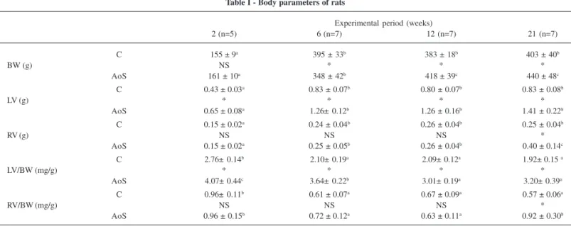
Corporal parameters are provided in table I. Body weight was similar between groups AoS and C in the period 2 weeks after surgery. Significant variations occurred between the groups: after 6 weeks body weight was greater in C; after 12 and 21 weeks, it was lower in the C groups in comparison with that in the AoS groups. Left ventricle weight was always greater in the AoS groups compared with that in the C groups. In the evolvement analysis, left ventricle weight increased significantly between 2 and 6 weeks in the 2 groups, C and AoS, and did not change in the other period of analysis. Right ventricle weight was greater in the AoS groups than in the C groups only in the 21-week period. In the evolvement analysis, RV weight had the same pattern as that of the left ventricle weight in the C groups and demonstrated the
follo-wing results in the AoS groups: AoS2<(AoS6=AoS12)<AoS21.
LV/BW ratio was greater in AoS in the 4 periods assessed. In
the C groups, the ratio was greater in C2 than at other times
and, in rats with AoS it had the following pattern: AoS2
>-AoS6> (AoS12=AoS21). The RV/BW ratio was greater in the
AoS group than in C only in the last period of evaluation. In
the C groups, this ratio was greater in C2 than in the other
pe-riods and, in AoS groups, it was similar in the 2- and 21-week periods, both greater than at 6 and 12 weeks.
Of the 12 animals in the AoS group, which evolved for 21 weeks, 3 had signs of heart failure, including tachypnea, pleural effusion, and ascites.
In the left ventricle histological evaluation, C groups had normal morphology. In the AoS groups, progressive
Table I - Body parameters of rats
Experimental period (weeks)
2 (n=5) 6 (n=7) 12 (n=7) 21 (n=7)
C 155 ± 9a 395 ± 33b 383 ± 18b 403 ± 40b
BW (g) NS * * *
AoS 161 ± 10a 348 ± 42b 418 ± 39c 440 ± 48c
C 0.43 ± 0.03a 0.83 ± 0.07b 0.80 ± 0.07b 0.83 ± 0.08b
LV (g) * * * *
AoS 0.65 ± 0.08a 1.26± 0.12b 1.26 ± 0.16b 1.41 ± 0.22b
C 0.15 ± 0.02a 0.24 ± 0.04b 0.26 ± 0.04b 0.25 ± 0.04b
RV (g) NS NS NS *
AoS 0.15 ± 0.02a 0.25 ± 0.05b 0.26 ± 0.04b 0.40 ± 0.14c
C 2.76± 0.14b 2.10± 0.19a 2.09± 0.12a 1.92± 0.15 a
LV/BW (mg/g) * * * *
AoS 4.07± 0.44c 3.64± 0.22b 3.01± 0.19a 3.20± 0.39a
C 0.96± 0.11b 0.61 ± 0.07a 0.67 ± 0.09a 0.57 ± 0.06a
RV/BW (mg/g) NS NS NS *
AoS 0.96 ± 0.15b 0.72 ± 0.12a 0.63 ± 0.11a 0.92 ± 0.30b
C- control; AoS- supravalvular aortic stenosis; BW- body weight; LV- left ventricle weight; RV- right ventricle weight. ANOVA and Tukey. a, b, c - groups that do not
for BW, the variable had similar patterns in the C and AoS groups, with a progressive reduction in their values during
evolvement assessment (P<0.05). LVSD was smaller in AoS6
than in C6. In the C group, this parameter did not change in
the experimental period, and in the AoS group, the 21-week period was significantly greater than that at 6 weeks. LVDT and LVDT/LVDD were greater in the AoS group in the 3 periods assessed and did not change with the age of the animal or the evolvement of the hypertrophic process. LA diameter was greater in the AoS group in the 3 periods, and
its greatest value was in AoS21 rather than in AoS6 and
AoS12. AO diameter was greater in AoS12 than in C12. The
LA/AO ratio was greater in group AoS in the 3 periods studied. In the 2 groups of animals, LA/AO did not change during evolvement, and LA/BW was greater in period 6 than in the 12- and 21-week periods.
Means and standard deviation of the heart rate (HR), and the systolic and diastolic function indexes are demons-trated in table III. Heart rate was greater in the AoS group in the 12- and 21-week periods, compared with the same period
in group C. In group C, HR was lower in C21 than in C6, and in
AoS this variable did not occur in the 3 periods analyzed.
DD was greater in AoS6 than in C6. In group C, this index did
not change with age, and in the AoS group a decrease oc-curred in its value in the 21-week period compared with that
in the 6- and 12-week periods. E/A ratio was greater in AoS12
and AoS21 compared with that in groups C and AoS6. In the
control group, E/A did not change with age.
Discussion
In this study, we assessed the cardiac function, mor-phology, and development of myocardial fibrosis in young rats undergoing induction of supravalvular aortic stenosis in different periods of follow-up.
AoS induction rapidly encourages left ventricle hy-pertrophy, which is evident in the early period of evaluation, with 2 weeks of follow-up. After 6 weeks, left ventricle weight or ventricular wall diastolic thickness did not increa-se, which could suggest that, after aortic stenosis induc-tion, left ventricular hypertrophy occurs precociously and hypertrophy of the myocytes was observed at 2-week
fol-low-up. Intramural branches of the coronary artery had me-dium lower hypertrophy that was discreet after 2 and 6 weeks, becoming evident after 12 weeks (fig. 1). Myocardial fibrosis, interstitial fibrosis, or peripheral arteritis was not observed in all periods studied.
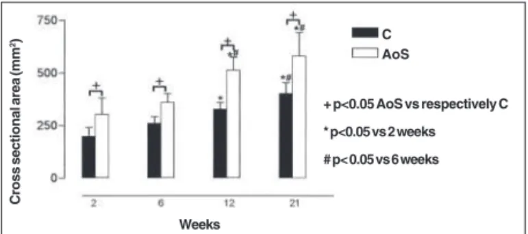
The cross sectional area of left ventricle myocytes was significantly greater in the AoS groups than in the C groups, in all periods studied. In both groups, a progressive increase of the myocyte cross sectional areas occurred during the evolvement process (fig. 2).
Myocardial concentration of hydroxyproline was greater in the AoS groups than in C groups, at 6 weeks of evolvement. In the C groups the concentration was similar in all periods of evaluation, and in AoS, a progressive in-crease in their values occurred with the evolvement of the hypertrophic process (fig. 3).
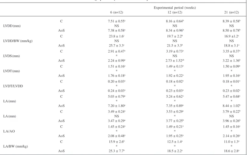
Mean and standard deviations of the left ventricle, left atrium, and aorta measures are presented in table II. LVDD was similar in the C groups and AoS in the 3 periods asses-sed and was greater in the 12- and 21-week periods, compa-red with that at 6 weeks, in both groups. When normalized
Fig. 1 - Medium layer hypertrophy of the intramural branch of the coronary artery in a rat with supravalvular aortic stenosis after 21 weeks. HE. 400X.
Fig. 2 - Myocyte cross sectional area of the left ventricle in the control groups (C) and supravalvular aortic stenosis (AoS) after 2, 6, 12, and 21 weeks of follow-up. Each column represents mean ± standard deviation +, *, # indicate significant dif-ferences between the groups according to the picture (ANOVA-two-way and Tukey test for P<0.05).
Cross sectional area (mm
2)
Weeks
+ p<0.05 AoS vs respectively C * p<0.05 vs 2 weeks # p< 0.05 vs 6 weeks
C AoS
Fig. 3 - Myocardial concentration of hydroxyproline (HOP) of the control groups (C) and supravalvular aortic stenosis (AoS) after 2, 6, 12, and 21 weeks of follow-up. Each column represents mean ± standard deviation. +, *, #, - indicate significant diffe-rences between the groups according to the picture (ANOVA-two-way and Tukey test for P<0.05).
HOP (µg/mg)
Weeks
+ p<0.05 AoS vs respectively C * p<0.05 vs 2 weeks # p< 0.05 vs 6 weeks
⊥ ⊥ ⊥ ⊥
then it becomes stable. However, morphometric evaluation demonstrated that the values of the cross sectional area of the myocytes increased progressively until 12 weeks, demonstrating the presence of additional hypertrophy during the evolvement of the process.
Echocardiographic study allowed the definition of left ventricular hypertrophy as concentric, characterized by in-creased wall thickness with normal or reduced dimensions
of the ventricular cavity. According to several authors 7,26,
animals start to develop heart failure in the 21st week after the induction of aortic stenosis. Thus, it would be expected that eccentric hypertrophy would be found during the last period of study. Because only 25% of our rats had heart fai-lure after the 21 weeks of evolvement, it is probable that the final evaluation was performed when most animals were in the compensated phases of left ventricular hypertrophy and, therefore, still having concentric hypertrophy. Howe-ver, the increase in right ventricle weight in the last
assess-Table II - Echocardiographic analysis of the structural parameters of the heart
Experimental period (weeks)
6 (n=12) 12 (n=12) 21 (n=12)
C 7.51 ± 0.55a 8.16 ± 0.64b 8.39 ± 0.58b
LVDD (mm) NS NS NS
AoS 7.38 ± 0.58a 8.34 ± 0.96b 8.50 ± 0.78b
C 23.8 ± 1.6c 19.7 ± 2.2b 16.9 ±1.2a
LVDD/BW (mm/kg) NS NS NS
AoS 25.7 ± 3.3c 21.5 ± 3.3b 18.8 ± 3.1a
C 2.91 ± 0.47a 3.19 ± 0.73a 3.35 ± 0.37a
LVDS (mm) * NS NS
AoS 2.24 ± 0.99a 2.73 ± 1.52ab 3.22 ± 1.36b
C 1.51 ± 0.16a 1.49 ± 0.13a 1.50 ± 0.09a
LVDT (mm) * * *
AoS 1.76 ± 0.18a 1.92 ± 0.22a 1.95 ± 0.16a
C 0.20 ± 0.03a 0.18 ± 0.02a 0.18 ± 0.01a
LVDT/LVDD * * *
AoS 0.24 ± 0.03a 0.23 ± 0.03a 0.23 ± 0.02a
C 5.03 ± 0.79a 5.24 ± 0.62a 5.47 ± 0.68a
LA (mm) * * *
AoS 7.20 ± 1.80a 7.35 ± 0.89a 8.44 ± 1.02b
C 3.49 ± 0.24a 3.53 ± 0.29a 3.79 ± 0.22b
LA (mm) NS * NS
AoS 3.47 ± 0.29a 3.77 ± 0.25b 3.96 ± 0.26b
C 1.45 ± 0.24a 1.49 ± 0.21a 1.45 ± 0.16a
LA/AO * * *
AoS 2.08 ± 0.48a 1.95 ± 0.25a 2.14 ± 0.26a
C 15.9 ± 2.6b 12.5 ± 1.4a 11.0 ± 1.3a
LA/BW (mm/kg) * * *
AoS 25.3 ± 7.7b 18.5 ± 2.2a 18.6 ± 2.8a
C- control; AoS- supravalvular aortic stenosis; LVDD and LVDS- left ventricle (LV) diastolic and systolic diameters; BW- body weight; LVDT: LV wall diastolic thickness; LA- left atrium diameter; AO- aorta diameter. Variance analysis of the multivariate profiles for dependent groups (MANOVA). a, b, c - groups that do not have
the same letters have statistically significant differences between the 3 evaluation periods (P<0.05); * P<0.05 vs respective control group; NS - nonsignificant.
Table III - Left ventricle functional evaluation with echocardiography
Experimental period (weeks)
6 (n=12) 12 (n=12) 21 (n=12)
C 281 ± 22a 265 ± 26ab 245 ± 15b
HR (bpm) NS * *
AoS 291 ± 21a 291 ± 22a 271 ± 29a
C 61.3 ± 5.4a 61.2 ± 6.6a 60.1 ± 2.9a
∆D (%) * NS NS
AoS 70.3 ± 10.8b 68.4 ± 12.2b 62.8 ±11.4a
C 1.64 ± 0.27a 1.61 ± 0.16a 1.72 ± 0.33a
E/A NS * *
AoS 2.07 ± 1.55a 4.20 ± 3.25b 4.71 ± 2.08b
C- control rats; AoS- rats undergoing supravalvular aortic stenosis; HR- heart rate; ∆D- shortening percentage; E/A- ratio between the initial filling flow velocity (E wave) and the atrial contraction (A wave) of the transmitral flow. Variance analysis of the multivariate profiles for dependent groups (MANOVA). a, b - groups that do
ment period demonstrates that the change from compensa-ted left ventricular hypertrophy to heart failure may have al-ready occurred. Histological analysis of the myocardium
confirmed the findings of other authors 7,26, who did not find
relevant tissue lesions, such as myocyte necrosis, extensive areas of myocardial fibrosis, or peripheral arteritis, after 21 weeks of aortic stenosis induction. In our study, in addition to myocyte cell hypertrophy, we observed the presence of vascular remodeling characterized by medium-layer hyper-trophy in the intramyocardial arteries, which was discreet in the first assessment periods and became evident after 12 weeks of follow-up. Thickening of the medium layer of the ar-teries is usually found in human beings with systemic
hy-pertension 40. This finding may be due to hypertrophy or
hyperplasia of the smooth muscle, or both; to an increase in water and/or collagen content of the vascular wall; and
thi-ckening of the basal membrane 40.
Although this model is not associated with severe
fi-brosis 7, myocardial concentration of hydroxyproline was
greater in the AoS groups at 6 weeks, and it increased gressively during the evolvement of the hypertrophic pro-cess. In the present study, we did not perform morphometric quantification of the collagen content and, therefore, we were not able to state whether the increase was due to the
interstitial collagen or perivascular. Weinberg et al 26, using
picrosirius red dye, verified the presence of a moderate in-crease in interstitial and perivascular collagen 21 weeks after the induction of aortic stenosis.
The analysis of left ventricle systolic function de-monstrated that in the initial phases of the hypertrophic pro-cess the shortening is increased. A decrease in contractile function of the chamber occurred over time, evidenced by the progressive increase in LVSD and by the reduction of
∆D. These indexes were significantly different between the
periods of 6 and 21 weeks after aortic stenosis. It is possible to detect the 3 evolvement phases of systolic function du-ring the hypertrophic process in this model: initially, the presence of supranormal performance, which is followed by
unchanged cardiac performance, and, finally, by depression of the contractile capacity of the heart. Similar results were observed by other authors who also assessed cardiac
func-tion sequentially with echocardiography 7,22.
Diastolic dysfunction may be detected in the 12th week of evolvement by the E/A ratio, which was significantly greater in the AoS groups than in the respective C groups. After this period, additional variation was not observed in the values of this ratio. However, the increase in LA diameter, verified in the 6th week, demonstrates that diastolic dysfunc-tion may have occurred earlier. Similar results were observed during the serial study of left ventricle function with
echocar-diography 22. E/A increase reflects the restrictive pattern of
ventricular filling alteration. Currently, this filling model has been attributed, in great part, to the increase in left atrial pres-sure and also to the increase in passive stiffness of the ventri-cular chamber, which may be due to the increase in wall
thickness or interstitial fibrosis, or both of these 22.
In conclusion, induction of supravalvular aortic steno-sis in young rats promotes early development of concentric left ventricular hypertrophy with cardiac function initially supranormal, followed by diastolic dysfunction in the 12th week of evolvement. Reduction of systolic ventricular per-formance occurred only in the late period after 21 weeks of AoS induction. Cardiac function alterations are followed by myocardial fibrosis without expressive histological lesions. These characteristics demonstrate that the model is suitable and potentially useful for studies on physiopatho-logy and treatment of cardiac hypertrophy due to chronic pressure overload both in the compensated stage and du-ring transition to heart failure.
Acknowledgments
For the valuable technical support of Vitor Marcos de Souza, José Carlos Georgette, and Mário Augusto Dallaqua and to the support of CNPQ/PIBIC.
1. Cicogna AC, Okoshi MP, Okoshi K. História natural da remodelação miocárdica: da agressão aos sintomas. Rev Soc Cardiol Estado de São Paulo 2000; 10:8-16. 2. Olivetti G, Cigola E, Maestri R, Lagrasta C, Corradi D, Quaini F. Recent advances
in cardiac hypertrophy. Cardiovasc Res 2000; 45:68-75.
3. Kaplan NM. Systemic hypertension: mechanisms and diagnosis. In: Braunwald E, Zipes DP, Libby P, eds. Heart Disease. A Textbook of Cardiovascular Medi-cine. Philadelphia: W.B. Saunders Company, 2001:941-71.
4. Okoshi MP, Matsubara LS, Franco M, Cicogna AC, Matsubara BB. Myocyte ne-crosis is the basis for fibrosis in renovascular hypertensive rats. Braz J Med Biol Res 1997; 30:1135-44.
5. Rossi MA, Peres LC. Effect of captopril on the prevention and regression of myo-cardial cell hypertrophy and interstitial fibrosis in pressure overload cardiac hy-pertrophy. Am Heart J 1992; 124:700-9.
6. Rodrigues MAM, Bregagnollo EA, Montenegro MR, Tucci PJF. Coronary vascu-lar and myocardial lesions due to experimental constriction of the abdominal aorta. Int J Cardiol 1992; 35:253-7.
References
7. Weinberg EO, Schoen FJ, George D, et al. Angiotensin-converting enzyme inhi-bition prolongs survival and modifies the transition to heart failure in rats with pressure overload hypertrophy due to ascending aortic stenosis. Circulation 1994; 90:1410-22.
8. Okamoto K, Aoki K. Development of a strain of spontaneously hypertensive rats. Jpn Circ J 1963; 27:282-93.
9. Bing OHL, Brooks WW, Robinson KG, et al. The spontaneously hypertensive rat as a model of the transition from compensated left ventricular hypertrophy to failure. J Mol Cell Cardiol 1995; 27:383-96.
10. Brooks WW, Bing OHL, Robinson KG, Slawsky MT, Chaletsky DM, Conrad CH. Effect of angiotensin-converting enzyme inhibition on myocardial fibrosis and function in hypertrophied and failing myocardium from the spontaneously hypertensive rat. Circulation 1997; 96:4002-10.
12. Cicogna AC, Padovani CR, Okoshi K, Aragon FF, Okoshi MP. Myocardial func-tion during chronic food restricfunc-tion in isolated hypertrophied cardiac muscle. Am J Med Sci 2000; 320:244-8.
13. Hirsch AT, Talsnec CE, Schunkert H, Paul M, Dzau VT. Tissue specific activation of cardiac angiotensin converting enzyme in experimental heart failure. Circ Res 1991; 69:475-82.
14. Kabour A, Henegar JR, Devineni VR, Janicki JS. Prevention of angiotensin II in-duced myocyte necrosis and coronary vascular damage by lisinopril and losartan in the rat. Cardiovasc Res 1995; 29:543-8.
15. Ratajska A, Campbell SE, Sun Y, Weber KT. Angiotensin II-associated cardiac myo-cyte necrosis: role of adrenal catecholamines. Cardiovasc Res 1994; 28:684-90. 16. Trippodo NC, Frohlich ED. Similarities of genetic (spontaneous) hypertension:
man and rat. Circ Res 1981; 48:309-19.
17. Cicogna AC, Padovani CR, Georgette JC, Aragon FF, Okoshi MP. Efeito da res-trição protéico-calórica sobre a função mecânica dos músculos cardíacos hiper-trofiados. Arq Bras Cardiol 1999; 72:431-5.
18. Cicogna AC, Robinson KG, Conrad CH, Squire R, Okoshi MP, Bing OHL. Par-ticipação do estado contrátil e do relaxamento miocárdico na disfunção ventricu-lar durante a transição hipertrofia-falência cardíaca. Arq Bras Cardiol 1997; 69:381-4.
19. Schunkert H, Dzau VJ, Tang SS, Hirsch AT, Apstein CS, Lorell BH. Increased rat cardiac angiotensin converting enzyme activity and mRNA expression in pres-sure overload left ventricular hypertrophy. Effects on coronary resistance, con-tractility, and relaxation. J Clin Invest 1990; 86:1913-20.
20. Feldman AM, Weinberg EO, Ray PE, Lorell BH. Selective changes in cardiac gene expression during compensated hypertrophy and the transition to cardiac decompensation in rats with chronic aortic banding. Circ Res 1993; 73:184-92. 21. Bruckschlegel G, Holmer SR, Jandeleit K, et al. Blockade of the renin-angio-tensin system in cardiac pressure-overload hypertrophy in rats. Hypertension 1995; 25:250-9.
22. Litwin SE, Katz SE, Weinberg EO, Lorell BH, Aurigemma GP, Douglas PS. Se-rial echocardiographic-doppler assessment of left ventricular geometry and function in rats with pressure-overload hypertrophy: chronic angiotensin-con-verting enzyme inhibition attenuates the transition to heart failure. Circulation 1995; 91:2642-54.
23. Turcani M, Rupp H. Development of pressure overload induced cardiac hypertro-phy is unaffected by long-term treatment with losartan. Mol Cell Biochem 1998; 188:225-33.
24. Condorelli G, Morisco C, Stassi G, et al. Increased cardiomyocyte apoptosis and changes in proapoptotic and antiapoptotic genes bax and bcl-2 during left ven-tricular adaptations to chronic pressure overload in the rat. Circulation 1999; 99:3071-8.
25. Schunkert H, Orzechowski HD, Bocker W, Meier R, Riegger GAJ, Paul M. The cardiac endothelin system in established pressure overload left ventricular hy-pertrophy. J Mol Med 1999; 77:623-30.
26. Weinberg EO, Lee MA, Weigner M, et al. Angiotensin AT1 receptor inhibition: effects on hypertrophic remodeling and ACE expression in rats with pressure-overload hypertrophy due to ascending aortic stenosis. Circulation 1997; 95:1592-600.
27. Gonçalves G, Okoshi K, Okoshi MP, et al. Efeito da inibição da enzima de con-versão da angiotensina sobre a hipertrofia ventricular esquerda induzida por so-brecarga pressórica. Rev Soc Cardiol Estado de São Palulo 1998; 8:2. 28. Bregagnollo EA, Okoshi K, Okoshi MP, Padovani CR, Cicogna AC. Efeitos do
lisinopril sobre as características da hipertrofia ventricular esquerda na vigência de sobrecarga pressórica persistente. Rev Soc Cardiol Estado de São Paulo 2001; 11:32.
29. Zierhut W, Zimmer H-G, Gerdes AM. Effect of angiotensin converting enzyme inhibition on pressure-induced left ventricular hypertrophy in rats. Circ Res 1991; 69:609-17.
30. Böcker W, Hupf H, Grimm D, Kurzidim K, Schunkert H. Effects of indapamide in rats with pressure overload left ventricular hypertrophy. J Cardiovasc Pharma-col 2000; 36:481-6.
31. Grimm D, Huber M, Jabusch HC, et al. Extracellular matrix proteins in cardiac fi-broblasts derived from rat hearts with chronic pressure overload: effects of beta-receptor blockade. J Mol Cell Cardiol 2001; 33:487-501.
32. Switzer BR. Determination of hydroxyproline in tissue. J Nutr Biochem 1991; 2:229-321.
33. Matsubara LS, Matsubara BB, Okoshi MP, Franco M, Cicogna AC. Myocardial fibrosis rather than hypertrophy induces diastolic dysfunction in renovascular hypertensive rats. Can J Physiol Pharmacol 1997; 75:1328-34.
34. Matsubara LS, Bojikian BB, Okoshi MP, Cicogna AC, Janicki JS. Alterations in myocardial collagen content affect rat papillary muscle function. Am J Physiol 2000; 279:1534-9.
35. Okoshi MP, Okoshi K, Dal Pai V, Dal Pai-Silva M, Matsubara LS, Cicogna AC. Mechanical, biochemical, and morphological changes in the heart from chronic food restricted rats. Can J Physiol Pharmacol 2001; 79:754-60.
36. Okoshi K. Estrutura e Função do Coração de Ratos Normotensos e Hipertensos Submetidos à Restrição da Ingestão Alimentar: Estudo In Vivo Pelo Ecocardio-grama e In Vitro do Coração Isolado. Botucatu: UNESP, 2000:106p. 37. Simone G, Devereux R, Camargo M, et al. In vivo left ventricular anatomy in rats
with two-kidney, one clip and one-kidney, one clip renovascular hypertension. J Hypertens 1992; 10:725-32.
38. Douglas PS, Katz SE, Weinberg EO, Chen MH, Bishop SP, Lorell BH. Hypertro-phic remodeling: gender differences in the early response to left ventricular pres-sure overload. J Am Coll Cardiol 1998; 32:1118-25.
39. Conrad CH, Brooks WW, Robinson KG, Bing OHL. Impaired myocardial func-tion in spontaneously hypertensive rats with heart failure. Am J Physiol 1991; 260:H136-45.