AR
TIGO ORIGINAL / ORIGINAL AR
TICLE
INTRODUCTION
Cirrhosis is the most advanced stage of ibrosis and is associated with the emergence of ibrotic septa and nodules, changes in hepatic blood low and the risk of liver failure. The hepatocyte dysfunction that results from hepatic disorder causes portal hypertension, which leads to the characteristic clinical manifesta-tions of cirrhosis that affect the liver as well as other organ systems(14,15,25,28).
One of the inherent complications of cirrhosis is protein-calorie malnutrition (PCM)(29). PCM is the loss of lean and fat mass in conjunction with meta-bolic alterations that compromise vital functions. The predominance of PCM in cirrhotic patients, regard-less of aetiology, ranges from 20% to 60% in patients with compensated cirrhosis and can reach 100% in candidates for orthotopic liver transplantation(9,26,37).
Thus far, there are no efficient, reliable or re-producible methods to assess the degree of liver involvement and to correlate it with the clinical and nutritional condition of a patient. The desired method should include parameters that identify metabolic
ALTERATION OF TASTE BUDS IN
EXPERIMENTAL CIRRHOSIS.
Is there correlation with human hypogeusia?
Sabrina Alves FERNANDES1, Silvia BONA2,3, Carlos Thadeu Schmidt CERSKI4,
Norma Possa MARRONI2,3,5,6 and Claudio Augusto MARRONI1,3,7
Received 27/6/2016 Accepted 9/8/2016
ABSTRACT - Background - The inherent complications of cirrhosis include protein-calorie malnutrition and micronutrient deiciencies. Changes in taste are detrimental to the nutritional status, and the mechanism to explain these changes is not well documented in the cirrhotic patients. Objective - To evaluate the taste buds of cirrhotic rats. Methods - Fourteen male Wistar rats were evaluated. After 16 weeks, the liver was removed to histologically diagnose cirrhosis, and blood was collected to perform liver integrity tests. The tongue was removed for histological examination and immunohistochemistry using antibodies against protein gene product PGP 9.5 and the sweet taste receptors T1R2 and T1R3. Morphological changes were determined by scanning electron microscopy. Serum zinc levels were measured. Results - The cirrhotic animals, but not the control animals, exhibited zinc deiciency. In both groups, there was positive immunoreactivity for type II and III cells and T1R2 receptors. The cirrhotic animals had no immunoreactivity for T1R3 receptors. Scanning electron microscopy analysis of the cirrhotic group revealed a uniform tapering of the gustatory papillae. Conclusion - In conclusion the experimental cirrhosis model mimicked the biochemical and histological parameters of human cirrhosis, therefore enabling a study of the gustatory papillae and taste buds.
HEADINGS - Liver cirrhosis. Taste buds. Malnutrition. Zinc. Microscopy, electron, scanning. Morphological and microscopic indings.
Declared conflict of interest of all authors: none Disclosure of funding: no funding received
1 Programa de Pós-Graduação em Hepatologia, Universidade Federal de Ciências da Saúde de Porto Alegre, RS, Brasil; 2 Programa de Pós-Graduação em Ciências
Médicas, Faculdade de Medicina, Universidade Federal do Rio Grande do Sul, RS, Brasil; 3 Laboratório de Hepatologia e Gastroenterologia, Hospital de Clínicas de Porto
Alegre, RS, Brasil; 4 Departamento de Patologia, Faculdade de Medicina, Universidade Federal do Rio Grande do Sul, RS, Brasil; 5 Programa de Pós-Graduação em Ciências
Biológicas: Fisiologia, Universidade Federal do Rio Grande do Sul, RS, Brasil; 6 Programa de Pós-Graduação em Bio-Saúde, Universidade Luterana do Brasil, RS, Brasil; 7 Grupo adulto de Transplante de Fígado, Santa Casa de Porto Alegre, RS, Brasil.
Correspondence: Claudio Augusto Marroni. Rua José Kanan Aranha, 102 - CEP: 91350-170 - Porto Alegre, RS, Brasil. Email: [email protected]
changes and simultaneously evaluate the common characteristics of this disease, such as oedema and ascites, that cause body asymmetry(2,31).
Bioelectrical impedance analysis (BIA) is a tool that utilises the phase angle (PA) to evaluate the pro-gress of liver cirrhosis in a practical way that meets the clinical requirements by reporting on body and cell structure, even in cases of body asymmetry. The PA enables the determination of cellular structure and function, thus indirectly improving the eficacy of nutritional interventions(1,13,17,31,32).
The human tongue is covered with gustatory papillae that have speciic functions (taste receptors and signalling). The papillae allows to differentiate the sweet, bitter, sour, salty and umami lavours. Papillae can be circumvallate, fungiform, iliform or foliate, and they are all distributed on the back of the tongue(18). In humans, changes in taste may exacerbate PCM related to cirrhosis(11).
It is possible to detect changes in tongue structure that may indicate the pathophysiological process of hypogeusia. There are four types of cells (I, II, III and IV) in the gustatory papillae. These cells, especially types II and III, are responsi-ble for the transduction of the taste signal to the brain(5,27,40). To differentiate the types of taste (bitter, sweet and umami), receptors are also present in the gustatory papillae (except for iliform papillae [FiP]) that enable the identiica-tion of changes in taste percepidentiica-tion(7,10).
The T1R family of hetero-oligomeric taste receptors comprises three speciic G protein-coupled receptors, T1R1, T1R2 and T1R3, that have distinct expression patterns. By studying these receptors, potential alterations in taste percep-tion can be identiied(22).
To date, there have been no morphological and/or histo-logical analyses of the tongues of cirrhotic patients, despite hypogeusia being a common complaint of these patients. Carbon tetrachloride (CCl4) is a hepatotoxic drug that is widely used in experimental studies of hepatic cirrhosis because its effects mimic human cirrhosis(12,23,34).
In this study, we hypothesised that morphological and/ or histological alterations occurred in the gustatory papillae of cirrhotic patients. However, performing tongue biopsies of cirrhotic and control patients was not feasible; therefore, we analysed the tongues ofcirrhotic rats, which exhibited morphological and histological structures similar to the human tongue.
In this study, we aimed to correlate the changes related to PCM and cellular deiciencies with the alteration of the gus-tatory papillae structure in CCl4-induced cirrhotic animals.
METHODS
Fourteen male Wistar rats weighing between 200 and 250 g from the State Foundation for the Production and Health Research (Fundação Estadual de Produção e Pesquisa em Saúde - FEPPS) were utilised. During the experiment, the animals were housed in the Animal Experimentation Unit of the Research Centre at the Teaching Hospital of Porto Alegre (Hospital de Clínicas de Porto Alegre - HCPA) in a 12-hour light/dark cycle (light from 7:00 to 19:00) and at a temperature of 22 ± 2°C. The animals were randomly divided into two experimental groups: control (CO) and cirrhotic (CCl4). Both groups had controlled access to food, and phenobarbital (en-zyme inducer) was added to the water (0.3 g/dL) to potentiate the CCl4 effect(4). The control group (CO) received 0.5 mL of mineral oil intraperitoneally (i.p.), and the CCl4 group received CCl4 (0.5 mL/kg i.p.) diluted in mineral oil at a 1:6 ratio. The drugs were administered on the following schedule: 10 doses at 5 day intervals, 10 doses at 4 day intervals and 7 doses at 3
day intervals. The animals were sacriiced 2 days after the last CCl4 dose, at the end of week 16(33).
All the procedures were performed according to the guide-lines recommended by the Research Ethics Committee of HCPA, and the animal care followed the recommendations of the Principles of Laboratory Animal Care formulated by the National Society for Medical Research and the Guide for the Care and Use of Laboratory Animals published by the National Institutes of Health(38). After 27 doses, the animals were anesthetised with an i.p. injection of xylazine (50 mg/kg) and ketamine (100 mg/kg). Blood samples were col-lected via the retro-orbital plexus to analyse the liver function(21). After sacriice, the abdominal region was shaved, and a midline laparotomy was performed to collect the liver and the tongue of the animals.
Serum biochemical analysis
Blood samples obtained from the retro-orbital plexus were utilised to evaluate serum aspartate aminotransfer-ase (AST), alanine aminotransferaminotransfer-ase (ALT) and alkaline phosphatase (ALP) levels, which were expressed in U/L and measured using routine laboratory methods at HCPA.
Bioelectrical impedance analysis
To measure total body resistance (R) and total body reactance (Xc), a phase-sensitive tetrapolar impedance ana-lyser (Biodynamics BIA 450E) was utilised with hypodermic needles as electrodes. The rats were anesthetised and placed in a prone position on a non-conductive surface to eliminate interference from the electrical induction. On the midline, source electrode 1 was placed at the anterior edge of the orbit, and source electrode 2 was placed 4 cm from the base of the tail. Detector electrode 1 was placed at the anterior opening of the ear, and detector electrode 2 was placed in the middle of the rat pelvis(19). The analysis was performed in two phases, the initial phase (time 1) and the inal phase (time 2).
Hepatic tissue lipid peroxidation measurements Frozen hepatic tissue from each rat was homogenised in ice-cold phosphate buffer (140 mM KCl and 20 mM phosphate, pH 7.4) and centrifuged at 3,000 rpm for 10 minutes. Oxidative stress was deter mined by measuring the concentration of aldehydic products (malondialdehyde [MDA]) using thiobarbituric acid reactive substances (TBARS). The absorbance (535 nm) of the supernatant was measured by spectrophotometry, and the values were expressed as nmol/mg protein(6).
Zinc measurements
The serum zinc concentration was measured using a Zinc Assay Kit (Abnova Corporation, Taipei City, Taiwan). The absorbance (425 nm) of the supernatant was measured by spectrophotometry, and the values were expressed as µg/dL.
Histology
for 24 hours. The blocks were dehydrated in a graded series of ethanol and embedded in parafin wax. Serial 3-mm sections were stained with picrosirius (liver tissue) and with H/E (tongue tissue). Five sections from each sample were analysed by two independent pathologists with no prior knowledge of the animal groups.
Scanning Electron Microscopy
The dorsal region of the tongue was collected and im-mersed in a ixative solution containing 4% glutaraldehyde. The samples were dehydrated in a graded acetone series: 30%, 50%, 70%, 95% and 100%. A CPD 030 Critical Point Dryer (Leica Microsystems, Buffalo Grove, IL) was utilised to remove moisture from the samples and to reach the critical point.
The metallisation was performed with a gold/palladium alloy. The samples were covered with an approximately 15-nm layer using a Metalizer Med 020 (Leica Microsystems). Images were captured with a Philips XL20 Scanning Electron Microscope at 40-800X magniication.
The taste buds were counted by pathologists who were blinded to the groups. The buds in each ield were counted, and the mean was calculated for each sample. Image-Pro Plus (Media Cybernetics, Bethesda, MD, USA) was utilised to deter-mine the diameter of the gustatory papillae and the taste buds.
PGP 9.5, T1R2 and T1R3
The expression of PGP 9.5, T1R2 and T1R3 in the tongue was determined by immunohistochemistry. Antigen recovery was performed using citrate butter at 100ºC, and endogenous peroxidase activity was blocked by incubating the slides at room temperature with absolute methanol containing 3% hydrogen peroxide. The slides were prein-cubated with 10% rabbit serum at room temperature to block potential undesirable reactions with the secondary antibody. The slides were incubated with polyclonal rabbit
antibodies against PGP 9.5, T1R2 or T1R3(Santa Cruz
Biotechnology, Santa Cruz, CA, USA) overnight at 4ºC followed by an incubation with the secondary antibody for 1 hour at room temperature. After 60 minutes at room temperature, the slides were treated with EnVision reagents and washed 3 times with hydrogen peroxide (in PBS). The nuclei were counterstained with hematoxylin. The primary antibody was diluted in PBS containing bovine serum albu-min; this buffer without a primary antibody was utilised as a negative control. The results were evaluated without prior knowledge of the groups using a microscope equipped with a digital camera to capture the images using Image-Pro Plus (Media Cybernetics).
Statistical analysis
The data were stored in Excel, and the statistical analyses were performed using SPSS (Statistical Package for Social Science) version 18.0. The results are expressed as the mean ± standard deviation (SD). Student’s t-test was utilised to compare the intragroup variables for paired samples and the intergroup variables for independent samples. A 5% level of signiicance was adopted (P≤0.05).
RESULTS
After 16 weeks, we obtained histological conirmation of cirrhosis in all the treated animals; there was no cirrhosis in the control animals. One animal was lost due to death before 16 weeks. The animals were randomized according to the phase angle.
In addition to the histological evidence, the cirrhotic animals displayed signiicant changes in liver function (AST, ALT and ALP), indicating the presence of liver damage. The zinc levels were lower in the CCl4 group than in the control group (Table 1).
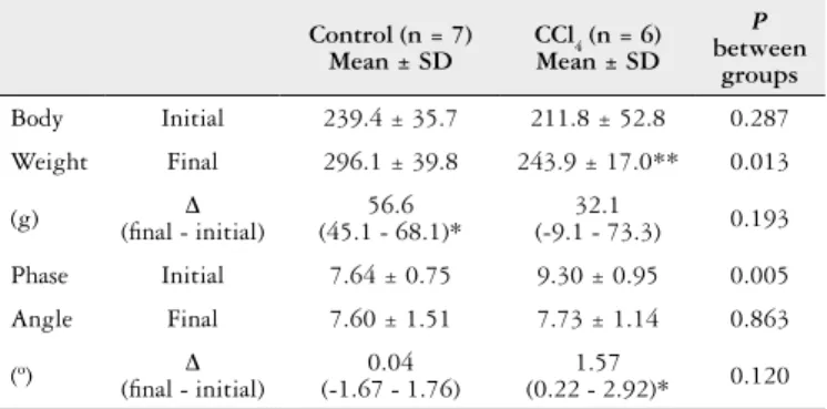
TABLE 2. Comparison of body weight and phase angle
Control (n = 7) Mean ± SD
CCl4 (n = 6) Mean ± SD
P between
groups
Body Initial 239.4 ± 35.7 211.8 ± 52.8 0.287
Weight Final 296.1 ± 39.8 243.9 ± 17.0** 0.013
(g) (inal - initial)∆ (45.1 - 68.1)*56.6 (-9.1 - 73.3)32.1 0.193
Phase Initial 7.64 ± 0.75 9.30 ± 0.95 0.005
Angle Final 7.60 ± 1.51 7.73 ± 1.14 0.863
(º) (inal - initial)∆ (-1.67 - 1.76)0.04 (0.22 - 2.92)*1.57 0.120
*P≤0.05. The results are expressed as the mean ± SD. ** Weight of animals with ascites.
TABLE 1. Zinc levels, liver integrity and lipid peroxidation analyses
Variables Control (n = 7) CCl4 (n = 6) P
Mean ± SD Mean ± SD
AST (U/L) 171 ± 32.2 1016 ± 305 <0.001
ALT (U/L) 57.9 ± 19.2 270 ± 81.2 0.001
ALP (U/L) 65.0 ± 26.2 386 ± 103 <0.001
Zinc (µg/dL) 48.6 ± 13.5 10.8 ± 3.5 <0.001
TBARS
(nmol/mg protein) 0.05 ± 0.00 0.09 ± 0.04 <0.05
The results are expressed as the mean ±SD. AST: aspartate aminotransferase; ALT: alanine aminotransferase; ALP: alkaline phosphatase; TBARS: substances reactive thiobarbituric acid.
In the lipid peroxidation analysis using TBARS, we observed an increase in lipid peroxidation in all the CCl4 -induced cirrhoticanimals (P=0.05).
Table 2 presents the body weight data for the control and cirrhotic (CCl4) animals at times 1 (initial) and 2 (inal) and the PA measured by bioelectrical impedance analysis at the same timepoints.
In analysing the initial (time 1) and inal (time 2) time-points, we discovered that the control group gained more weight than the CCl4 group (Table 2).
Histological analysis of the rat tongues
The initial histological analysis of the liver (Figure 1A and 1B) structure was performed using Picrosirius staining. The histological analysis of the rat tongue structure was performed using H/E. (Figure 2A and 2B). This revealed a signiicant reduction in FiP in the cirrhotic animals com-pared with the control animals (Figure 2A and 2B).
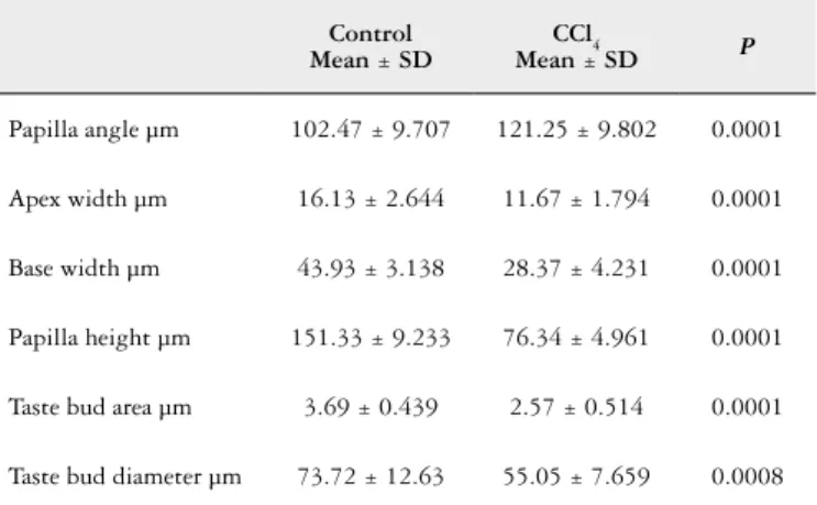
SEM analysis of the morphological structure of the tongue revealed that tapering of the gustatory papillae occurred, par-ticularly in the base diameter and the apical region, in the CCl4 group (Figures 3A and 3B). Compared with the control animals, the taste buds in the cirrhotic animals displayed morphological changes (Table 3).
In the animals in the cirrhosis group, some tongues had only one taste bud per ield, whereas others contained no taste buds. In the control group, the presence of taste buds per ield ranged between 2 and 6.
The immunohistochemical analysis of the innervation of the gustatory papillae using a PGP 9.5 antibody revealed that the control and CCl4 animals had fungiform papillae (FuP) and FiP in the dorsal region of the tongue.
An immunohistochemical analysis of the T1R receptor family (T1R2 and T1R3) in the gustatory papillae of rats was performed to determine the potential for the sweet taste.
T1R2 receptor immunoreactivity was observed in both groups.
There was positive immunoreactivity for T1R3 receptor in the control animals but not in the cirrhotic animals.
FIGURE 1. Hostological analysis of the liver by pricrosirius. (A) Control
group, exhibited normail structure. (B) Cirrhosis group had an abnormal structure.
Figure 1A
Figure 1B
FIGURE 3. Morphological analysis of the gustatory papillae by SEM. (A)
Morphological analysis of the papillae in the control group: intact papillae (red arrow) and normal taste buds (*) (500X). (B) FiP of the reduced diameter in the apical region (red arrow) and a smaller, exposed taste bud (*) (500X).
Figure 3A
Figure 3B
FIGURE 2. Histological analysis of the rat tongues by H/E staining. (A) FiP in
the control group exhibited normal structure; the arrow indicates a FiP (100X magniication). (B) FiP in the cirrhosis group had an abnormal structure, and there were fewer papillae; the arrow indicates a FiP (100X magniication).
Figure 2A
DISCUSSION
The classic CCl4-induced cirrhosis model is widely ac-cepted in the literature because it has similar characteristics to those of human cirrhosis.
AST and ALT are enzymes that are sensitive to hepa-tocellular injury. The release of large amounts of these en-zymes into the bloodstream is associated with centrilobular necrosis, degeneration and decreased liver function. In our study, we observed a significant increase in the liver en-zyme levels in cirrhotic animals, conirming what has been previously reported(4,8).
Zinc is hepatoprotective and required for ALP activity. Zinc is critical to a large number of structural proteins, en-zymatic processes, and transcription factors. Zinc deiciency can result in a spectrum of clinical manifestations, such as poor of appetite, loss of body hair, altered taste and smell, testicular atrophy, cerebral and immune dysfunction, and diminished drug elimination capacity. These are common symptoms in patients with chronic liver diseases, especially liver cirrhosis(16). Reinforcing the characteristics of this metal, low zinc levels have been observed in cirrhotic animals(39), and this was conirmed in the present study.
The CCl4-induced cirrhosis model involves various signal-ling pathways that result in liver injury; this was conirmed in this study by the TBARS values that indicated a relation-ship between CCl4-induced changes in the liver parenchyma and the formation of reactive oxygen species (ROS) by the hepatic microsomal system, potentially through the forma-tion of trichloromethyl (CCl3•) and trichloromethyl peroxide (CCl3OO•) radicals. This oxidative process causes cellular damage and may increase ion permeability and membrane and structural disintegration(20), which can be measured by bioelectrical impedance analysis (the PA).
The reduced PA in the cirrhotic animals was accompanied by an increase in liver enzyme levels and in lipid peroxida-tion. The body structure of the cirrhotic animals changed as they lost weight.
The cirrhotic animals exhibited body asymmetry result-ing from the development of ascites, a symptom that is
characteristic of cirrhosis. This change in body composition dificult the determination of the actual nutritional status of the analysed organism when conventional methods, such as anthropometric measurements, are utilised that are based primarily on the amount of body fat(19).
The PA has been used as a prognostic factor that assesses cell functionality, and it is associated with other indices that are indicative of the health status. Currently, there have been no studies using this method to evaluate animals.
Cells in the nerve plexus in taste buds are divided based on function into type I, II, III and IV cells. Only type II and III cells are responsive to stimuli and are called “light” cells(35). Type II cells receive a stimulus and transmit the signal to type III cells, which form a synapse. In 1982, Nagy et al. demonstrated that there are nerve ibres in the fungiform and circumvallate papillae of rats and that it is possible to record them(30).
We conirmed these indings by identifying nerve ibres in the FuP in both animal groups. Yee et al. reported that PGP 9.5 immunoreactivity in the gustatory papillae indicated the presence of type III cells(40).
Five types of taste have been described; in this study, because of the disease characteristics, we chose to analyse the sweet taste because it is linked to a higher percentage of daily caloric intake. The sweet taste is present in all types of gustatory papillae, and its response to stimuli is similar in different mammals(3).
The sweet taste can be detected via the combination of two receptors (T1R2 and T1R3) that determine the function of the heteromeric T1R receptor. T1R recep-tors are G protein-coupled receprecep-tors (GPCRs); class C GPCRs have a long N-terminus, similar to metabo-tropic glutamate receptors, GABAB receptors and calcium signal receptors(36).
Hoon et al. reported in 1997 that the majority of T1R1 receptors are expressed in FuP and are rare in circumval-late papillae. In contrast, T1R2 are poorly expressed in FuP but are expressed in all circumvallate papillae. T1R3 are expressed in both papillae types(22). We did not observe T1R3-immunoreactive cells, which would mediate the sweet taste, in the cirrhosis group.
In 2003, Kim et al. compared the signalling activity of circumvallate papillae and FuP and determined that FuP can play the same role in responding to sweet stimuli(24).
In our study, using various techniques, we observed thinning of FiP in the cirrhosis group, which would impair the salty taste. The salty taste is detected directly by Na+ permeability in the apical region of the papilla, causing cell depolarisation to perceive the taste. When there is a thinning of the papillae or they have a decreased diameter, sodium comes into contact with other structures in the dorsal region of the tongue, causing a gradual loss of papillary structure because of a continuous imbalance(3).
Sweet taste aversion and salty taste intolerance are the two symptoms related to taste that are commonly mentioned by cirrhotic patients, and the changes in the gustatory papillae of cirrhotic animals may play a role in dysgeusia.
TABLE 3. Quantitative analysis of taste bud structure
Control Mean ± SD
CCl4
Mean ± SD P
Papilla angle µm 102.47 ± 9.707 121.25 ± 9.802 0.0001
Apex width µm 16.13 ± 2.644 11.67 ± 1.794 0.0001
Base width µm 43.93 ± 3.138 28.37 ± 4.231 0.0001
Papilla height µm 151.33 ± 9.233 76.34 ± 4.961 0.0001
Taste bud area µm 3.69 ± 0.439 2.57 ± 0.514 0.0001
CONCLUSION
The experimental cirrhosis model induced by the intra-peritoneal injection of CCl4 mimicked the biochemical and histological parameters of human cirrhosis. However, we know that xenobiotics may alter the conformity of some cells, these may be the taste buds. In the present study, it was possible to determine that cirrhotic rats present signiicant alterations in the gustative papillae mainly related to sweet and salt taste. Further studies are needed, such as cirrhosis by ligation of bile duct, to associate this like change to cirrhosis.
ACKNOWLEDGMENTS
We would like to gratefully acknowledge the Postgradu-ate Programme in Hepatology at the Federal University of Health Sciences of Porto Alegre (Universidade Federal de Ciências da Saúde de Porto Alegre - UFCSPA), the Incen-tive Fund for Research and Events (Fundo de Incentivo à
Fernandes SA, Bona S, Cerski CTS, Marroni NP, Marroni CA. Alteração de paladar em um modelo experimental de cirrose. Existe correlação com hipogeusia humana? Arq Gastroenterol. 2016,53(4):278-84.
RESUMO - Contexto - As complicações inerentes de cirrose incluem a desnutrição proteico-calórica e deiciências de micronutrientes. Alterações no paladar são prejudiciais para o estado nutricional e o mecanismo para explicar essas mudanças não é bem documentada nos pacientes cirróticos. Objetivo - Avaliar as papilas gustativas de ratos cirróticos. Métodos - Foram avaliados 14 ratos Wistar machos. Após 16 semanas, o fígado foi removido para diagnosticar histologicamente cirrose, e o sangue foi colhido para efetuar testes de integridade hepática. A língua foi removida para exame histológico e imuno-histoquímica utilizando anticorpos contra o gene da proteína PGP 9.5 e os receptores de sabor doce T1R2 e T1R3. As alterações morfológicas foram determinadas por microscopia eletrônica de varredura e os níveis de zinco no soro foram medidos. Resultados - Os animais cirróticos, em relação aos animais controle, apresentaram deiciência de zinco signiicativa. Em ambos os grupos, houve imunorreatividade positiva para o tipo II e células III e receptores T1R2. Os animais cirróticos não tinham imunoreactividade para receptores T1R3. Microscopia eletrônica de varredura do grupo cirrótico revelou um ailamento uniforme das papilas gustativas. Conclusão - O modelo de cirrose experimental imitou os parâmetros bioquímicos e histológicos de cirrose humana, portanto, permitindo um estudo das papilas gustativas e paladar.
DESCRITORES - Cirrose hepática. Papilas gustativas. Desnutrição. Zinco. Microscopia eletrônica de varredura. Achados morfológicos e microscópicos. Pesquisa e Eventos/Hospital das Clínicas de Porto Alegre - FIFE/HCPA (12-0139)), the Coordination of Improve-ment of Higher Education Personnel (Coordenação de Aperfeiçoamento de Pessoal de Nível Superior - CAPES), the National Council of Scientiic and Technological Devel-opment (Conselho Nacional de Desenvolvimento Cientíico Tecnológico - CNPQ), the Foundation for Research Support of the State of Rio Grande do Sul (Fundação de Amparo à Pesquisa do Estado do Rio Grande do Sul - FAPERGS) and Lutheran University of Brazil (Universidade Luterana do Brasil - ULBRA).
Authors’ contributions
REFERENCES
1. Baumgartner RN, Chumlea WC, Roche AF. Bioelectric impedance phase angle and body composition. Am J Clin Nutr. 1988;48:16-23.
2. Baumgartner RN, Heymsield SB, Lichtman S, Wang J, Pierson RN. Body com-position in elderly people: effect of criterion estimates on predictive equations. Am J Clin Nutr. 1991;53:1345-53.
3. Behrens M, Meyerhof W. Gustatory and extragustatory functions of mammalian taste receptors. Physiol Behav. 2011;105:4-13.
4. Bona S, Filippin LI, Di Naso FC, de David C, Valiatti B, Isoppo Schaun M, Xavier RM, et al. Effect of antioxidant treatment on ibrogenesis in rats with carbon tetrachloride-induced cirrhosis. ISRN Gastroenterol. 2012;2012:762920. 5. Breslin PA, Huang L. Human taste: peripheral anatomy, taste transduction, and
coding. Adv Otorhinolaryngol. 2006;63:152-90.
6. Buege JA, Aust SD. Microsomal lipid peroxidation. Methods Enzymol. 1978;52:302-10.
7. Chaudhari N, Roper SD. The cell biology of taste. J Cell Biol. 2010;190:285-96. 8. Cremonese RV, Pereira-Filho AA, Magalhães R, de Mattos AA, Marroni CA,
Zettler CG, Marroni NP. Experimental cirrhosis induced by carbon tetrachloride inhalation: adaptation of the technique and evaluation of lipid peroxidation. Arq Gastroenterol. 2001;38:40-7.
9. D’Amico G, Garcia-Tsao G, Pagliaro L. Natural history and prognostic indi-cators of survival in cirrhosis: a systematic review of 118 studies. J Hepatol. 2006;44:217-31.
10. DeFazio RA, Dvoryanchikov G, Maruyama Y, Kim JW, Pereira E, Roper SD, Chaudhari, N. Separate populations of receptor cells and presynaptic cells in mouse taste buds. J Neurosci. 2006;26:3971-80.
11. Doty RL. Handbook of Olfaction and Gustation. New York: Dekker; 1995. 12. Fang HL, Lin WC. Lipid peroxidation products do not activate hepatic stellate
cells. Toxicology. 2008;253:36-45.
13. Fernandes SA, Bassani L, Nunes FF, Aydos ME, Alves AV, Marroni CA. Nu-tritional assessment in patients with cirrhosis. Arq Gastroenterol. 2012;49:19-27. 14. Friedman SL. Liver ibrosis -- from bench to bedside. J Hepatol. 2003(Suppl
1):S38-53.
15. Friedman SL. Mechanisms of hepatic fibrogenesis. Gastroenterology. 2008;134:1655-69.
16. Grüngreiff K, Reinhold D, Wedemeyer H. The role of zinc in liver cirrhosis. Ann Hepatol.2016;15:7-16.
17. Gupta D, Lis CG, Dahlk SL, King J, Vashi PG, Grutsch JF, Lammersfeld CA. The relationship between bioelectrical impedance phase angle and subjective global assessment in advanced colorectal cancer. Nutr J. 2008;7:19.
18. Guyton AC, Hall JE. Tratado de Fisiologia Médica. 10 ed. Rio de Janeiro: Guanabara Koogan; 2002.
19. Hall CB, Lukaski HC, Marchello MJ. Estimation of rat body composition using tetrapolar bioelectrical impedance analysis. Nutr Rep Int. 1989;39:627-33. 20. Halliwell B, Gutteridge J. Free radicals in biology and medicine. 3 ed. New York:
Oxford; 1999.
21. Halpern BN, Pacaud A. Technique of obtaining blood samples from small laboratory animals by puncture of ophthalmic plexus. C R Seances Soc Biol Fil. 1951;145:1465-6.
22. Hoon MA, Adler E, Lindemeier J, Battey JF, Ryba NJ, Zuker CS. Putative mam-malian taste receptors: a class of taste-speciic GPCRs with distinct topographic selectivity. Cell. 1999;96:541-51.
23. Jaramillo-Juárez F, Rodríguez-Vázquez ML, Rincón-Sánchez AR, Consolación Martínez M, Ortiz GG, Llamas J, et al. Acute renal failure induced by carbon tetrachloride in rats with hepatic cirrhosis. Ann Hepatol. 2008;7:331-8. 24. Kim MR, Kusakabe Y, Miura H, Shindo Y, Ninomiya Y, Hino A. Regional
ex-pression patterns of taste receptors and gustducin in the mouse tongue. Biochem Biophys Res Commun. 2003;312:500-6.
25. Kukner A, Tore F, Firat T, Terzi EH, Oner H, Balaban YH, Ozogul C. The pre-ventive effect of low molecular weight heparin on CCL₄-induced necrosis and apoptosis in rat liver. Ann Hepatol. 2010;9:445-54.
26. Lowell JA. Nutritional assessment and therapy in patients requiring liver trans-plantation. Liver Transpl Surg. 1996;5(Suppl 1):79-88.
27. Margolskee RF. Molecular mechanisms of bitter and sweet taste transduction. J Biol Chem. 2002;277:1-4.
28. Mattos AA, Dantas-Corrêa EB. Tratado de Hepatologia. 1 ed. Rio de Janeiro: Rubio; 2010.
29. Milke García MP. Nutritional support in the treatment of chronic hepatic en-cephalopathy. Ann Hepatol. 2011;10(Suppl 2):S45-9.
30. Nagy JI, Goedert M, Hunt SP, Bond A. The nature of the substance P-containing nerve ibres in taste papillae of the rat tongue. Neuroscience. 1982;7:3137-51. 31. Norman K, Stobäus N, Pirlich M, Bosy-Westphal A. Bioelectrical phase angle
and impedance vector analysis - Clinical relevance and applicability of impedance parameters. Clin Nutr. 2012;31:854-61.
32. Paiva SI, Borges LR, Halpern-Silveira D, Assunção MC, Barros AJ, Gonzalez MC. Standardized phase angle from bioelectrical impedance analysis as prognostic factor for survival in patients with cancer. Support Care Cancer. 2010;19:187-92. 33. Pavanato A, Tuñón MJ, Sánchez-Campos S, Marroni CA, Llesuy S, González-Gal-lego J, Marroni N. Effects of quercetin on liver damage in rats with carbon tetrachloride-induced cirrhosis. Dig Dis Sci. 2003;48:824-9.
34. Pérez Tamayo R. Is cirrhosis of the liver experimentally produced by CCl4 and adequate model of human cirrhosis? Hepatology. 1983;3:112-20.
35. Pumplin DW, Yu C, Smith DV. Light and dark cells of rat vallate taste buds are morphologically distinct cell types. J Comp Neurol. 1997;378:389-410. 36. Renwick AG, Molinary SV. Sweet-taste receptors, low-energy sweeteners, glucose
absorption and insulin release. Br J Nutr. 2010;104:1415-20.
37. Rivera Irigoin R, Abilés J. Nutritional support in patients with liver cirrhosis. Gastroenterol Hepatol. 2012;35:594-601.
38. Rozmiarek H. Current and future policies regarding laboratory animal welfare. Invest Radiol. 1987;22:175-9.
39. Weismann K, Christensen E, Dreyer V. Zinc supplementation in alcoholic cirrho-sis. A double-blind clinical trial. Acta Med Scand. 1979;205:361-6.