Materials Research, Vol. 11, No. 1, 93-96, 2008 © 2008
*e-mail: [email protected]
Characterization of Antiseptic Apatite Powders
Prepared at Biomimetics Temperature and pH
Soumia Belouafaa, Hassan Chaaira*, Hayate Loukilib, Khalid Diguaa, Brahim Sallekc
a
Laboratoire de Génie des Procédés et de Dépollution,
Facultés des Sciences et Techniques de Mohammedia, B. P. 146, Mohammedia 20 800, Morocco
b
Laboratoire des Matériaux, Catalyse et Environnement,
Facultés des Sciences et Techniques de Mohammedia, Morocco
c
Laboratoire de Génie des Procédés, Faculté des Sciences de Kenitra, Morocco
Received: October 28, 2007; Revised: March 4, 2008
Antiseptic apatite-based calcium phosphates were prepared as the single-phase powders. Phosphocalcic oxygenated apatites were synthesized from calcium salts and orthophosphate dissolved in oxygenated water solution at 30%, under the biomimetic conditions of 37 °C and pH 7.4. The characterization and chemical analysis of the synthesized biomimetic apatite powders were performed by scanning electron microscopy (SEM), powder X ray diffraction (XRD), Fourier-transformed infrared spectroscopy (FT-IR) and chemical analysis. The obtained materials are a calcium deficient apatites with different morphologies.
Keywords: calcium phosphate, oxygenated apatite, precipitation, biomimetic conditions
1. Introduction
Phosphocalcic oxygenated apatites are among the most promis-ing calcium phosphate apatites because of there antiseptic properties which make them able of limiting the proliferation of micro-organisms at the site of implantation1. These properties are due to the oxygenated
species (peroxide ions: O22– and/or molecular oxygen: O
2) contained
in the channels of the apatitic structure1,2. These species were
liber-ated in the living environment either by progressive dissolution of the material, or by chemical exchange with the living environment1. The
peroxide ions thus liberated act in situ to destroy the micro-organisms with a well known effectiveness for these species1. The molecular
oxygen acts in a specific manner on anaerobic micro-organisms while locally increasing the partial pressure of oxygen1.
Phosphocalcic oxygenated apatite powders have generally been synthesized by using aqueous solutions1-6.
In this study, phosphocalcic oxygenated apatite powders were prepared at the physiological conditions of pH 7.4 and 37 °C using calcium salts and phosphoric acid as Ca and P precursors. Their characteristic were discussed and compared.
2. Experimental
The preparation of phosphocalcic oxygenated apatites were performed by precipitation reaction with calcium and phosphate solutions:
The calcium solution (1 M) was prepared by dissolution of cal-cium salt (CaCO3, Ca(NO3)2 or CaCl2) in oxygenated water (30%). The phosphate solution (0.6 M) was prepared by adding phos-phoric acid (84%) in oxygenated water (30%).
The synthesis method consists in putting the calcium solution into 1 L capacity reactor maintained at 37 °C. The pH was adjusted to 7.4 by manual addition of NH4OH solution (d = 0.92). Then the phosphoric acid solution was poured into the reactor all at once. The reacting medium was kept under agitation for 4 hours at the pH value of 7.4. At the end, the suspension was vacuum filtered, washed with distilled water and air dried.
X ray diffraction analysis was carried out by means of a SEIFERT XRD 3000 P using CuK radiation.
For infrared absorption analysis, 1 mg of the powered samples was carefully mixed with 300 mg of KBr and palletised under vacuum. The pallets were analysed using a Perkin Elmer 1600 FTIR spectrophotometer.
Scanning electron microscopy was used for morphological in-vestigation by means of SEM, Cambridge 360.
Calcium, phosphorus and oxygenated species contents were determined by wet chemical methods:
Calcium was titrated by complexometry7. The error on the calcium
content is around 0.5%.
Phosphorus content was analysed by colorimetry8. The accuracy
of this dosage was determined with a relative error of 0.5%. Molecular oxygen was determined by measuring the volume displaced during the acid dissolution of powder Using asbestos sodé to adsorb the CO2 released1. The same dosage was achieved without
using asbestos sodé; the quantity in carbonate ions is determined by the difference between the two volumes. Uncertainty in these dos-age is about 2%.
Peroxide ions were titrated by manganimetry9. The relative error
on this dosage is approximately 1%.
3. Results and Discussion
The prepared material presents a yellowish colouring character-istic of phosphocalcic oxygenated apatites3,10,11.
The X ray diffraction data of as-dried powder (Figure 1) shows its poor crystallinity. Its diffractogram closely resembles that of bone12,13.
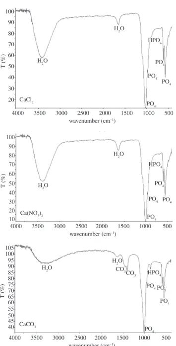
The infrared spectra (Figure 2) confirm the presence of PO43– (1096-1036 cm–1 and 606-562 cm–1), OH– (3566 cm–1), H
2O
(1640 cm–1 and 3430 cm–1) and HPO 4
2– (874 cm–1) which exists in
94 Belouafa et al. Materials Research
oxygenated apatite prepared from CaCO3 present of the bands ascrib-able to the carbonate ions CO32– (1412 and 1465 cm–1) which do not
disappear after calcination of this apatite at 900 °C for 2 hours (Fig-ure 3). This proves that these carbonate ions are of type B15. These ions can be to come only from calcium carbonate used like calcium precursor. The Figure 3 shows yet the absorption band corresponding to the OH– vibrationnelle mode (3566 cm–1) and the OH– bending
de-formation mode (633 cm–1) which exist in HAP16 and a characteristic
band (985 cm–1) of β-TCP15. The thermal decomposition reaction has
been proposed to occur in according to the reaction observed in the case of non-stiochiometric phosphocalcic HAP17:
Ca10–x(HPO4)x(PO4)6–x(OH)2–x→
(1–x)Ca10(PO4)6(OH)2 + 3xCa3(PO4)2 + xH2O (1)
The presence of HAP18 and β-TCP19 is confirmed by XDR
(Figure 4).
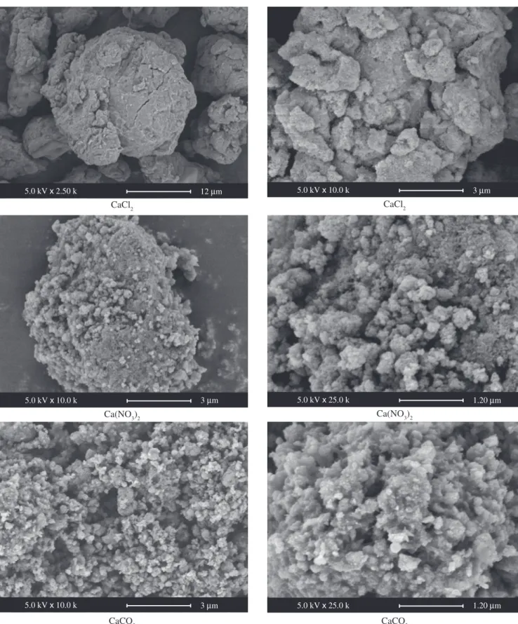
The aspect of the synthesized powders suggests the typical apatite appearance as shown in Figure 5. These photomicrographs suggest porous aggregates of particles prepared from CaCO3, very compact grains of particles prepared from CaCl2, intermediate aspect of par-ticles prepared from Ca(NO3)2. The porosity of the apatite prepared from the CaCO3 can be due to release of CO2 during the attack acid of CaCO3 by hydrogen peroxide.
The variety of morphological states of these biomaterials broad-ened their field of biomedical application. As the porous apatites
CaCl2
CaCO3
Ca(NO3)2
Angle (2 )
Figure 1. XRD patterns of obtained powders.
T (%) 100 90 80 70 60 50 40 30 20
H2O
H2O
PO4 PO4 PO4 PO4 HPO4 HPO4
4000 3500 3000 2500 2000 1500 1000 500
wavenumber (cm–1)
CaCl2 100 90 80 70 60 50 40 30 20 10
H2O
H2O
PO4 PO4 PO4 PO4 HPO4 HPO4
4000 3500 3000 2500 2000 1500 1000 500
wavenumber (cm–1)
Ca(NO3)2
T (%) 105 95 85 75 65 55 50 60 70 80 90 100 45 40
H2O
H2O
PO4
PO4PO
4
PO4
PO4
HPO4
HPO4
4000 3500 3000 2500 2000 1500 1000 500
wavenumber (cm–1)
CaCO3
T (%)
CO3 CO3
Figure 2. FT-IR spectra of obtained powders.
T (%)
OH– OH–
CO3
PO43–
du β-TCP
(cm–1)
Figure 3. FT-IR spectra of carbonated oxygenated apatite after calcination at 900 °C for 2 hours.
* * * * * * * * HAP * β-TCP 002 300
25 30 35 40 45 50
Vol. 11, No. 1, 2008 Characterization of Antiseptic Apatite Powders Prepared at Biomimetics Temperature and pH 95
Figure 5. Scanning electron microscopy of as-dried powder. 12 m
5.0 kV x 2.50 k 5.0 kV x 10.0 k 3 m
3 m
5.0 kV x 10.0 k 5.0 kV x 25.0 k 1.20 m
3 m
5.0 kV x 10.0 k 5.0 kV x 25.0 k 1.20 m
exhibit strong bonding to the bone; the pores provide a mechanical interlock leading to a firm fixation of the material20. Bone tissue grows
well into the pores, increasing strength of the apatite implant. It is realized that dimension and morphology of pores are crucial factors for an excellent osteointegraton. As for dense apatite, they are used
for the formation of ceramic blocks with different forms and for the recovery of implants21.
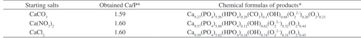
The chemical analysis (Table 1) shows that the obtained materi-als are a calcium deficient apatites with Ca/P ratio and following chemical formulas:
CaCl2 CaCl2
Ca(NO3)2 Ca(NO3)2
96 Belouafa et al. Materials Research
4. Conclusions
Single-phase phosphocalcique oxygenated apatite powders were synthesized by a novel chemical precipitation technic at the physi-ological conditions of pH 7.4 and 37 °C. The produced powders were shown to have poor crystallinity and various compositions and mor-phological states what widened their biomedical applicability.
References
1. Ledard C. Benque E. Lacout JL. Rey C. Biomatériaux de comblement osseux ou dentaire, et procédés de préparation. Patent FR 2652748; 1989.
2. Simpson DR. Substitutions in apatite. I. Potassium-bearing apatite. Am. Mineral. 1968; 53: 432-444.
3. Rey C. Etude des relations entre apatites et composés moléculaires; Thèse d’Etat I.N.P: Toulouse 1984.
4. Belouafa S. Chaair H. Digua K. Sallek B. Mountacer H. Nouveau procède de synthèse d’une apatite carbonatée à caractère antiseptique. Phosphorus, Sulfur and Silicon. 2005; 180: 2679-2687.
5. Belouafa S. Chaair H. Digua K. Oudadesse H. Sallek B. Mountacer H. Utilisation des plans d’experiences pour la modelisation de l’élaboration d’un phosphate de calcium de proprietes antiseptiques à usage biomedical.
Phosphorus, Sulfur and Silicon. 2006; 181: 337-349.
6. Belouafa S. Chaair H. Digua K. Sallek B. Essaadani A. Oudadesse H. Synthesis, Characterization and thermal behaviour of a phosphocal-cic oxygenated apatite. Journal of Advanced Materials. 2007; 2: 139-142.
7. Meyer JL. Eanes ED. The maturation of crystalline calci-. um phosphates in aqueous suspensions at physiologic pH. Calcified Tissue Res. 1977; 23: 259-269.
8. Gee A. Dietz VR. Determination. of phosphate by differential
spectro-photometry. Anal. Chem. 1953; 25: 1320-1324.
9. Trombe JC, Montel G. Some features of the incorporation of. oxygen in different oxidation states in apatite lattice. J. Inorg. Nucl. Chem. 1978; 40: 15-21
10. Vignoles-Montrejaud, M. Contribution a l’étude des apatites carbonattes de type B. Toulouse, France: These d’Etat, INP; 1984.
11. Trombe JC. Annal. Chim. 1973; 8: 335.
12. LeGeros RZ. Biological and synthetic apatites. In: PW Brown and B. Constantz, Editors, Hydroxyapatite and related materials, CRC Press, Boca Raton. 1994; 3.
13. Peters F. Schwarz K. Epple M. The structure of bone studied with syn-throtron X-ray diffraction, X-ray absorption spectroscopy and thermal analysis. Thermochim. Acta. 2000; 361: 131-138.
14. Yubao L. Kelein CPAT. Xingdong Z. de Groot K. Formation of a bone apatite-. Like layer on the surface of porous HA. Ceramics. Biomaterials. 1994; 15: 835-840.
15. El Feki H. Khattech I. Jemal M. and Rey C. Decomposition thermique d’hydroxyapatites carbonatées sodées: Thermal decomposition of carbon-ated hydroxyapatites containing sodium ion. Thermochimica Acta. 1994; 237: 99-110.
16. Yubao L. Kelein CPAT. Xingdong Z. de Groot K. Formation of a bone apatite-. Like layer on the surface of porous HA. Ceramics. Biomaterials. 1994; 15: 835-840.
17. Destainville A. Champion E. Bernache-Assollant D. Laborde E. Synthesis, characterization and thermal behavior of apatitic tricalcium phosphate.
Materials Chemistry and Physics. 2003; 80: 269-277.
18. File N° 73-0293. International Center for Diffraction Data, ICDD.
19. File N° 70-2065. International Center for Diffraction Data, ICDD. 20. Daculci G. Passuti N. Martin S. Macroporous calcium phosphate ceramic
for long bone surgery in humans and dogs. Clinical and histological study.
Biomed. Mater. Res. 1990; 24(3): 379-396.
21. Daculci G. Legros J.P. Three-dimensional defects in hydroxyapatite of biological interest. Biomed. Mater. Res., 1996; 4: 495-501.
Table 1. Results of the chemical analysis of prepared apatites.
Starting salts Obtained Ca/P* Chemical formulas of products* CaCO3 1.59 Ca9.27(PO4)5.56(HPO4)0.29(CO3)0.1(OH)0.68(O22–)
0.20(O2)0.21
Ca(NO3)2 1.60 Ca9.57(PO4)5.88(HPO4)0.12(OH)0.62(O22–)
0.32(O2)0.44
CaCl2 1.60 Ca9.59(PO4)5.82(HPO4)0.18(OH)0.74(O22–)
0.31(O2)0.45
*: ∆(Ca/P) = ± 0.01; ∆(Ca) = ± 0.005; ∆(PO4) = ± 0.005; ∆(HPO4) = ± 0.01; ∆(CO3) = ± 0.02; ∆(O22–) = ± 0.01; ∆(O