in the diagnosis. Starting in the 1980’s, ultrasound became a procedure indispensable to the clinical practice in the field of tocogynecology, modifying concepts and procedures, and bringing an important contribution to this specialty. The advent of endocavitary transducers, amplitude and spectral color Doppler, as well as the increasing improvement in the imaging quality, has contributed to this progress. Over the last ten years, much has been studied, published and discussed about the role of the three-dimensional ultrasonography. The authors review the topic and emphasize the importance of this method as a diagnostic modality. Keywords: Uterine malformations; Müllerian malformations; Three-dimensional ultrasonography.
Ultra-sonografia tridimensional em ginecologia: malformações uterinas.
As malformações uterinas são achados pouco comuns na clínica ginecológica. As estatísticas nesta área são muito falhas. Corrobora, ainda, a falta de uniformização na terminologia empregada e as dificuldades diagnósticas. A partir da década de oitenta, a ultra-sonografia tornou-se um procedimento indispensável à prática toco-ginecológica, contribuindo e modificando conceitos e procedimentos dentro dessa especialidade. O advento dos transdutores endocavitários, a análise com Doppler colorido de amplitude e espectral, assim como a melhoria crescente da qualidade de imagem contribuíram para isso. Nos últimos dez anos muito se tem pesquisado, publicado e discutido sobre o papel da ultra-sonografia tridimensional. Os autores fazem uma revisão do tema e ressaltam a importância dessa metodologia como modalidade diagnóstica.
Unitermos: Malformações uterinas; Malformações müllerianas; Ultra-sonografia tridimensional. Resumo
* Study developed at EURP – Escola de Ultra-sonografia e Reciclagem Médica de Ribeirão Preto, Ribeirão Preto, SP, Brazil. 1. PhD, Professors at EURP – Escola de Ultra-sonografia e Re-ciclagem Médica de Ribeirão Preto and Faculdade de Medicina de Ribeirão Preto (Unaerp), Ribeirão Preto, SP, Brazil.
2. MDs at EURP – Escola de Ultra-sonografia e Reciclagem Médica de Ribeirão Preto, Ribeirão Preto, SP, Brazil.
3. Student in the Program of Post-graduation at EURP – Es-cola de Ultra-sonografia e Reciclagem Médica de Ribeirão Pre-to, Ribeirão PrePre-to, SP, Brazil.
Mailing address: Prof. Dr. Adilson Cunha Ferreira. Rua Ma-noel Ache, 980, Ed. Van Gogh, ap. 222, Jardim Irajá. Ribeirão Preto, SP, Brazil, 14020-590. E-mail: [email protected]
Received December 20, 2004. Accepted after revision July 1, 2005.
INTRODUCTION
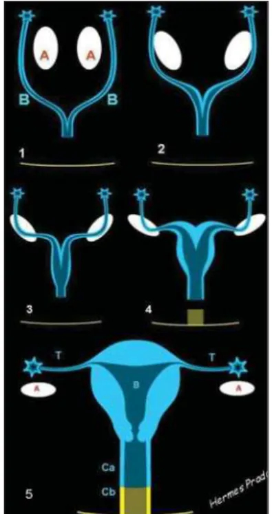
Uterine malformations are secondary to failure in development, reabsorption or fusion of Müllerian ducts. Around the sixth week of the embryogenesis, an invagina-tion of the coelomic lining epithelium forms a depression creating a sulcus, whose borders fuse to form the lateral Müllerian ducts (or paramesonephric ducts) (Figure 1). The Müllerian ducts initially are formed in the upper dorsal wall of the coelomic cavity and progress caudally to enter the pelvis where they incline towards the
cen-Figure 1. Scheme showing the embryological de-velopment and sequence (1 to 4) of Müllerian ducts fusion (B). (A) indicates the ovaries. Number 4 demonstrates the formation of the uterine body after fusion. 5B, uterine body; T, Fallopian tubs; Ca, proximal third of the vagina; Cb, distal third.
ter, fusing medially. Farther on, the caudal progress results in a contact of these fused ducts with the urogenital sinus. The proxi-mal segments of the uterovaginal canal originated from coelomic epithelium re-main unfused and open into the peritoneal cavity to form the Fallopian tubes. The upper portion of the vagina is, therefore, considered to have Müllerian origin, and the lower portion as originating from the urogenital sinus. He whole lining epithe-lium (uterus and tubes) originates from the coelomic epithelium. This is the reason for uterine malformations being denominated Müllerian malformations or anomalies(1).
In the past, the uterus only could be clinically evaluated by means of a physical examination. Several methods have been introduced for gynecological evaluation. As an example, we can mention; radiologi-cal examinations(2) by means of
hysteros-alpingography(3–5) (Figure 2), surgical
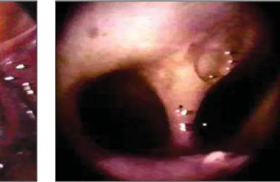
pro-cedures — laparotomy, laparoscopy, and most recently, videolaparoscopy (Figure 3) — and hysteroscopy(6) (Figure 4).
has brought the greatest contribution as non-invasive method for evaluation of the uterus and its attachments(7), initially as a
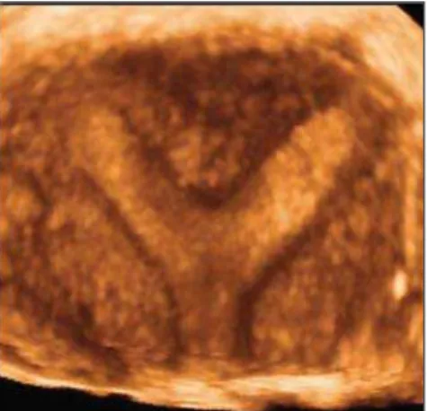
two-dimensional (2D) modality with an abdominal approach (Figure 5), and later with a transvaginal approach (Figure 6). Most recently, ultrasonography has been added of the three-dimensional (3D) pro-cessing(8–11) in both modalities — the
multiplanar (Figure 7) and the volumetric ones (Figures 8, 9 and 10). Magnetic reso-nance imaging also has been utilized in many services(12).
DISCUSSION
Uterine malformations are unusual find-ings in the gynecological clinical practice. This abnormality is a reflection of an array of presentations, associated with the fact that the diagnosis of the majority of mal-formations does not occur before a gesta-tion or are diagnosed only after manifesta-tion of an obstetric problem(13–15). Statistics
in this area are flawed, and there is a lack of standardization of the pertinent termi-nology, besides the difficulty in the diag-nosis(16). Patients with these alterations
fre-quently are oligosymptomatic or even as-ymptomatic, with preserved menstrual, sexual and even reproductive functions. So, frequently presented case reports and casuistics reflect particularities of certain groups of women with obstetric complica-tions, patients of infertility and sterility services, or cases of medical urgency re-sulting from menstrual flow obstruction(17).
Presently, 2D ultrasound, and especially 3D ultrasound are indispensable diagnos-tic tools for evaluation of uterine
malfor-pian tubes are observed. alization excludes this diagnostic hypothesis. The (see Figure 3). final diagnosis was of partial septate uterus.
Figure 7. Normal uterus on 3D multiplanar ultrasound. Observe the secretory pattern of the endometrium on the three views.
Figure 5. Longitudinal view of a normal uterus on 2D transabdominal ultrasound. Observe the ad-equately filled bladder, and the secretory pattern of the endometrium.
mations, allowing accurate diagnoses, most of times more specific than a simple de-scription of a septate uterus, characterizing the abnormality and providing information to assist in the definition of therapeutic regimen and reproductive prognosis(18–21).
The appropriate classification of Mül-lerian anomalies is important, the most rel-evant one being proposed by Jarcho in 1946, and later adapted by Zanetti et al., based on the embryonal development. Later on, this classification was modified by Butram and Gibbons, in 1975, and is currently adopted by the American Society of Fertility.
A better evaluation with 2D ultrasound is achieved by the association between transabdominal and transvaginal ap-proaches. The first one allows a better vi-sualization of the uterine fundus, and analysis of the bladder and ureteral jets. The second allows a more detailed analy-sis of the cervix (cervices) and endometrial cavity (cavities).
The main view for correctly diagnosing the type of the malformation is the coronal view, sometimes difficult to be obtained with the 2D technique, but feasible, pro-vided some technical prerequisites are met: the bladder must be almost emptied; the transducer must be placed transversely to the patient’s abdomen with slight, cranial movements.
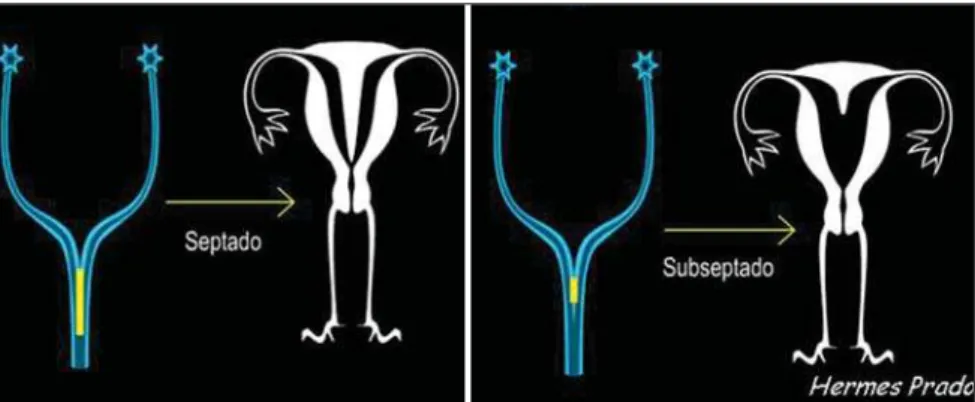
Several anomalies may originate from an incomplete fusion the Müllerian ducts: clefts in the uterine funds, but not in the cervix, a complete division of the uterus by
Figure 8. Coronal view of a septate uterus on 3D volumetric ultrasound. Observe the secretory pat-tern of the endometrium.
Figure 10. Coronal view of a septate uterus on 3D volumetric ultrasound with digital subtraction of the septum.
Figure 9. Coronal view of a septate uterus on 3D volumetric ultrasound with digital subtraction of the endometrium.
Figure 11. Double cavity uterus. Final diagnosis: septate uterus. Evaluation by 2D transabdominal ultrasound.
a septum into two endometrial cavities, forming the called septate uterus. The sep-tum may be partial or complete, with vari-able extent from the uterine fundus towards the cervix (Figures 11, 12, 13, 14 and 15).
Sometimes, the differential diagnosis with bicornuate uteri is difficult, especially if the sonographic evaluation is restricted to the transvaginal approach. Also, it may be as-sociated with a longitudinal or oblique vaginal septum.
The complete non-fusion of Müllerian ducts originates an anomaly previously called complete double uterus with double cervix, currently denominated didelphys uterus (Figures 16 and 17), with each
en-dometrial cavity ending in a solitary fallo-pian tube. Such anomalies are perfectly compatible both with normal fertility and menstrual cycles, but sometimes they may result in significant clinical problems(20).
Figure 12. Double cavity uterus. Final diagnosis: septate uterus. Evaluation by 2D transabdominal ultrasound. Observe the difference in relation to Figure 11. In this image, the transducer was slightly more cranially inclined.
Figure 16. Schemes showing failures in complete (didelphys uterus) and partial (bi-cornuate uterus) fu-sion of the Müllerian ducts.
Figure 18. Bicornuate uterus with a ten-week ges-tation. Transabdominal evaluation. Observe decidua in the non-pregnant uterine cavity.
Figure 17. Double cavity uterus. Final diagnosis: didelphys uterus. Transabdominal 2D ultrasound is the method of choice for the correct diagnosis of this anomaly. Observe the complete separation between uterine bodies.
Pregnancy in a half of a septate, bicornu-ate (Figures 18, 19 and 20) or didelphys uterus may be associated with bleeding of the non-pregnant half of the uterus. In rare cases, the pregnancy may occur in a half of the uterus after the other is already
preg-nant; this is called superfetation. The 2D ultrasound is sufficient for an adequate evaluation and diagnosis.
Bicornuate uteri (Figures 21 and 22) is the result of an incomplete fusion of the uterovaginal horns at the level of the
fun-dus; that is to say, a partial fusion has oc-curred, resulting in two uterine fundi (horns) presenting different fusion degrees, symmetrically or not, most often at the level of the uterine isthmus, and, therefore, frequently presenting a single cervix. This
septate uterus. Evaluation by multiplanar, transvagi-nal 3D ultrasound. Observe the symmetrical en-dometrial cavities.
Figure 20. Bicornuate uterus with a five-week ges-tation. Transvaginal evaluation.
type of anomaly may be confused with sep-tate uterus so currently 3D ultrasound is a valuable diagnostic method.
The small sized uterus is subdivided into hypoplastic and infantile uterus. The uterine hypoplasia (Figure 23) is found in an array of endocrine disorders, with a 1:1 uterine body/cervix ratio. In the infantile uterus, the uterine body/cervix ratio is 2:1. In the majority of cases, hypoplastic/infan-tile uterus occurs because of an ovarian or hypophyseal hypofunction.
An aspect that should not be disre-garded during an ultrasound examination in a patient with uterine malformation is the evaluation of the renal lodges, considering the frequent association of uterine anoma-lies with urinary tract malformations such as renal agenesis (Figure 24) and/or ecto-pia(23). Li et al. have found renal agenesis
in 17 (29.8%) of 57 patients. No other re-nal anomaly has been found. Rere-nal agen-esis was the most frequent association in patients with didelphys uterus (13/16 cases), uterine agenesis (2/5 cases) and unicornuate uterus (2/7 cases). All the 11 cases of obstructed didelphys uterus were associated with homolateral renal agenesis towards the transverse septum of the ob-structed hemivagina. So, they have con-cluded that renal agenesis is more fre-quently found in didelphys uterus than in other types of uterine malformations(23).
The uterus and vagina agenesis is
de-nominated Mayer-Rokitansky-Kuster-Hauser syndrome and results from dyspla-sia of the Müllerian ducts, with absence of the normal uterus and part or the whole vagina. The diagnosis is basically clinical, however, the 2D ultrasound may confirm a clinical suspicion. The 3D ultrasound pre-sents no advantage over the 2D ultrasound in these cases.
Although there are several types of uter-ine malformations, almost all of them can be diagnosed as follows: arcuate uterus, septate uterus (partial or complete), bicor-nuate uterus, unicorbicor-nuate uterus, didelphys uterus, agenesis, hypoplasia, and infantile uterus.
With the exception of arcuate uterus, considered as a variant of the normal uterus, the most usual uterine anomalies, frequently resulting in diagnostic difficulty are the septate and bicornuate uterus(9).
The term “arcuate” refers to cases where there is a minimal alteration of the uterine cavity, with a convex or flat uterine fundus. The endometrial cavity may present a mini-mal, residual septum in the fundal region. There is no need for correction.
Regardless the type of uterine malfor-mation, both 2D and 3D ultrasound should be performed during the second phase of the menstrual cycle for a better visualiza-tion of the endometrium, and therefore a better definition of the uterine cavity. Dur-ing the first phase of the menstrual cycle,
Figure 21. Double cavity uterus. Final diagnosis: bicornuate uterus. Transvaginal 3D multiplanar ultra-sound. Observe asymmetrical endometrial cavities.
Figure 22. Bicornuate uterus. Coronal, 3D multi-planar evaluation showing asymmetrical cavities.
Figure 23. Very reduced volume of the uterus. Fi-nal diagnosis: hypoplastic uterus. Observe the al-most imperceptible presence of the uterus on the transabdominal study.
dominal and transvaginal approaches may be adopted, the latest being preferable. The transrectal modality is an excellent alterna-tive in the unfeasibility of the transvaginal approach. With the multiplanar 3D ultra-sound, longitudinal, axial and coronal planes can be concomitantly evaluated, the coronal plane being indispensable to an appropriate diagnosis of Müllerian ducts anomalies.
Although magnetic resonance imaging is considered by some authors(12) as the
method of choice for uterine anomalies evaluation, with up to 100% efficacy, this has not been a routine practice because of its high cost.
For the majority of authors, the ultra-sound is the primary method for evaluation of Müllerian abnormalities. Fedele et al., studying 43 infertile patients diagnosed with double uterus by hysterosalpingogra-phy, have submitted them to subsequent ultrasound and laparoscopy/hysteroscopy to evaluate the ultrasound capacity to cor-rectly demonstrate a malformed uterus. The ultrasound visualization was adequate in 39 cases (90.7%): one of two cases of didelphys uteri, all the 11 bicornuate uteri, all the four complete septate uteri, and all the 22 partial septate uteri with 92.3% sen-sitivity, and 100% specificity. Therefore, a differential and accurate diagnosis of “double uterus or endometrial cavity” is possible with this technique(21).
2. Bracci M, Busilacchi P, Ciccognani G, Lorenzoni A, Serri L. Abnormalities of the female genital tract. Comparison of radiographic and echograph-ic pechograph-ictures. Radiol Med (Torino) 1988;75:181– 191.
3. Matheus M, Franceschini AS, Sala MA, Barrio-novo N. Histerossalpingografia: estudo
retrospec-tivo de 535 casos.J Bras Ginecol 1986;96:123–
127.
4. Goldberg JM, Falcone T, Attaran M. Sonohystero-graphic evaluation of uterine abnormalities noted on hysterosalpingography. Hum Reprod 1997;12: 2151–2153.
5. De Meo I. Hysterosalpingography in the diagno-sis of uterine malformations. Minerva Ginecol 1983;35:127–130.
6. Valli E, Zupi E, Marconi D, et al. Hysteroscopic findings in 344 women with recurrent spontane-ous abortion. J Am Assoc Gynecol Laparosc 2001;8:398–401.
7. Grimbizis GF, Camus M, Tarlatzis BC, Bontis JN, Devroey P. Clinical implications of uterine mal-formations and hysteroscopic treatment results. Hum Reprod Update 2001;7:161–174. 8. Jurkovic D, Gruboeck K, Tailor A, Nicolaides KH.
Ultrasound screening for congenital uterine anomalies. Br J Obstet Gynaecol 1997;104:1320– 1321.
9. Raga F, Bonilla-Musoles F, Blanes J, Osborne NG. Congenital mullerian anomalies: diagnostic accu-racy of three-dimensional ultrasound. Fertil Steril 1996;65:523–528.
10. Chan L, Uerpairojkit B, Reece EA. Diagnosis of congenital malformations using two-dimensional and three-dimensional ultrasonography. Obstet Gynecol Clin North Am 1997;24:49–69. 11. Lev-Toaff AS, Pinheiro LW, Bega G, Kurtz AB,
Goldberg BB. Three-dimensional multiplanar sonohysterography: comparison with conven-tional two-dimensional sonohysterography and x-ray hysterosalpingography. J Ultrasound Med 2001;20:295–306.
12. Fedele L, Dorta M, Brioschi D, Massari C,
Can-sound; a pilot study. Fertil Steril 1996;66:848– 850.
17. Jones HW Jr. Reproductive impairment and the malformed uterus. Fertil Steril 1981;36:137–148. 18. Nicolini U, Bellotti M, Bonazzi B, Zamberletti D, Candiani GB. Can ultrasound be used to screen uterine malformations? Fertil Steril 1987;47:89– 93.
19. Woodward PJ, Sohaey R, Wagner BJ. Congeni-tal uterine malformations. Curr Probl Diagn Radiol 1995;24:178–197.
20. Heinonen PK, Savolainen A, Pystynen P. Septate uterus and habitual abortion: a case report illus-trating successful outcome of pregnancy after second metroplasty. Eur J Obstet Gynecol Reprod Biol 1986;23:233–238.
21. Fedele L, Ferrazzi E, Dorta M, Vercellini P, Can-diani GB. Ultrasonography in the differential di-agnosis of “double” uteri. Fertil Steril 1988;50: 361–364.
22. Woelfer B, Salim R, Banerjee S, Elson J, Regan L, Jurkovic D. Reproductive outcomes in women with congenital uterine anomalies detected by three-dimensional ultrasound screening. Obstet Gynecol 2001;98:1099–1103.
23. Li S, Qayyum A, Coakley FV, Hricak H. Associa-tion of renal agenesis and mullerian duct anoma-lies. J Comput Assist Tomogr 2000;24:829–834. 24. Wu MH, Hsu CC, Huang KE. Detection of con-genital mullerian duct anomalies using three-di-mensional ultrasound. J Clin Ultrasound 1997; 25:487–892.
25. Hosli IM, Tercanli S, Herman A, Kretschmann M, Holzgreve W. In vitro volume measurement by three-dimensional ultrasound: comparison of two different systems. Ultrasound Obstet Gynecol 1998;11:17–22.