177
Radiol Bras. 2011 Mai/Jun;44(3):177–182
Collaborative environment for nuclear medicine training
*
Ambiente colaborativo para formação de pessoal em medicina nuclear
Cláudia Régio Brambilla1, Gabriel Goulart Dalpiaz2, Ana Maria Marques da Silva3, Neivo da Silva Júnior4, Lucia Maria Martins Giraffa5, Tiago Coelho Ferreto6, Cesar Augusto Fonticielha De Rose7, Vinicius Duval da Silva8
Objective: To validate the proposal for development of a virtual collaborative environment for training of nuclear medicine personnel. Materials and Methods: Organizational assumptions, constraints and functionalities that should be offered to the professionals in this field were raised early in the development of the environment. The prototype was developed in the Moodle environment, including data storage and interaction functionalities. A pilot interaction study was developed with a sample of specialists in nuclear medicine. Users’ opinions collected by means of semi-structured questionnaire were submitted to quantitative and content analysis. Results: The proposal of a collaborative environment was validated by a community of nuclear medicine professionals and considered as an aid in the training in this field. Suggestions for improvements and new functionalities were made. There is a need to establish a program for education of moderators specifically for this environment, considering the different interaction characteristics as the online and conventional teaching methods are compared. Conclusion: The collaborative environment will allow the exchange of experiences and case discussions among professionals from institutions located in different regions all over the country, enhancing the collaboration among them. Thus, the environment can contribute in the early and continued education of nuclear medicine professionals.
Keywords: Collaborative environment; Nuclear medicine; Medical education; Distance education.
Objetivo: Validar a proposta do desenvolvimento de um ambiente colaborativo virtual para formação de pessoal em medicina nuclear. Materiais e Métodos: No desenvolvimento inicial do ambiente foram levantadas as premissas, restrições e funcionalidades que deveriam ser oferecidas aos profissionais da área. O protótipo foi desenvolvido no ambiente Moodle, incluindo funcionalidades de armazenamento de dados e interação. Um estudo piloto de interação no ambiente foi realizado com uma amostra de profissionais especialistas em medicina nuclear. Análises quantitativas e de conteúdo foram realizadas a partir de um questionário semiestruturado de opinião dos usuários. Resultados: A proposta do ambiente colaborativo foi validada por uma comunidade de profissionais que atuam nesta área e consi-derada relevante visando a auxiliar na formação de pessoal. Sugestões de melhorias e novas funcionalidades foram indicadas. Observou-se a necessidade de estabelecer um programa de formação dos moderadores no ambiente, visto que são necessárias características de interação distintas do ensino presencial. Conclusão: O ambiente colaborativo poderá permitir a troca de experiências e a discussão de casos entre profissionais localizados em instituições de dife-rentes regiões do País, possibilitando uma aproximação e colaboração entre esses profissionais. Assim, o ambiente pode contribuir para formação inicial e continuada de profissionais que atuam em medicina nuclear.
Unitermos: Ambiente colaborativo; Medicina nuclear; Educação médica; Ensino a distância. Abstract
Resumo
* Study developed at Pontifícia Universidade Católica do Rio Grande do Sul (PUCRS), Porto Alegre, RS, Brazil. Financial sup-port: Fundo Regional para a Inovação Digital na América Latina e Caribe – FRIDA, Project B80.
1. Medical Physicist, Master in Health Sciences, Pontifícia Universidade Católica do Rio Grande do Sul (PUCRS), Porto Ale-gre, RS, Brazil.
2. Graduate Student of Engineering, Developer of Research Projects for Pontifícia Universidade Católica do Rio Grande do Sul (PUCRS), Porto Alegre, RS, Brazil.
3. PhD of Physics, Director of the Department of Physics – Pontifícia Universidade Católica do Rio Grande do Sul (PUCRS), Porto Alegre, RS, Brazil.
4. PhD, Nuclear Physician at Hospital São Lucas – Pontifícia Universidade Católica do Rio Grande do Sul (HSL-PUCRS), Porto Alegre, RS, Brazil.
5. PhD, Titular Professor at School of Information Technology, Researcher for the Program of Mastership in Education, Sciences
Brambilla CR, Dalpiaz GG, Marques da Silva AM, Silva Júnior N, Giraffa LMM, Ferreto TC, De Rose CAF, Da Silva VD. Collaborative environment for nuclear medicine training. Radiol Bras. 2011 Mai/Jun;44(3):177–182.
and Mathematics and Program of Post-graduation at School of Education of Pontifícia Universidade Católica do Rio Grande do Sul (PUCRS), Porto Alegre, RS, Brazil.
6. PhD, Assistant Professor, Instituto de Informática da Pon-tifícia Universidade Católica do Rio Grande do Sul (PUCRS), Porto Alegre, RS, Brazil.
7. PhD, Professor at Instituto de Informática da Pontifícia Universidade Católica do Rio Grande do Sul (PUCRS), Porto Ale-gre, RS, Brazil.
8. PhD, Professor, Department of Pathology and Radiations, School of Medicine, Pontifícia Universidade Católica do Rio Grande do Sul (Famed-PUCRS), Porto Alegre, RS, Brazil.
Mailing Address: MSc. Cláudia Régio Brambilla. Rua Major Ismael Alves, 74, Centro. Gravataí, RS, Brazil, 94010-350. E-mail: [email protected]
Received December 14, 2010. Accepted after revision May 6, 2011.
INTRODUCTION
Traditional ways for medical learning in nuclear medicine generally comprise dis-cussions over clinical image banks, moni-toring of specialists in the images analysis, classroom or distance learning courses, and studies on the available literature(1). Within
small number of training centers in the country?
Currently, the availability of digital and virtual technologies allows the access to virtual environments where instructional contents and learning support platforms are available(1). Some of those are targeted at
medical teaching. However, normally such contents are offered in the form of open or restricted access online courses, such as the case of the initiative from the American Association of Physicists in Medicine (AAPM), together with the Radiological Society of North America (RSNA), in which modules were developed for the education of residents in radiology or nuclear medicine(2,3). The self-explanatory
modules were developed by interdiscipli-nary specialist groups, comprising at least a physicist and a radiologist.
The virtual medical learning environ-ments usually utilize static and sequential contents, without incorporating interaction capabilities. In such environments, interactivity is usually restricted to indi-vidual users browsing, without allowing interaction between users or the insertion of questionings or new contents into the environment.
The available information and commu-nication technologies can be utilized to en-hance the understanding of the processes involved in medical images acquisition and processing. The present study presents a proposal of development and validation of a virtual collaborative environment that allows interactivity, coupled with compu-tational power, for the education of nuclear medicine professionals.
MATERIALS AND METHODS
Initially, exploratory meetings were held with multi-disciplinary teams com-prising medical physicists, nuclear medi-cine physicians and computer scientists, in order to identify the organizational, envi-ronmental and external assumptions and re-strictions to the collaborative environment. Aspects related to infrastructure of partner institutions and hospitals as well as matters related to data accessibility and security were also addressed by the information technology team. From that preliminary study, the following requirements for the
development of the collaborative environ-ment were defined:
– provide the users with a friendly in-terface, allowing the interaction among users and access to dynamic contents;
– allow users to interact in discussion forums;
– provide contents of interest to nuclear medicine, such as image banks and litera-ture-based support contents;
– allow user-friendly submission of nuclear medicine computer simulations, by means of the utilization of high-perfor-mance computing resources.
Based on the social constructivist learn-ing theory(4) that permeates the proposal of
collaborative environments, materials were made available in the environment, relying on the assumption that the construction and expansion of the environment´s resources would be accomplished by the collabora-tion among users.
For the construction of a collaborative environment prototype and subsequent validation, the myocardial perfusion study was selected as the initial application be-cause of its high demand at nuclear medi-cine centers. Discussion topics, such as to-mographic images acquisition and recon-struction parameters were selected as dis-cussion starters in the environment, as well as clinical case studies. Free digital image visualization and processing tools were also made available. Concomitantly, a pro-totype was developed for the submission of computer simulations by utilizing the re-sources of a high-performance laboratory. The computer simulation of images by means of the Monte Carlo method is widely utilized to simulate the effects produced by parameter changes in the acquisition of nuclear medicine images(5–7). In order to
create a simulated images bank, a nuclear medicine equipment was modeled by uti-lizing the resources available at the GATE package (geant4 application for tomo-graphic emission), which simulates PET and SPECT systems(8,9).
As the contents of the collaborative en-vironment is dynamic and dependent on the users’ interest, a limited number of images and documents was initially made avail-able. The management of the contents and security of the collaborative environment was carried out by an anonymous
admin-istrator that authorized user registrations under pre-established rules. Experienced professionals in the field of nuclear medi-cine acted as moderators, coordinating the discussions and selecting themes to be dis-cussed in the forums.
The users sample for validation of the proposed collaborative environment was intentionally recruited, and the users were nuclear medicine specialists in order to interact in the collaborative environment. A pilot study was developed with 10 medi-cal physicists and 5 nuclear physicians. At the end of the interaction, the users filled out a semi-structured questionnaire (Likert scale) evaluating the relevance and ease on different aspects of functionalities and in-teraction with the environment, indicating suggestions for improvements in the pro-posal. The interaction test was carried out over a one-month period.
The analysis was initially performed on the closed part of the questionnaire, allow-ing the evaluation of available function-alities in the environment by means of the opinion from users after the interaction test (Likertscale).
A contents analysis based on Moraes & Galiazzi was also performed in the open sections of the questionnaire. Such an analysis approach has an operations cycle that initiates by the unitarization of mate-rials in the textual “corpus”, moving to the categorization of analysis units. From such a process new comprehensions emerge, constituted by the self-organization of the results interpretation text(10).
RESULTS
Development of the environment
Such categories are divided into sub-cat-egories with the themes and discussion top-ics. At all the sub-categories, discussion forums are enabled to allow themes discus-sions among users. The moderators and users can utilize the image banks and sup-port documentation in the discussions, be-sides adding other materials and new ques-tions. A prototype of computational simu-lations submission in the collaborative en-vironment is currently under development, which will allow the choice of parameters for the simulation of nuclear medicine studies by means of the GATE application. Figure 2 depicts a diagram demonstrat-ing the process of simulation submission in the environment.
Clinical image banks, both experimen-tal and simulated, as well as free softwares for images processing and visualization in the environment are accessed at Menu> Im-ages.
Validation of the collaborative environment proposal in the pilot study
The interaction sample in the pilot test of the nuclear medicine collaborative en-vironment prototype comprised 15 sub-jects, with 10 of them being medical physi-cists (67%) and 5, nuclear medicine phy-sicians (33%). Among the subjects, 67% were male individuals (6 medical physicists and 4 nuclear medicine physicians) and 33% were female individuals (4 medical physicists and one nuclear medicine phy-sician). The mean graduation time was 9.4 years (median of 6 years). In the sample, 67% of the participants in the interaction had completed post-graduation, with a mean post-graduation time of 3.5 years. Mean time in the current position or func-tion in the sample was 6.27 years (median of 5 years). Mean age in the sample was 33.87 years (median of 32 years).
Data corresponding to the responses to the questionnaire are presented below, and evaluate the relevance of the following topics: Menu Items; Image Banks; Possi-bility of Contributions by the Users (cases, questions and materials); Document Banks (articles, support materials, dissertations and theses, published papers and reports); Availability of Free Softwares (images vi-sualization and processing); Interaction
with the Collaborative Environment per-formed in the discussion forums (quality of the discussions and moderators’ perfor-mance); and Possibility of Submitting Nuclear Medicine Simulations by means of the Collaborative Environment (prototype). Figure 3 shows the result of users’ opin-ions with respect to relevance of the catego-ries on the evaluation questionnaire.
As regards Evaluation of Materials Available in the Environment, the quanti-tative data represented by the first five
cat-egories on Figure 3, show that all the users considered as being relevant the items available in the menu, the image banks and the possibility provided to users to contrib-ute for the environment with cases, ques-tions and materials. As regards the quality of the available materials, some users con-sidered them of little relevance. One of the users suggested:
“the inclusion of other topics, not only those related with the area of image quality.” (FM-01).
Figure 1. Homepage of the Collaborative Environment in Nuclear Medicine.
The free softwares were considered of little relevance by one of the users, and some comments on the complexity in their utilization were reported.
As far as the Discussion Forums are concerned, whose quantitative data are rep-resented by the sixth category on Figure 3, which analyzes the quality of discussions and moderators’ performances, two partici-pants rated them as little or no relevant at all. Even among those who considered the discussion forums as relevant, the partici-pants in the interaction test presented sev-eral suggestions, as the following user opinion transcripts demonstrate:
“...the moderator did not provide an at-tractive and interesting environment, which caused a small number of partici-pations... the moderator should pro-mote discussions from the beginning and also encourage access to the website…” (FM-04).
“...I did not get the answers I needed from the moderator in the single case in which I participated...” (MN-01). As regards the possibility of Submission of Simulations by means of the collabora-tive environment, the quantitacollabora-tive data rep-resented by the seventh category on Figure 3, all users considered such functionality as relevant or very relevant.
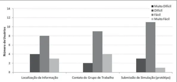
The qualitative data on the Interface Re-sources are represented by the three catego-ries on Figure 4, evaluating the
organiza-tion of the elements in the collaborative environment, according to the following requirements: Localization of the Data (icons and files); Contact with the Research Group and prototype of the Submission of Simulations (icons for modeling, submis-sion of simulations and simulation status progress bar).
As regards Localization of Data, some of the users considered it to be difficult. From an excerpt from a user’s opinion, one notices the anxiety towards the initial inter-action with the environment:
“For several moments I was disoriented with respect to the website routine, and had some operational difficulties, that I was finally able to overcome. Posting as well as task execution could be made a little easier...the difficulties were not lasting, but they were a hindrance…”
(MN-02).
As regards the Contact with the Research Team, in spite of some reports of difficulties in contacting the team, no e-mails were re-ceived by the group contact available in the environment. No postings on the environ-ment’s doubts forum were made on this theme. No justification was found in the open questions, thus making the comprehen-sion of such difficulty, quite troublesome. As regards the interface of the Submis-sion of Simulations interface prototype, the users did not offer criticism or suggestions, a fact that also impairs the proper
under-standing of the reasons leading to the op-tion by difficulties with such an interface. The analysis of the contents of the open questions on the last section of the ques-tionnaire on the Utilization of this Environ-ment by a Supervisor and his Team for Continued Education is presented next. Such analysis results from the opinions on the utilization of the environment in the field of nuclear medicine, both for initial and continued education purposes.
The users suggest that the collaborative environment in nuclear medicine be uti-lized for case discussions and for the dem-onstration of artifacts on images, providing the possibility of clarifying doubts related to relevant themes. In the opinions, the tool is considered as potentially useful in the evaluation/discussion of difficult cases and in the discussion of conflicting opinions in medical diagnosis. Additionally, this tool allows the solution of daily problems that can be discussed/solved by the group, by means of interaction and exchange of ex-periences among users in the virtual com-munity. The availability of forums and chats allows the exchange of knowledge and experiences, particularly in relation to different levels of competence in the field of nuclear medicine. The users point out that by means of the collaboration among users, it will be possible to generate data and image banks for reference regarding pathologies, false-positive and
tive results. Thus, as the number of active participants in the collaborative environ-ment grows, the data banks can be updated and increased by means of the insertion of rare cases, reducing the loss of data/cases due to geographical limitations. Multicter studies may also benefit from this en-vironment by means of the ease of access to images and exchange of opinions on cases. The virtual models for the simulation of medical images could be utilized for research in the field of nuclear medicine.
In the users opinion, with the high de-mand and data flow in the current labor market, the professionals updating and ca-pacitation are of utmost importance, as are the rapid and efficient solving of cases. The professionals face time limitations for studying, besides increase in workloads and information flow. The environment could be utilized for continued individual learning at one’s own rhythm.
One observed that most of the subjects that effectively interacted in the environ-ment became familiar with the social constructivist approach inherent to collabo-rative environments, providing significant contributions.
DISCUSSION
This study presents the development and validation of a collaborative environ-ment in nuclear medicine for personnel education, by means of a pilot interaction study with experienced users acting in the field. The functionalities required for the
implementation of a collaborative environ-ment that allowed primary and continued learning by nuclear medicine professional groups were analyzed.
By analyzing the profile of users that considered the quality of available materi-als as being of little relevance, one observes that such users were those with a longer experience in the field, a fact that may have generated little motivation/interest towards the subjects discussed in the environment, as well as those users that were interested in other specific areas of nuclear medicine. As regards the option for initially mak-ing available a limited number of materi-als in the environment meets the recom-mendations from specialists in collabora-tive environments found in the literature(4),
in order to encourage the users to contrib-ute with the environment construction. Aretio et al.(4) highlight the need to
gradu-ally increase the complexity of the collabo-rative environments, starting them only with the baseline materials.
Considering the limitations of the free softwares that were available in the envi-ronment, it would be interesting to develop software for images visualization and pro-cessing similar to those utilized in the workstations used in the clinical nuclear medicine routine as the present study pro-posal is expanded. Another possibility would be entering in an agreement with the software developers in order to provide the environment with a suitable version of their own image processing software to be used in the collaborative environment.
Although all the moderators that acted in the environment were experienced pro-fessionals in the field of nuclear medicine, they were not initially trained to participate in virtual collaborative environments. Af-ter the evaluation, such training proved to be a necessity. Another important aspect was the short time span of the interaction test (one month). A longer period of inter-action with the environment would allow a greater participation of users, greater use of the available materials and a more effec-tive contribution from users.
In spite of some criticism, the function-alities available in the collaborative envi-ronment were generally accepted. The pre-sented suggestions, such as the online chat, inclusion of new topics and materials can be easily implemented on account of functionalities available at Moodle.
It should be highlighted that on account of a research strategy option, no preferen-tial path was indicated to users at the be-ginning of the interaction, thus letting us-ers feel free to define their own “pathways” in the environment, which allows the evaluation of the need for the implemen-tation of guidance in the website and ini-tial instructions in the environment for new users. The aim was assessing whether the environment’s interface would be suscep-tible to cognitive understanding without any initial help. The users’ opinions suggest the need for the planning of a map of the environment, explaining on how the user can interact by means of a summary of the features with multimedia explanations.
After the assessment of users’ percep-tions on the environment’s interface re-sources, one realized that users accepted the available resources and functionalities. The criticism and suggestions present good indications for improvements on the pro-posal, particularly with respect to the need of providing more information on the pos-sibilities in the initial interaction to pro-mote a greater familiarization with the col-laborative environment.
CONCLUSION
The developed environment was evalu-ated as relevant by a community of nuclear medicine professionals, with respect to its usefulness in assisting the education of personnel. The available functionalities, the materials and discussion topics were considered as being relevant by most of the
users. It is possible to conclude that the method utilized in the virtual environment may allow the exchange of experiences and case discussions among professionals lo-cated in institutions from different regions of the country, which would enable greater professional proximity and collaboration among such professionals.
REFERENCES
1. Wallis JW, Miller MM, Miller TR, et al. An internet-based nuclear medicine teaching file. J Nucl Med. 1995;36:1520–7.
2. RSNA/AAPM Online Physics Modules. [cited 2009 Dec 10]. Available from: http://www.aapm. org/education/webbasedmodules.asp
3. Diagnostic radiology residents physics curricu-lum – AAPM Subcommittee of the Medical Phy-sics Education of Physicians Committee - May 2009. [cited 2009 Dec 10].Available from: http:// w w w. a a p m . o r g / e d u c a t i o n / d o c u m e n t s / Curriculum.pdf
4. García Aretio L, Ruíz Corbella M, Domínguez Figaredo D. El profesor y el formador en los
siste-mas digitales de enseñanza y aprendizaje. In: García Aretio L, Ruíz Corbella M, Domínguez Fi-garedo D, editores. De la educación a distancia a la educación virtual. Barcelona: Ariel; 2007. p. 163–86.
5. Brambilla CR. Impacto da determinação da pro-fundidade renal na quantificação renal absoluta
em estudos de cintilografia plana com 99m
Tc-DMSA [trabalho de conclusão de curso]. Porto Alegre, RS: Pontifícia Universidade Católica do Rio Grande do Sul; 2007.
6. Silva AM. Reconstrução quantitativa de SPECT: avaliação de correções [tese]. São Paulo, SP: Uni-versidade de São Paulo; 1998.
7. Zaidi H. Relevance of accurate Monte Carlo mod-eling in nuclear medical imaging. Med Phys. 1999;26:574–608.
8. Strulab D, Santin G, Lazaro D, et al. GATE (geant4 application for tomographic emission): a PET/ SPECT general-purpose simulation platform. Nucl Phys B (Proc Suppl). 2003;125:75–9. 9. De Beenhouwer J, Staelens S, Kruecker D, et al.
Cluster computing software for GATE simula-tions. Med Phys. 2007;34:1926–33.