PREVALENCE OF MORPHOLOGICAL ALTERATIONS OF THE
STYLOID PROCESS IN PATIENTS WITH TEMPOROMANDIBULAR
JOINT DISORDER*
Simone Maria Ragone Guimarães1
, Antonio Carlos Pires Carvalho2
, Josemar Parreira Guimarães3
, Marden Batista Gomes4
, Mariana de Mello Melquíades Cardoso4
, Henrique Nogueira Reis4
OBJECTIVE: To assess radiographically the prevalence of morphological alterations of the styloid process in patients with temporomandibular joint disorders. MATERIALS AND METHODS: We have analyzed 1,500 panoramic x-ray films of temporomandibular joint from both male and female patients with no age limit, who were attended in the Service of Temporomandibular Joint Disorder of Faculty of Odontology at Universidade Federal de Juiz de Fora, MG, Brazil, in the period between 1997 and 2003. RESULTS: Eighty-three (5.53%) of the analyzed patients (74 female and 9 male), predominantly in the age range between 41 and 50 years (32.5%), presented with, at least, one of the joint sides with morphological alteration of the styloid process. As far as the type of styloid process in concerned, 113 elongated, 21 pseudoarticulated and 19 segmented were observed. Also, it was noticed that the morphological alterations of the styloid process develop in a symmetrical fashion. CONCLUSION: In individuals with temporomandibular joint disorder, styloid process alterations occur symmetrically and in a different way for each patient, independently from sex and age. Keywords: Styloid process; Temporomandibular joint disorder; Radiography.
Prevalência de alteração morfológica do processo estilóide em pacientes com desordem temporomandibular. OBJETIVO: Avaliar, radiograficamente, a prevalência de alterações morfológicas do processo estilóide em pacientes com desordens temporomandibulares. MATERIAIS E MÉTODOS: Foram analisadas 1.500 radio-grafias panorâmicas da articulação temporomandibular de pacientes de ambos os sexos e sem limitação de idade, que foram atendidos pelo Serviço de Desordem Temporomandibular da Faculdade de Odontologia da Universidade Federal de Juiz de Fora, MG, no período de 1997 a 2003. RESULTADOS: Oitenta e três (5,53%) dos pacientes da amostra apresentaram pelo menos um dos lados da articulação com alteração morfológica do processo estilóide, sendo 74 do sexo feminino e 9 do sexo masculino, concentrados na faixa dos 41 a 50 anos de idade (32,5%). Com relação ao tipo morfológico do processo estilóide, verificaram-se 113 alonga-dos, 21 pseudo-articulados e 19 segmentados. Constatou-se, também, que as alterações morfológicas do processo estilóide desenvolvem-se de forma simétrica. CONCLUSÃO: Em pacientes com desordem tempo-romandibular as alterações do processo estilóide ocorrem de forma diferenciada e de maneira simétrica em cada paciente, independentemente do sexo e da idade.
Unitermos: Processo estilóide; Desordem temporomandibular; Radiografia. Abstract
Resumo
* Study developed at Faculdade de Odontologia da Universi-dade Federal de Juiz de Fora Service of Diagnosis and Guidance for Patients with Temporomandibular Joint Disorders, Juiz de Fora, MG, as a part of requirements for conclusion of Masters Program in Sciences – Radiology – at Faculdade de Medicina da Universi-dade Federal do Rio de Janeiro.
1. Surgeon-Dentist, Specialist and Master in Radiology, Pro-fessor of Radiology at Faculdade de Odontologia da Universidade Federal de Juiz de Fora.
2. MD, Doctor in Radiology, Adjunct Professor at Faculdade de Medicina da Universidade Federal do Rio de Janeiro.
dibular dysfunctions, temporomandibular disorders and Eagle syndrome, also called styloid process syndrome or carotid artery syndrome, deserve to be mentioned(1).
The Eagle syndrome includes anatomi-cal variants of the styloid process and/or stylohyoid ligament which may cause clini-cal manifestations similar to those from temporomandibular disorders. On its turn, temporomandibular disorders are
associ-ated with many complex temporomandibu-lar joint structural and functional charac-teristics and present symptoms similar to those of the Eagle syndrome(2).
Several symptoms have been attributed to the Eagle syndrome, including cervical pain, otalgia, pain and foreign body sensa-tion in the throat, pain on rotasensa-tion of the neck, headache, cervicofacial pain, pain during deglutition, shoulders pain, among others(3). Amongst the symptoms presented
by patients with temporomandibular disor-der, the following present more frequently: arthralgia, joint clicking, headache, otalgia, muscular pain, sonitus, limited mouth motions, limited excursive motion, and others(4).
The styloid process is a slender, cylin-drical bone (protrusion)(5). It is situated 3. Surgeon-Dentist, Master and Doctor in Orthodontics,
Co-ordinator for the TMJ Service at Faculdade de Odontologia da Universidade Federal de Juiz de Fora.
4. Surgeon-Dentists, Members of the Interdisciplinary Team of TMJ Service at Faculdade de Odontologia da Universidade Fed-eral de Juiz de Fora.
Mailing address: Dra. Simone Maria Ragone Guimarães. Ave-nida Rio Branco, 2406/1103, Centro. Juiz de Fora, MG, Brazil. 36016-200. E-mail: [email protected]
Received September 27, 2005. Accepted after revision March 20, 2006.
INTRODUCTION
cranioman-above the common carotid artery, between the external and internal branches, imme-diately proximal to the internal jugular vein and the facial nerve(1). The styloid process
length may vary, but the mean length is 25 mm; also, it may be absent(6), as well as
present elongated.
Langlais et al.(7) have classified the
sty-loid process elongation and/or stylohyoid ligament calcification into three types, ac-cording to their radiographic appearance: elongated — the styloid process and the stylohyoid ligament appear like a continu-ous, 2.5–3.2 cm-length structure; pseudo-articulated — the process seems to be joined to the stylomandibular or stylohyoid ligament by means of a single pseudo-joint, typically situated above the mandible angle; segmented — the styloid process and ligaments consist of several mineral-ized segments.
The differential diagnosis between Eagle syndrome and temporomandibular joint dysfunction may be performed on the basis of clinical history, digital styloid pro-cess palpation in the area of the tonsillar fossa, local anesthesia infiltration(5,8), as
well as visualization of the styloid process on x-ray or computed tomography(9).
Be-sides palpation, mouth opening test, intra-and extra-oral inspections, intra-and algesic area observation(1), in 1937, Eagle(10) suggested
the diagnostic confirmation through the classical triad of foreign body sensation in the throat, palpable stiffness in the tonsil-lar region, and radiographic image with styloid process elongation.
Imaging studies like panoramic, modi-fied panoramic, transfacial, transcranial, transorbital x-rays, and tomography are the basis for the temporomandibular disorders diagnosis(3). However, several authors(3,5, 11,12) recommend the panoramic x-ray for
visualization and analysis of the styloid process as well as its variations in size and shape. Arellano(13) has asserted that the
panoramic x-ray presents limitations like images amplification, overlapping and unclearness.
Therefore, by associating temporoman-dibular disorders with Eagle syndrome, this study is intended to radiographically evalu-ate patients with temporomandibular joint disorders for the presence of morphologi-cal alterations of the styloid process and/or
stylohyoid ligament likely to influence the diagnosis of this joint disorder.
MATERIALS AND METHODS
This study has encompassed the analy-sis of 1,500 temporomandibular joint x-rays of both male and female patients with no age range limit, attended at Faculdade de Odontologia da Universidade Federal de Juiz de Fora – Service of Diagnosis and Guidance for Patients with Temporoman-dibular Disorders (Juiz de Fora, MG, Bra-zil) between August/1977 and March/2003. All the studies were performed in a Roto-graph Plus with fixed 10 mA and (Dabi Atlante) model appliance, according to the patient’s physical complexion (varying between 60 kVp and 85 kVp). A Kodak ®
film with a curved 12.7 x 30.5 cm.
intensi-fying screen was employed. The film pro-cessing was manually made employing the time-temperature method.
The x-ray images have been analyzed and those presenting at least an one-sided morphological alteration of the styloid pro-cess were selected. The morphological types (variations) have been evaluated and classified according Langlais et al. crite-ria(7) as follows: normal, elongated,
seg-mented and pseudoarticulated (Figure 1). The Cramer’s V correlation coefficient was utilized for evaluating the association between nominal, qualitative variables (categorical).
RESULTS
From the group of 1,500 x-rays, 83 pa-tients (5.53%) presented with at least an
Figure 1. Scheme for classification of morphological alterations of the styloid process(7)
(modified by the author).
1 – Normal 2 – Elongated
3 – Pseudoarticulated 4 – Segmented
Mandible
one-sided morphological alteration of the styloid process, with a female prevalence (74 patients), i.e. 89.2% of the sample, and in only 9 male patients. In spite of this dis-tribution by sex, there was not a predomi-nance (in %) of a sex in relation to the other, although the sample had a higher number of women.
The distribution by age range can be seen on Table 1, where a higher number of patients (32.5%) in the age range between 41 and 50 years is shown. There was no pa-tient with less than 10 years of age present-ing morphological alteration of the styloid process.
Table 1 Study sample distribution by age range (n = 83).
Age range
1 (up to 10 anos)
2 (11 to 20 anos)
3 (21 to 30 anos)
4 (31 to 40 anos)
5 (41 to 50 anos)
6 (51 to 60 anos)
7 (61 to 82 anos)
n Frequency (n) 0 5 12 14 27 13 12 83 (%) 0 6 14.5 16.9 32.5 15.7 14.5 100
Table 2 Styloid process morphological alterations in the study sample (n = 83).
Styloid process morphological type
1 – Normal
2 – Elongated
3 – Pseudoarticulated
4 – Segmented
Total Right-sided Left-sided % 12.0 65.1 14.5 8.4 100.0 N 10 54 12 7 83 % 3.6 71.1 10.8 14.5 100.0 N 3 59 9 12 83 Total frequency (n) 13 113 21 19 166
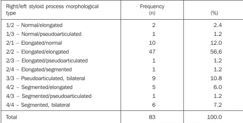
Table 3 Bilateral styloid process morphological type classification (n = 83).
Right/left styloid process morphological type
1/2 – Normal/elongated
1/3 – Normal/pseudoarticulated
2/1 – Elongated/normal
2/2 – Elongated/elongated
2/3 – Elongated/pseudoarticulated
2/4 – Elongated/segmented
3/3 – Pseudoarticulated, bilateral
4/2 – Segmented/elongated
4/3 – Segmented/pseudoarticulated
4/4 – Segmented, bilateral
Total Frequency (n) 2 1 10 47 1 1 9 5 1 6 83 (%) 2.4 1.2 12.0 56,6 1.2 1.2 10.8 6.0 1.2 7.2 100.0
In spite the fact of several studies per-forming the measurement of the styloid process, this has not been the aim of the present study, since the analyzed x-ray im-ages (modified, panoramic for temporo-mandibular joint) presented distortions and amplification in their processing(13).
How-ever, in several published studies(3,5,11,12)
the panoramic radiography has been uti-lized for observation and process analysis, because it is an easy-to-perform examina-tion, besides being widely available for patients. For Arellano(13), Basekim et al.(6)
and Heitz et al.(15), computed tomography
is the most effective method for evaluating the styloid process length and morphologi-cal alterations, since this examination pro-vides 2D and 3D reconstructions of the whole temporomandibular joint(16).
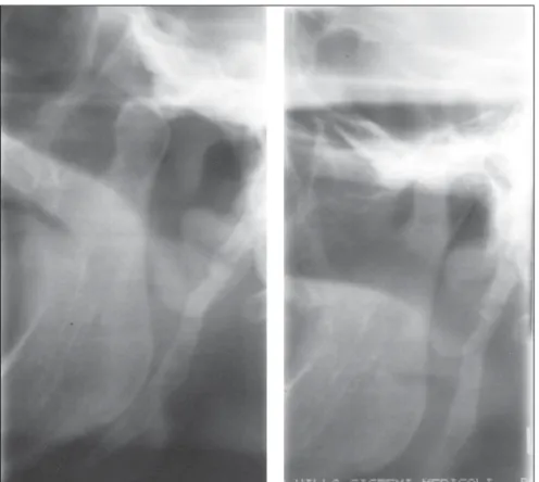
How-ever, based on the results of the present study, one may consider that the modified radiography for the temporomandibular joint and the panoramic x-ray are good imaging methods for visualizing morpho-logical alterations of the temporomandibu-lar joint, including the styloid process. The great limitation of this examination is the
impossibility of measuring the process ex-tent, however this has not been a determin-ing factor in the diagnosis of the syn-drome(17) (Figure 2).
In the whole sample of the present study, it was found that 15.7% of the pa-tients were male, and 84.3%, female, with the majority of them (65.2%) in the age range between 21 and 50 years. Both re-sults confirm the reports from Okeson(14)
and Saad & Barros(1) about
temporoman-dibular disorders.
Among the patients with temporoman-dibular disorder, 5.53% presented at least one side of the temporomandibular joint with morphological alteration of the styloid process(18). However, Zaki et al.(19) have
found, also in patients with temporoman-dibular disorder, an even more significant incidence (27%). For Eagle(17), in 4% of the
patients with morphological alteration of the styloid process, only 4% presented symptoms compatible with the Eagle syn-drome.
In absolute numbers, the incidence of morphological alterations of the styloid process was higher in women (74) when The general classification of the styloid
process morphological pattern can be seen on Table 2, the elongated type being the most frequent (n = 113), with 59 right-sided, and 54 left-sided.
As regards the symmetry (Table 3), it was found that 56% (n = 47) of morpho-logical alterations of the styloid process are elongated, bilateral (2/2). The second more incident were the right-sided elongated and left-sided normal types (2/1), with 12% (n = 10) of the sample. The third more inci-dent type was the pseudoarticulated bilat-eral one (3/3), in nine patients (10.8%).
DISCUSSION
Many studies(1,2,4,14) have been
compared with men (9). However, once the V Cramer test was applied, it was found that the prevalence is similar for both fe-male and fe-male patients. This result is dif-ferent from the findings of Gozil et al.(20),
Eagle(10) and Aral et al.(21), who have
re-ported a higher male incidence, and also differ from those of Hernandez et al.(22),
who have found a higher female incidence. In the present study, 32.5% of the pa-tients presented with morphological alter-ations of the styloid process, and the ma-jority of patients were in the age range be-tween 41 and 50 years for both sexes. In the other age ranges (21–30 years, 31–40 years, 51–60 years and 61–82 years) the mean in-cidence was 15%. This result is in disagree-ment with the studies of Zaki et al.(19) and
Aral et al.(21) (31–40 years), Basekim et al.(6) (34 years) and Gozil et al.(20) (31–50
years). In the present study, only five pa-tients (6% of the sample) in the age range between 11 and 20 years presented with such alteration. Also in this study, in a to-tal of 1,500 patients, there was no patient less than 10 years old with morphological alteration of the styloid process. However,
in the referred scientific literature(8) there
is a case report on a ten-year old patient. With this great variation in the presence of morphological alterations of the styloid process in several age ranges, and based on several studies results(6,18,21,23), including
the present ones, it should be considered that the styloid process elongation or sty-lohyoid complex mineralization develops in a particular fashion for each individual, independently from sex and age (23).
Most of the studies found in the litera-ture about morphological alterations of the styloid process are clinical case reports(8,10– 12,15,21,22,24) and rather than evaluating the
styloid process morphology, they analyze the symptomatology and the presence of the process elongation.
Based on the studies of Langlais et al.(7),
the 83-patient sample was studied for the morphological type of each styloid process. The elongated pattern was the most fre-quent one (113), and the other morphologi-cal type (normal, pseudoarticulated and segmented) have not demonstrated signifi-cant differences, confirming the findings of several authors(6,20,23). Similarly to the study
of Aral et al.(21), in the present study there
was no predominance of one of the sides in the morphological alteration of the sty-loid process.
It is important to highlight the relevance of the diagnosis in the craniomandibular dysfunctions by means of clinical and im-aging studies(6,9,13,14,16), aiming at
establish-ing the final diagnosis, since the treatment for temporomandibular disorders is prima-rily symptomatic, with conservative and reversible therapies(19), and the treatment
for Eagle syndrome involves surgical ex-cision(5,20,22,24,25), with intra- or extra-oral
approach(24) for the elongated styloid
pro-cess or the calcified stylohyoid ligament removal. However, Camarda et al.(25) have
asserted that many patients may tolerate the symptoms of the elongated styloid process, without being submitted to surgery.
In agreement with several authors(1,5,24),
it is believed that, in cases of orofacial pains, there is a necessity of interdiscipli-nary intervention and cooperation of radi-ologists, otorhinolaryngradi-ologists, surgeon-dentists, neurologists, orthopedists, oph-thalmologists, phonoaudiologists, physio-therapist and psychologists(16), each
profes-sional acting in his/her specialty for defi-nition of the diagnosis and appropriate therapeutical conduct.
CONCLUSIONS
Radiographically seen, the prevalence of morphological alteration of the styloid process in patients with temporomandibu-lar disorder was of 5.53% (in 9 (10.8%) male and 74 (89.2%) female patients). A higher frequency was found in the age range between 41 and 50 years (32.5%).
The morphological types of the tempo-ral bone styloid process radiographically seen were: 113 elongated, 21 pseudoarticu-lated and 19 segmented, totaling 153 sty-loid processes of 83 patients, 13 being con-sidered as normal. Also, it was found that there is a correlation between the right and the left patterns, i.e., the morphological alteration is symmetrical.
REFERENCES
1. Saad PA, Barros JJ.In: Barros JJ, Rode SM. Tra-tamento das disfunções craniomandibulares – ATM. 1ª ed. São Paulo: Livraria Editora Santos, 1995;341–345.
2. Ash MM, Ramfjord SP, Schimideserder J. Oclu-são. 1ª ed. São Paulo: Livraria Editora Santos, 1998;55.
3. Rodrigues JRC, Chilvarquer F. Avaliação clínica e radiográfica de pacientes portadores de apófise estilóide alongada e calcificada e com disfunção dolorosa da ATM. Anais SBPqO 1991;7:57. 4. Rocha APF, Nardelli MR, Rodrigues MF.
Epide-miologia das desordens temporomandibulares: estudo da prevalência da sintomatologia e sua inter-relação com a idade e o sexo do pacientes. Rev Serviço-ATM-FO/UFJF 2002;2:1:5–9. 5. Noronha MJR, Gandelmann I, Araújo Jr GP,
Shu-nemann WG. Alongamento do processo estilói-de. Síndrome de Eagle. Rev Bras Otorrinolarin-gol 1987;53:60–63.
6. Basekim CC, Mutlu H, Gungor A, et al.
Evalua-tion of styloid process by three-dimensional com-puted tomography. Eur Radiol 2005;15:134–139. 7. Langlais RP, Langland OE, Nortjé CJ. Diagnos-tic imaging of the jaws. 1st ed. Baltimore: Wil-liams & Wilkins, 1995;620–622.
8. Galán IR, Galán CR, Diez FA. Sindrome estilo-hióideo en la infancia. An Esp Pediatr 1999;50: 507–508.
9. Lee HA. Three-dimensional computed tomogra-phy imaging of Eagle’s syndrome. Am J Otolaryn-gol 2004;25:109.
10. Eagle WW. Elongated styloid process. Report of two cases. Arch Otolaryngol 1937;25:584–587. 11. Reis SSPM, Carvalho PL, Reis HSM. Processo estilóide alongado – relato de dois casos. J Bras Oclus ATM & Dor Orofac 2001;1:296–300. 12. Slavin KV. Eagle syndrome: entrapment of the
glossopharyngeal nerve? Case report and review of the literature. J Neurosurg 2002;97:216–218. 13. Arellano JCV. Metodologia no diagnóstico da disfunção da articulação temporomandibular. J Bras Oclus ATM & Dor Orofac 2002;2:78–86. 14. Okeson JP. Fundamentos de oclusão e desordens
temporomandibulares. 4ª ed. São Paulo: Artes
Médicas, 2000;119–121.
15. Heitz C, Rocha JRM, Souza PHC, Costa NP. Mineralização atípica do complexo estilóide: re-lato de caso. Rev Bras Prótese e Implante 1999;6: 31–37.
16. Sá ACD, Zardo M, Paes Jr AJO, et al.
Alongamen-to do processo estilóide (síndrome de Eagle): re-lato de dois casos. Radiol Brasil 2004;37:385– 387.
17. Eagle WW. Elongated styloid process; symptoms and treatment. Arch Otolaryngol 1958;67:172– 176.
18. Goldstein GR, Scopp IW. Radiographic interpre-tation of calcified stylomandibular and stylohy-oid ligaments. J Prosthet Dent 1973;30:330–334.
19. Zaki HS, Greco CM, Rudy TE, Kubinski JA. Elongated styloid process in a temporomandibu-lar disorder sample: prevalence and treatment out-come. J Prosthet Dent 1996;75;399–405. 20. Gozil R, Yener N, Çalguner E, Araç M, Tunç E,
Bahcelioglu M. Morphological characteristics of styloid process evaluated by computed axial to-mography. Ann Anat 2001;183:527–535. 21. Aral IL, Karaca I, Gungor N. Eagle’s syndrome
masquerading as pain of dental origin. Case re-port. Aust Dent J 1997;42:18–19.
22. Hernandez C, Rodriguez MM, Sano R, Vargas S, Monasterio MV. Síndrome de Eagle: a propósito de un caso. Acta Otorrinolaringológica 2000;12: 57–62.
23. Diamond LH, Cottrell DA, Hunter MJ, Papa-george M. Eagle’s syndrome: a report of 4 patients treated using a modified extraoral approach. J Oral Maxillofac Surg 2001;59:1420–1426. 24. Trevisan DRS. Análise dos aspectos do complexo
estilohióideo na radiografia panorâmica. J Bras Oclus ATM & Dor Orofac 2002:2:105–115. 25. Camarda AJ, Deschamps C, Forest D. I.