rev bras hematol hemoter.2 0 1 4;3 6(4):300–301
Revista Brasileira de Hematologia e Hemoterapia
Brazilian Journal of Hematology and Hemotherapy
w w w . r b h h . o r g
Images in Clinical Hematology
Mycobacterium leprae
in bone marrow
Leonardo Rodrigues de Oliveira
∗, André Luiz Maltos
Universidade Federal do Triângulo Mineiro (UFTM), Uberaba, MG, Brazil
a r t i c l e
i n f o
Article history:
Received 4 March 2014 Accepted 17 March 2014 Available online 29 May 2014
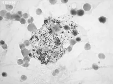
The case of a 54-year-old man, who was referred to eval-uate a consumptive syndrome with anemia and fever of unknown origin, is reported. A physical examination revealed madarosis, nodular lesions on the ears, nasal soft tissue collapse, subcutaneous nodules on arms and generalized lymphadenopathy. Laboratory tests showed anemia (Hb 8.8 g/dL, mean corpuscular volume 82.4 fL, mean corpuscular hemoglobin 24.5 pg), normal reticulocyte count (0.5%), throm-bocytosis (482×109/L), normal serum ferritin (268 ng/mL) and elevated C-reactive protein (124 mg/L). Serologies (viral hep-atitis and human immunodeficiency virus) were non-reagent. Lepromatous leprosy was confirmed by staining for acid-fast bacilli using samples from the ear and subcutaneous nodules and bone marrow smears. Bone marrow was hyper-cellular with myeloid hyperplasia (myeloid–erythroid ratio 7:1) but without dysplasia.Mycobacterium lepraewas detected lying free and in foamy histiocytes named Virchow cells (Figures 1 and 2).1,2A multidrug therapeutic regimen
(clofaz-imine, dapsone, rifampicin) was established with progressive improvement.
∗ Corresponding author at: Universidade Federal do Triângulo Mineiro – UFTM, Central de Quimioterapia, Rua Getúlio Guarita, s/n, 38080-125 Uberaba, MG, Brazil.
E-mail address: [email protected] (L.R. de Oliveira).
Figure 1 – Large cells (histiocytes) with their abundant cytoplasm filled with acid-fast bacilli (Mycobacterium leprae) in bone marrow (Ziehl-Neelsen stain) magnification 1000×.
http://dx.doi.org/10.1016/j.bjhh.2014.05.010
rev bras hematol hemoter.2 0 1 4;3 6(4):300–301
301
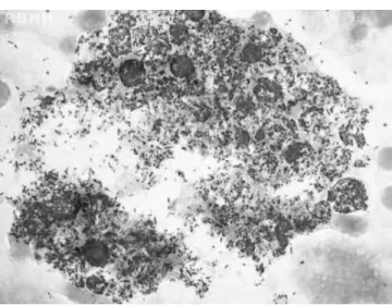
Figure 2 – Large cells (histiocytes) with their abundant cytoplasm filled with acid-fast bacilli (Mycobacterium leprae) in bone marrow (Fite-Faraco stain) magnification 1000×.
Conflicts of interest
The authors declare no conflicts of interest.
r e f e r e n c e s
1. Singh N, Bhatia A, Lakra A, Arora VK, Bhattacharya SN. Comparative cytomorphology of skin, lymph node, liver and bone marrow in patients with lepromatous leprosy. Cytopathology. 2006;17:257–61.
2. Suster S, Cabello-Inchausti B, Robinson MJ.