Rev Bras Anestesiol CLINICAL INFORMATION 2012; 62: 2: 274-280
274 Revista Brasileira de Anestesiologia
Vol. 62, No 2, March-April, 2012
Received from Universidade de Brasília (UnB), Brazil. 1. Anesthesiologist
2. MSc; Associate Professor, Universidade de Brasília (UnB); Scientific Director of the Brazilian Society of Anesthesiology; Head of CET/SBA, UnB
Submitted on December 8, 2010 Approved on June 19, 2011 Correspondence to: Dra. Silvia Piccolo Daher SGAN 608, módulo F Asa Norte
70850080 Brasília, DF, Brazil E-mail: [email protected]
CLINICAL INFORMATION
Anesthesia in Patient with Shrinking Lung Syndrome:
Case Report
Silvia Piccolo-Daher
1, Edno Magalhães, TSA
2Summary: Piccolo-Daher S, Magalhães E – Anesthesia in Patient with Shrinking Lung Syndrome: Case Report.
Background and objectives: The incidence of pulmonary involvement in systemic lupus erythematosus (SLE) may be presented as a syndrome called shrinking lung syndrome (SLS). SLS has quite a controversial pathophysiology, which can induce to a mechanical ventilation dependency. Due to its rarity, there is a limited number of publications on the subject. The objective of this report is to present the case of a patient with SLS who underwent incisional hernia repair under epidural anesthesia.
Case report: Female patient with SLE, hypertensive and obese, diagnosed with SLS 18 years ago. She was dependent on nocturnal oxygen at home, had dyspnea on minimal exertion and spirometry with severe restrictive ventilatory defect.In a previous post-operative period under general anesthesia, she remained on mechanical ventilation for 9 days with difficult weaning. She underwent incisional hernia repair for 3 hours under thoracic epidural anesthesia without any pre- or post-operative respiratory complication.
Conclusions: Shrinking lung syndrome is a rare disease that requires a prior knowledge of the clinical and laboratory history of the patient by the anesthesiologist. The thoracic epidural anesthesia technique proved to be a satisfactory option for this patient, with highly satisfactory respiratory evolution.
Keywords: Anesthesia, epidural; General Surgery; Lupus Erythematosus, systemic; Respiratory Muscles; Ventilation/complications.
©2012 Elsevier Editora Ltda. All rights reserved.
INTRODUCTION
Systemic lupus erythematosus (SLE) is an autoimmune con-nective tissue disease, characterized by the formation of auto-antibodies and immune complexes, with various clinical and immunological manifestations 1. Pulmonary involvement can
occur between 60% and 80% of the cases 2, and may affect
the upper airways, pleura, parenchyma and pulmonary
ves-sels in different ways 3. Some pleuropulmonary
manifesta-tions include pleuritis, pneumonitis, fibrosis, alveolar hemor-rhage, bronchiolitis obliterans, hypertension, and pulmonary thromboembolism. More rarely, it may arise as the shrinking lung syndrome (SLS), a rare manifestation of SLE, character-ized by unexplained dyspnea, restrictive pattern on spirom-etry, and elevation of the diaphragm, in the absence of paren-chymal lung diseases 4.
Although there are some studies in literature describing the SLS, none of them consider the anesthetic management of these patients. The purpose of this report is to describe a case of a patient with SLS who underwent thoracic epidural anes-thesia for incisional hernia repair.
CASE REPORT
Female patient, 54 years old, who underwent surgery for su-praumbilical incisional hernia three months after a left adrena-lectomy surgery (incidentaloma).
Diagnosed with rheumatoid arthritis for 33 years and SLE for 22 years, the patient was taking prednisone (5 mg.d-1);
had had an episode of acute myocardial infarction 20 years before with no sequelae; and was diagnosed with SLP for 18 years. Currently, she depends on nocturnal oxygen (2 L.
min-1) at home. She presented class II obesity,
hyperten-sion taking propranolol (80 mg.d-1), and diabetes mellitus
taking acarbose (150 mg.d-1). She was also taking AAS
(100 mg.d-1), discontinued during the previous two weeks
and atorvastatin (20 mg.d-1). The patient reported dyspnea
ANESTHESIA IN PATIENT WITH SHRINKING LUNG SYNDROME: CASE REPORT
Revista Brasileira de Anestesiologia 275
Vol. 62, No 2, March-April, 2012
At physical examination, the patient presented good gen-eral condition; was lucid and oriented, afebrile, acyanotic, anicteric, hydrated, and ruddy (weight 90 kg, height 1.60 m; BMI = 35 kg.m-2).
Respiratory system showed universally audible breath sounds with rales at the right base of the hemithorax. Car-diovascular system showed a regular heart rhythm in two stages, with normal sounds and no murmurs; blood pressure of 110x70 mmHg and heart rate at 64 bpm. Abdomen was distended with hernial ring of ± 4 cm in the right supraumbilical
region; airway Malampatti III; good oral opening (4 cm), with tracheostomy scar.
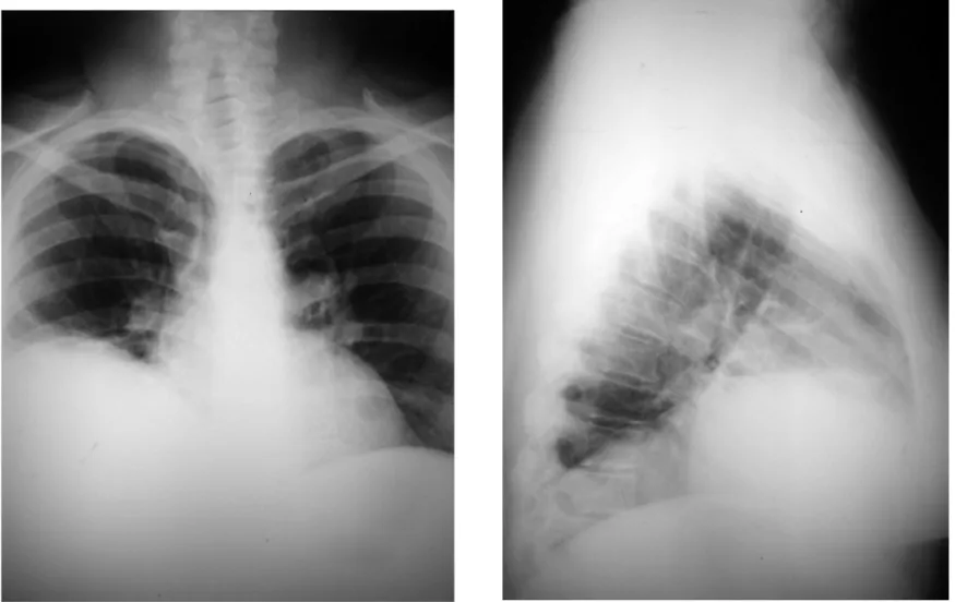
Blood count showed no changes. Arterial blood gas mea-surements were: pH 7.29; pCO2 45.2 mmHg; pO2 49.4 mmHg;
HCO3 20.4. Echocardiogram showed ejection fraction of 69%
and holosystolic murmur of mitral regurgitation without sys-temic repercussions. Chest X-ray showed right-sided dia-phragmatic elevation (Figure 1). Chest tomography revealed subsegmental atelectasis in the middle and lower lobes of right lung, bilateral pleural thickening, elevated right hemi-diaphragm, and pericardial thickening. Spirometry showed a severe restrictive ventilatory defect with moderate reduction of the alveolar-capillary oxygen diffusion. The patient was
pre-viously evaluated by the cardiology and pulmonary teams and referred to the procedure. Physical status classification: ASA PS (P) III.
In the operating room, all airway material was prepared and tested, including the available laryngeal mask. After perform-ing venous puncture, the patient was properly monitored with noninvasive blood pressure, oxygen saturation, electrocardio-gram, and urinary catheter. Oxygen saturation was 94%, but after using nasal cannula the oxygen saturation level was be-tween 96% and 98%. Midazolam 2.5 mg and hydrocortisone 300 mg were administered. For epidural anesthesia induction, the loss of resistance technique was used with median punc-ture at T10-T11 and infusion with a test dose of 3 mL of 2% lidocaine with vasoconstrictor, without hemodynamic effects. The epidural catheter was placed and then 15 mL of 0.75%
ropivacaine and 50 µg fentanyl were administered. Blood
pressure was measured and an invasive arterial blood gas sample was collected. The surgery was uneventful, and the patient remained hemodynamically stable and adequately se-dated.
At the end of surgery, which lasted three hours, a new epi-dural dose of 10 mL 0.125% ropivacaine with 2 g dipyrone and 8 mg ondansetron was administered intravenously for
PICCOLO-DAHER, MAGALHÃES
276 Revista Brasileira de Anestesiologia
Vol. 62, No 2, March-April, 2012
operative analgesia and prevention of nausea and vomiting. Total urine output was 250 mL. The patient was transferred to the Intensive Care Unit (ICU) hemodynamically stable, awake, lucid and cooperative, receiving oxygen by Venturi mask (10 L.min-1) and monitored. In the ICU, the patient remained
without complaints, receiving dipyrone 2 g every 4 hours, with-out requiring other medications for pain or vomiting. She was discharged from ICU after 24 hours, transferred to the ward, and discharged from hospital on the third postoperative day.
DISCUSSION
The pathogenesis of SLS is controversial 3,5. It has been
suggested that there is a weakness of respiratory muscles, mainly diaphragmatic, due to a myopathy unrelated to corti-costeroids 5. This hypothesis is not always accepted by some
researchers who suggest chest wall restriction as the main cause 6. Most authors agree that SLS is a heterogeneous
en-tity relating to multiple pathogenic processes 5. The treatment
remains empirical and involves the use of corticosteroids, im-munosuppressive agents, xanthines, and inhaled beta-ago-nist (due to the positive inotropic effect of this substance on beta-receptors of diaphragmatic muscle) 2-5,7. The prognosis
is generally good, with improvement and stabilization of pul-monary symptoms. However, there is a report on death of a patient who remained dependent on mechanical ventilation, despite high doses of corticosteroids 5.
The patient in this study had a restrictive pattern on spirometry and lung volumes reduced, without pleuropulmo-nary diseases justifying the dyspnea and oxygen. The epidu-ral anesthesia was aimed at preventing early ministration of mechanical ventilation in this patient, allowing surgery under spontaneous breathing. The aim was also to avoid possible prolonged mechanical ventilation and to ensure adequate control of postoperative analgesia.
The decision on the choice of anesthesia is influenced by the surgical time and field, in addition to the patient’s clinical conditions. The effects of epidural anesthesia on pulmonary function seem to be beneficial 8,9. Both local anesthetics and
opioids help diaphragmatic function, improving lung volume and capacity and reducing postoperative complications 9,10. If
the spread of local anesthetic reaches the cervical roots (C5 to C3), respiratory function may be compromised by block-ing intercostal nerves and the partial or total blockage of the phrenic nerve. Changes in lung volumes and capacities after epidural block should not be forgotten because they directly alter the respiratory reserve.
Ropivacaine is a local anesthetic belonging to the pipe-coloxylididegroup, noted by the lower intensity of motor block and reduced potential for cardiovascular toxicity compared to bupivacaine 8,9. It is known that the thoracic epidural block
with concentrated solutions of bupivacaine causes a moder-ate reduction in vital capacity and in maximal inspiratory flow, probably due to a certain degree of relaxation of the intercos-tal muscles 8,9. The use of less concentrated solutions of a
lo-cal anesthetic such as ropivacaine may reduce the possibility of significant impairment of respiratory muscles 9.
Considering the high incidence of hypotension and brady-cardia resulting from blockade of sympathetic cardioaccelera-tory fibers, this technique should be performed with extreme caution. Proper selection of patients, accurate monitoring of vital functions, knowledge of the technique chosen and its physiological implications are of vital importance.
CONCLUSION
Revista Brasileira de Anestesiologia 279 Vol. 62, No 2, Março-Abril, 2012
ANESTESIA NO PACIENTE COM SÍNDROME DO PULMÃO ENCOLHIDO: RELATO DE CASO
REFERÊNCIAS/REFERENCES
1. Hines RL, Marschall KE – Skin and Musculoskeletal Diseases em: Stoelting’s Anesthesia and Co – Existing Diseases, Quinta Edição, Philadelphia, Elsevier Inc., 2008.
2. Oud KTM, Bresser P, ten Berge RJM et al. – The shrinking lung syn-drome in systemic lupus erythematosus: improvement with corticos-teroid therapy. Lupus, 2005;14:959-63.
3. Costa CA, Junior DOC, Jezler S et al. – Síndrome do pulmão encolhi-do no lúpus eritematoso sistêmico. J Bras Pneumol, 2004;30(3):260-63.
4. Hoffbrand BI, Beck ER – Unexplained dyspnoea and shrinking lungs in systemic lúpus erythematosis. Br Med J, 1965;5445:1273-1277. 5. Warrington KJ, Moder KG, Brutinel WM – The shrinking lung syndrome
in systemic lupus erythematosus. Mayo Clin Proc, 2000;75:467-72. 6. Ernest D, Leung A – Ventilatory failure in shrinking lung syndrome is
associated with reduced chest compliance. Intern Med J, 2010;40: 66-79.
7. Toya SP, Tzelepis GE – Association of the shrinking lung syndrome in systemic lupus erythematosus with pleurisy: a systematic review. Semin Arthritis Rheum, 2008;38:30-37.
8. Novaes MVM, Francisco CRL, Pimenta KB et al. – Estudo compara-tivo entre bupivacaína a 0,25% e ropivacaína a 0,2% em anestesia peridural para cirurgia torácica. Rev Bras Anestesiol, 2001;51(6):493-502.
9. Nociti JR, Serzedo PSM, Zuccolotto EB et al. – Ropivacaína em blo-queio peridural torácico para cirurgia plástica. Rev Bras Anestesiol, 2002;52(2):156-165.