Received from HM Ankara Numune Training and Research Hospital, Turkey.
1. Anaesthesia and Reanimation Department, Ankara Numune Training and Research Hospital, Turkey
Submitted on May 13, 2011. Approved on June 19, 2011.
Correspondence to: Dr. Dilsen Ornek
HM Ankara Numune Training and Research Hospital Ulku Mahallesi Talatpasa Bulvari No: 5
Altindag, Ankara 06100, Turkey E-mail: [email protected]
SCIENTIFIC ARTICLE
Cigarette Smoking and the Effect of Dexmedetomidine and
Fentanyl on Tracheal Intubation
Kemal Gulsoy
1, Serpil Deren
1, Semih Baskan
1, Dilsen Ornek
1, Bayazit Dikmen
1Summary: Gulsoy K, Deren S, Baskan S, Ornek D, Dikmen B – Cigarette Smoking and the Effect of Dexmedetomidine and Fentanyl on Tracheal Intubation.
Background and objectives: To compare the effect of dexmedetomidine and fentanyl on hemodynamic changes in chronic male smokers.
Methods: This is a prospective, randomized, blinded study. Were selected 60 chronic male smokers (aged 16 to 60 years). The patients were randomly divided into two groups: Group D (n = 30) received 1 µg.kg-1 dexmedetomidine and Group F (n = 30) received 3 µg.kg-1 fentanyl in 150 mL of normal saline, beginning 10 minutes before anesthesia induction. Before intubation, the heart rate and blood pressure of patients were measured. After anesthesia induction for endotracheal intubation, heart rate and blood pressure values were measured at 1, 3, and 5 minutes after intubation.
Results: Heart rate was low in Group D before anesthesia induction, intubation, and at the 1st and 3rd minutes after intubation. Systolic arterial pressure was low in Group F before intubation. Although diastolic arterial pressure was lower before anesthesia induction and at 5 minutes after intubation in both groups, it was already low in Group F before intubation. Whereas the mean arterial pressure was low in Group D before anes-thesia induction, it was low in Group F before intubation. The values for rate-pressure product were low in Group D before induction and at 1 and 3 minutes after intubation.
Conclusions: Dexmedetomidine, which was applied via infusion at a loading dose of 1 µg.kg-1 10 minutes before anesthesia induction in chronic male smokers, better suppressed increases in heart rate and rate-pressure product at 1 and 3 minutes after intubation compared to the group receiving 3 µg.kg-1 fentanyl.
Keywords: Dexmedetomidine; Hemodynamics; Intratracheal intubation; Monitoring, Intraoperative; Smoking.
©2012 Elsevier Editora Ltda. All rights reserved.
INTRODUCTION
Cigarette smoke contains various chemical substances, in-cluding nicotine and tar, most of which are carcinogens. Grassi et al. 1 suggested that smoking increases sympathetic activation due to increased release of catecholamines and the delay in nicotine clearance from the neuroeffector junction. Because smokers experience a high hemodynamic response to anesthesia induction and intubation, serious problems may develop in chronic smokers. The cardiovascular system func-tion varies between genders because the autonomic nervous system is influenced by hormonal and developmental differ-ences between genders. For this reason, response to tracheal intubation is more evident in males 2-10. Long-term exposure to cigarettes causes an increased response to the mechanical
stimulation caused by laryngoscopy and intubation. Hemody-namic response, which develops secondary to laryngoscopy and tracheal intubation, include heart rate, catecholamine level, blood pressure, and rate-pressure product 2-10.
Anesthesiologists have focused on controlling secondary responses to laryngoscopy and intubation. For this purpose, several drugs were used, including lidocaine, opioids, sodium nitroprusside, nitroglycerine, α-2 agonists, β blockers, and
calcium channel blockers 1-11.
Although opioid analgesics are widely used to minimize acute increases in blood pressure and heart rate during an-esthesia induction, they have side effects, such as respira-tory depression, muscular rigidity, and delayed recovery from anesthesia. Opioid analgesia also causes sympatholysis and an increased vagal activity as a result of the central effect of dexmedetomidine, which is a selective α-2 agonist. It also
further increases the sympatholytic effect due to its peripheral potency 1-11.
MATERIAL AND METHODS
After approval by the local Research Ethics Committee and af-ter obtaining informed consent from all participants, the study was conducted with 60 ASA I-II male patients (aged 18-60 years) who were chronic cigarette smokers. All patients had a body mass index of 30 kg.m-2; were all normotensive and re-ceiving no medication; with Mallampati classification I-II; and requiring endotracheal intubation involving elective surgical interventions. To qualify as chronic smokers, patients had to have smoked for two years at least, and a minimum consump-tion of 10 cigarettes.day -1. Patients with hypertension, hypov-olemia, history of opioid or sedative drugs, morbid obesity, or ischemic heart disease, and those in whom a difficult intuba-tion was anticipated were excluded from the study. Patients in whom more than one laryngoscopy and intubation attempt had been made or whose laryngoscopy and intubation opera-tion took more than 20 seconds were also excluded.
Patients were divided into two similar groups (30 sub-jects each) and assigned to receive either dexmedetomidine (Group D) or fentanyl (Group F). A non-invasive blood pres-sure (NIBP), electrocardiogram (ECG), and pulse oximetry (SpO2) monitoring (Datex Ohmeda ADU S/5, Finland) were performed before the patient was taken to the operating room. Afterwards, a 20 gauge intravenous catheter was used to gain vascular access, and Hartmann’s solution 5 mL.kg-1.h-1 was administered. Ten minutes before anesthesia induction, Group D (n = 30) was given 1 µg.kg-1 dexmedetomidine, whereas
Group F (n = 30) was given 3 µg.kg-1 fentanyl in 150 mL of
normal saline.
After pre-oxygenation for 3 minutes, anesthesia was pro-vided to both groups with 4-7 mg.kg-1 thiopental until loss of the eyelash reflex occurred, after which 0.1 mg.kg-1 vecuro-nium was administered. Endotracheal intubation was per-formed by an anesthesiologist (blinded to the drugs used in infusion); a size 3-4 Macintosh laryngoscope blade and 8-8.5 mm (internal diameter) disposable tracheal tube was used. The patient’s lungs were ventilated with 2% inhaled sevoflu-rane and 60% nitrous oxide in 40% oxygen via a circle system, and the ventilator frequency was adjusted to a range of 12 to 15 breaths.min-1 to maintain end-tidal carbon-dioxide concen-tration between 4.5-5.0 kPa. Hemodynamic parameters were measured as basal values following the first measurement, post-infusion, pre-intubation, and at one, three, and five min-utes post-intubation. The measurements were also recorded by an anesthesiologist who was unaware of the drugs being used.
The sample size was calculated based on the assump-tion that a 20% difference in heart rate was significant. In accordance with the power calculation method, 18 patients per group should demonstrate a 20% difference in heart rate value at α = 0.05 and power of 90%. The primary endpoint
was an increase in heart rate after tracheal intubation in both groups. Data analysis was conducted using the SPSS 15.0
software (SPSS Inc., Chicago, IL, USA), and data distribu-tion was controlled using the Levene test. The main variables were percentage heart rate, blood pressure, and rate-pressure product as compared with the baseline figures. Rate-pressure product was obtained by multiplying systolic blood pressure and heart rate. In the intergroup evaluations, an independent t test was used to analyze the normally distributed data, and the Mann-Whitney U test was used for the data that were not distributed normally. A chi-square test was used to evaluate the non-parametric data. In the intragroup evaluations, paired t tests were used. A probability of p < 0.05 was established as significant.
RESULTS
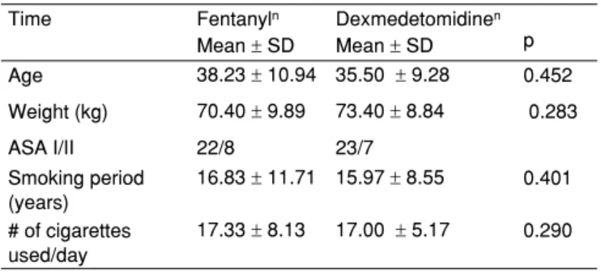
Patient characteristics are shown in Table I. There was no difference between groups regarding age, weight, ASA clas-sification, length of time as smokers, or number of cigarettes consumed daily (Table I).
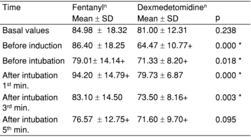
Heart rates were low in Group D before anesthesia induc-tion (p = 0.000) and before intubainduc-tion (p = 0.18), as well as at one (p = 0.000) and three (p = 0.003) minutes after intubation (Table II, Figure 1). Systolic arterial pressure was low in Group F before intubation (p = 0.018); (Table III; Figure 2). Whereas
Table I – Demographic Characteristics of the Study Groups Time Fentanyln Dexmedetomidinen
Mean ± SD Mean ± SD p
Age 38.23 ± 10.94 35.50 ± 9.28 0.452
Weight (kg) 70.40 ± 9.89 73.40 ± 8.84 0.283
ASA I/II 22/8 23/7
Smoking period (years)
16.83 ± 11.71 15.97 ± 8.55 0.401
# of cigarettes used/day
17.33 ± 8.13 17.00 ± 5.17 0.290
n = 30.
Table IV – Diastolic Pressure Comparisons between Study Groups Time Fentanyln Dexmedetomidinen
Mean ± SD Mean ± SD p
Basal values 80.47 ± 10.46 79.04 ± 8.23 0.548 Before induction 79.83 ± 10.60 71.63 ± 9.35 + 0.002 * Before Intubation 68.33 ± 9.51 + 76.37± 12.24 0.006 * After intubation
1st min
89.83 ± 12.24 + 88.07 ± 11.94 + 0.577
After intubation 3rd min
77.07 ± 10.80 72.83 ± 11.26 + 0.143
After intubation 5th min
69.27 ±10.18 + 64.23 ± 9.14 + 0.049 *
n = 30; + intragroup comparisons, level of significance p < 0.05; * significant at p < 0.05.
Figure 1 – Intergroup Heart Rate.
* Significantatp < 0.05; ** Significant at p <0.001. 140
120
100
80
60
40
20
0
Min/rate
T0 T1 T2 T3 T4 T5
Time
Fentanyl Dexm.
Heart Rate
** *
**
*
Table III – Intergroup Systolic Arterial Pressure
Time Fentanyln Dexmedetomidinen Mean ± SD Mean ± SD p
Basal values 130.67 ± 16.15 128.57 ± 20.05 0.675 Before induction 130.01± 17.50 121.33 ± 15.73 + 0.056 Before intubation 106.25± 14.62+ 117.87 ± 19.68 + 0.012 * After intubation
1st min.
134.16 ± 19.20 137.04 ± 17.32 + 0.537
After intubation 3rd min.
117.48± 14.11+ 116.28± 16.77 + 0.761
After intubation 5th min.
105.01 ± 13.29 + 104.08 ± 12.65 + 0.862
n = 30; + intragroup comparisons level of significance p < 0.05; * significant at p < 0.05.
Figure 3 – Diastolic Arterial Pressure. * Significant at p < 0.05 level.
0
mmHg
T0 T1 T2 T3 T4 T5
Time
Fentanyl Dexm.
SAP
140
120
100
80
60
40
20 180
160
*
Figure 2 – Systolic Arterial Pressure. * Significant atp < 0.05.
120
100
80
60
40
20
0
mmHg
T0 T1 T2 T3 T4 T5
Time
Fentanyl Dexm.
DAP
* * *
Table II – Intergroup Heart Rates
Time Fentanyln Dexmedetomidinen Mean ± SD Mean ± SD p Basal values 84.98 ± 18.32 81.00 ± 12.31 0.238 Before induction 86.40 ± 18.25 64.47 ± 10.77+ 0.000 * Before intubation 79.01± 14.14+ 71.33 ± 8.20+ 0.018 *
After intubation 1st min.
94.20 ± 14.79+ 79.73 ± 6.87 0.000 *
After intubation 3rd min.
83.10 ± 14.50 73.50 ± 8.16+ 0.003 *
After intubation 5th min.
76.57 ± 12.75+ 71.60 ± 9.70+ 0.095
upper airway epitheliumcharacteristics15,16.For this reason, laryngoscopy and tracheal intubation can also induce more hemodynamic changes by possibly triggering the same path-way. It has also been reported that chronic smokers are more prone to coronary artery disease than nonsmokers. Increases in rate-pressure product can even further increase both the myocardial oxygen requirement and ischemia in patients with ischemic heart disease 17,18.Cuvas
’ş et al. 19 suggested that determining the smoking history of the patient is important for preoperative evaluation and that chronic smokers are particu-larly high-risk patients. It has been emphasized that ischemic heart disease and increased post-intubation myocardial oxy-gen requirement increases the cardiac risk19.
Dexmedetomidine is an α-2 agonist that decreases
sympa-thetic activity and reduces pain20,21.There is report on the use of dexmedetomidine instead of fentanyl22,23.The use of dex-medetomidine in anesthesia regimes to deepen the effect of Table V – Intergroup Mean Arterial Pressure
Time Fentanyln Dexmedetomidinen Mean ± SD Mean ± SD p
Beginning 97.23 ± 11.24 95.58 ± 11.16 0.572
Before induction 96.50 ± 11.95 88.26 ± 10.7 + 0.007 * Before intubation 80.97 ± 10.52 + 90.22 ± 14.21 0.006 *
After intubation 1st min
104.622 ± 13.63 + 104.42 ± 13.20 + 0.954
After intubation 3rd min
90.56 ± 11.09 + 87.33 ± 12.51 0.294
After intubation 5th min
81.11 ± 10.58 + 77.56 ± 9.81 + 0.182
n = 30; + intragroup comparisons level of significance p < 0.05; * level of sig-nificance p < 0.05.
Figure 4 – Mean Arterial Pressure. * Significant atp < 0.05 level.
140
120
100
80
60
40
20
0
mmHg
T0 T1 T2 T3 T4 T5
Time
Fentanyl Dexm.
AAP
* *
0
mmHg* rate/min
T0 T1 T2 T3 T4 T5
Time
Fentanyl Dexm. 12000
10000
8000
6000
4000
2000 14000
16000 Pressure-Velocity Product
** *
*
Figure 5 – Rate-Pressure Product.
* Significant at p < 0.05; ** Significant at p < 0.001.
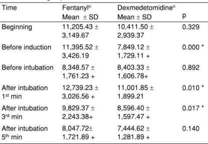
Table VI – Intergroup Rate-Pressure Product
Time Fentanyln Dexmedetomidinen Mean ± SD Mean ± SD p
Beginning 11,205.43 ±
3,149.67
10,411.50 ±
2,939.37 0.329
Before induction 11,395.52 ±
3,426.19
7,849.12 ±
1,729.11 +
0.000 *
Before intubation 8,348.57 ±
1,761.23 +
8,403.33 ±
1,606.78+ 0.892 After intubation
1st min
12,739.23 ±
3,026.56 +
11,001.85 ±
1,899.21
0.010 *
After intubation 3rd min
9,829.37 ±
2,243.38+
8,596.40 ±
1,597.47 +
0.017 *
After intubation 5th min
8,047.72±
1,721.89 +
7,444.62 ±
1,281.89 +
0.140
n = 30; + intragroup comparisons level of significance p < 0.05; * level of sig-nificance p < 0.05.
DISCUSSION
There was an evident increase in plasma norepinephrine and epinephrine levels, heart rate, and blood pressure among young chronic smokers after laryngoscopy and endotracheal intubation8-11.These hemodynamic changes can be reduced by blocking the α- and β-adrenergic receptors12-14.It seems
that the mechanisms responsible for the increase in blood pressure and heart rate are adrenergic in nature.
anesthesia has increased among anesthesiologists and inten-sive care doctors24.However, it has been reported that high concentrations of dexmedetomidine increase the anterior and posterior weight of the heart25.Blood pressure decreases with the central effect of the α2-adrenoreceptor agonists and as a
result of the increase in norepinephrine synthesis in peripheral presynaptic α2-receptors 26.Additionally, α2-adrenoreceptor
agonists create peripheral vasoconstriction by directly activat-ing the α2-receptors in vascular smooth muscles27.For this
reason, the hemodynamic effects of α2-adrenoreceptor
ago-nists are a result of their central sympatholytic and peripheral vasoconstrictive effects.
The most important issue related to the use of dexme-detomidine is its hemodynamic side effects. Although brady-cardia and hypotension are the most common side effects, dexmedetomidine also presents a biphasic, dose-dependent, blood pressure response similarly to other α2-adrenoreceptor
agonists. High doses when first administered result in a tem-porary blood pressure increase and reflex decrease in heart rate. This increase in blood pressure in the beginning may be related to dexmedetomidine rate of infusion and its high con-centration28.The hypertensive effect that is visible when the dose is first administered is followed by temporary sympathet-ic depression 29.Özköse et al. 30 infused dexmedetomidine at a dose of 1 µg.kg-1 over 10 minutes and reported no biphasic
effect on blood pressure. In the study, it was not observed any biphasic effect because the dose was administered at a lower concentration and infused more slowly than has been previ-ously reported.
The increase in blood pressure began after approximately 15 seconds and reached its maximum level after 30-45 sec-onds31.It was possible to concluded that evaluations during the first minute after administration, in which the greatest de-gree of blood pressure change occurs, are important. In the group receiving dexmedetomidine, according to the basal values recorded, an increase of 7% in systolic pressure, 1% in diastolic blood pressure, and 9% in mean blood pressure occurred in the first minute after intubation. Apart from that, it was observed that the hemodynamic response is stabilized. In a study by Aantaa et al. 32, dexmedetomidine was adminis-tered 15 minutes before anesthetic induction at doses of 0.35-0.67 mcg.kg-1, and they reported a mean decrease in systolic and diastolic blood pressure. These decreases were not dose-dependent and reached maximum level after 10 minutes 32.
Bradycardia resulting from dexmedetomidine may initially be triggered by baroreflex activity; however, subsequent de-creases in heart rate primarily derive from central sympathetic depression 29.
Özköse et al. 30 administered a 1 µg.kg-1 dose of dexme-detomidine over 10 minutes and observed atropine-requiring bradycardia in 4 of 20 patients. Aho et al. 33 reported that the use of 2.4 µg.kg-1 intramuscular dexmedetomidine resulted in
endotracheal intubation in 8 of 20 patients who underwent gy-necological laparoscopy.In the study, heart rates decreased by 20% after the administration of dexmedetomidine. How-ever, no bradycardia requiring treatment was observed. There was no statistically significant change in heart rates
com-pared with the basal values measured one minute after intu-bation. The fact that no bradycardia requiring treatment was observed, even though the same doses as Özköse et al. 30 suggests were used, these results may be related to the high sympathoadrenal response of chronic male smokers.
Because tachycardia, which develops after tracheal intu-bation, is more closely related to myocardial ischemia than hypertension, another parameter used for monitoring the heart’s workload is the rate-pressure product. Rate-pressure product, which is frequently utilized as an indicator of the myo-cardial oxygen requirement, is a numeric value obtained by multiplying heart rate by systolic blood pressure. Its upper lim-it is 12000-15000, which is the crlim-itical threshold and indicates cardiac ischemia34,35.
In a study of normotensive patients with coronary artery disease, Gobel et al. 36 emphasized that rate-pressure product is a significant parameter for determining oxygen demand and consumption by the heart during exercise. Kaplan et al. 37 sug-gested that the rate-pressure product causes ischemic elec-trocardiographic changes when it is over 12000 in patients with history of coronary artery surgery.Willigers et al. 34,38 re-ported in their study that dexmedetomidine has the potential to suppress some of the cardiovascular and neuroendocrine changes caused by sympathetic stimulation and to decrease systolic pressure and heart rate, which are indications that myocardial oxygen is required. They also found that the rate-pressure product decreases as a result39.In a study using 22 µg.kg-1 single-dose dexmedetomidine, Lawrence et al. 28
did not observe any statistically significant change in the dex-medetomidine group regarding heart rate or rate-pressure product after endotracheal intubation as compared to the be-ginning of the process. They reported that dexmedetomidine decreased the hemodynamic response associated with tra-cheal intubation.In the study, no changes were observed in heart rate or rate-pressure product after dexmedetomidine in-fusion in the first minute of intubation. We calculated the rate-pressure product as 11011 when the response to tracheal intubation was the highest. This value was lower than 12000, which is the upper limit for ischemia. Willigers et al. 34 also showed that dexmedetomidine decreases the rate-pressure product at approximately 26% after infusion. In the study, the rate-pressure product decreased at approximately 24%.
Low-dose fentanyl is widely used to increase the response to laryngoscopy and tracheal intubation. Salihoğlu et al. 31 compared the effects of fentanyl, alfentanil, and remifentanil in morbidly obese patients. They observed that a 1 µg.kg-1
dose of fentanyl, which is expected to decrease the hemody-namic response to intubation, decreased blood pressure and heart rate. No significant side effects were observed31.In their study, Katoh et al. 40 found that 2 µg.kg-1 of fentanyl prevents an increase in blood pressure and heart rate during tracheal intubation.Cuvas
that 3 µg.kg-1 fentanyl prevented increases in blood pressures
and heart rate in the first minute after intubation 42.Adachi et al. 43 suggested that fentanyl suppresses the response to intubation more effectively than the hemodynamic response associated with laryngoscopy.Iyer et al. 44 suggested that a 3 µg.kg-1 dose of fentanyl prevents mean blood pressure
in-creases compared to high-dose induction values; a 10 µg.kg-1 dose is necessary to prevent an increase in heart rate. How-ever, the use of fentanyl in high doses causes adverse side ef-fects such as hypotension, post-operative prolonged respira-tory depression, nausea, vomiting, and muscle rigidity. In the study, it was observed that 3 µg.kg-1 fentanyl did not prevent
increases in heart rate or rate-pressure product one minute
after intubation. One minute post-intubation, the rate-pressure product value obtained for the fentanyl group was 12,746. This value is near the upper limit for myocardial ischemia. This re-sult suggests that chronic smokers may exhibit an increased sympathoadrenal response and that fentanyl used to prevent increased heart rate is inadequate.
REFERÊNCIAS/REFERENCES
1. Grassi G, Seravalle G, Calhoun DA et al. – Mechanisms responsible for sympathetic activation by cigarette smoking in humans. Circula-tion, 1994;90:248-253.
2. Kovac AL – Controlling the hemodynamic response to laryngoscopy and endotracheal intubation. J Clin Anesth, 1996;8:63-79.
3. Shribman AJ, Smith G, Achola KJ – Cardiovascular and catecholamine responses to laryngoscopy with and without tracheal intubation. Br J Anaesth, 1987;59:295-299.
4. Finfer SR, MacKenzie SI, Saddler JM, Watkins TG – Cardiovascular responses to tracheal intubation: a comparison of direct laryngoscopy and fiberoptic intubation. Anaesth Intensive Care, 1989;17:44-48. 5. Bishop MJ, Harrington RM, Tencer AF – Force applied during tracheal
intubation. Anesth Analg, 1992;74:411-414.
6. Slogoff S, Keats AS – Does perioperative myocardial ischemia lead to postoperative myocardial infarction? Anesthesiology, 1985;62:107-114.
7. Jee D, Moon HL – Gender may affect the hemodynamic response to induction and intubation in young adults. J Clin Anesth, 2003;16;563-567.
8. Che S – Actions of nicotine and smoking on circulation. Pharmacol Ther, 1982;17:129-141.
9. Groppelli A, Giorgi DMA, Omboni S, Parati G, Mancina G – Persitent blood pressure increase induced by heavy smoking. J.Hypertens, 1992;10:495-499.
10. Baer L, Radichevich I – Cigarette smoking in hypertensive patients. Am J Med. 1985;78:564-568.
11. Trap-Jensen L, Carlen JE, Svensen TL, Christensen NS – Cardio-vascular and adrenergic effects of smoking during immediate non-se-lective beta-adrenoreceptor blockade in hummans Eur J.Clin Invest, 1979;9;181-183.
12. Trap-Jensen J – Effects of smoking on the heart and peripheral circu-lation Am Heart J, 1988;115:258-263.
13. Winniford MD – Smoking and cardiovascular function. J Hypertens, 1990;9(suppl 5):17-23.
14. Groppelli A, Giorgi DMA, Omboni S, Parati G, Mancina G – Blood pressure and heart rate response to repeated smoking before and after betablockade and selective alpha 1-inhibation function J Hyper-tens, 1990;8(suppl 5):35-40.
15. Laxton CH, Milner Q, Murphy PJ – Haemodynamic changes after tra-cheal intubation in cigarette smokers compared with non-smokers. Br J Anaesth, 1999;82(3):442-443.
16. Erskine RJ, Murphy PJ, Langton JA – Sensitivity of upper airway reflexes in cigarette smokers: effect of abstinence. Br J Anaesth, 1994;73:298-302.
17. Fitz-Henry J, Curran J, Griffiths D – Smokers and haemodynamic re-sponse to desflurane. Anaesthesia, 1999;54:800-803.
18. McBride PE – The health consequences of smoking-cardiovascular disease. Med Clin North Am, 1992;76:333-353.
19. Cuvas
’ş O, Er A, Ikeda OC, Dikmen B, Başar H – Cigarette smoking and the haemodynamic response to tracheal intubation. Anaesthesia, 2008;1365-2004.
20. Jaakola ML, Kanto J, Scheinin H, Kallio A. – Intramuscular dexme-detomidine premedication - an alternative to midazolam-fentanyl-combination in elective hysterectomy? Acta Anaesthesiol Scand, 1994;38:238-243.
21. Jaakola ML – Dexmedetomidine premedication before intravenous regional anesthesia in minor outpatient hand surgery. J Clin Anesth, 1994;6:204-211.
23. Hofer RE, Sprung J, Sarr MG, Wedel DJ – Anesthesia for a patient with morbid obesity using dexmedetomidine without narcotics. Can J Anaesth, 2005;52:176-180.
24. Paris A, Tonner PH – Dexmedetomidine in anaesthesia. Curr Opin Anaesthesiol, 2005;18;412-418.
25. Ebert TJ, Hall JE, Barney JA, Uhrich TD, Colinco MD – The effects of increasing plasma concentrations of dexmedetomidine in humans. Anaesthesiology, 2000;93:382-394.
26. Flacke JW – α2-adrenergic agonists in cardiovascularanesthesia J Cardiothorac Vasc Anesth, 1992;6:344-359.
27. Chen DG, Dai XZ, Zimmerman BG, Bache RJ – Postsynaptic α1 and α2 - adrenergic mechanism in coronary vazoconstruction J Cardio-vasc Pharmacol, 1988;11:61-67.
28. Lawrence CJ, De Lange S – Effects of a single perioperative dexme-detomidine dose on isoflurane requiretmens and perioperative hae-modynamic stability. Anaesthesia, 1997;52:736-744.
29. Xu H, Aibiki M, Seki K, Ogura S, Ogli K – Effects of dexmedetomidine, an α2 adrenoceptor agonist,on renal sympathetic nerve activity,blood pressure,heart rate and central venous pressure in urethane-anesthe-tized rabbits. J Auton Nerv Syst, 1998;71:48-54.
30. Özköse Z, Demir FS, Pampal K, Yardım S – Hemodynamic and anes-thetic advantages of dexmedetomidine, α2 agonist for surgey in prone position. Tohoku J Exp Med, 2006;210:153-160.
31. Salihoğlu Z, Demiroluk S, Demirkiran, Kose Y – Comparison of effects of remifentanil, alfentanil and fentanyl on cardiovascular responses to tracheal intubation inmorbidly obese patients. Eur J Anaesthesiol, 2002;19(2):125-128.
32. Aantaa RE, Kanto JH, Scheinin M, Kallio AMI, Scheinin H – Dexme-detomidine Premedication for Minor Gynecologic Surgery. Anesth An-alg, 1990;70:407-413.
33. Aho M, Scheinin M, Lehtinen AM, Erkola O, Vuorinen J, Korttila K – Intramuscularly administered dexmede tomidine attenuates hemo-dynamic and stress hormone responses to gynecologic laparoscopy. Anesth Analg, 1992;75:932-999.
34. Willigers HM, Prinzen FW, Roekaerts HJ – Comparison of the effects of dexmedetomidine and esmolol on myocardial oxygen consumption in dogs. E J A, 2004;21(12):957-966.
35. Kayhan Z – Kardiyovasküler Sistem ve Anestezi. Klinik Anestezi, 3th edition, Logos Press: p. 308-10, Istanbul, 2004.
36. Gobel FL, Norstrom LA, Nelson RR – The rate-pressure product as an index of myocardial oxygen consumption during exercise in patients with angina pectoris. Circulation, 1978;57(3):549-556.
37. Kaplan JD, Schuster DP – Physigolic consequences of tracheal intu-bation.Clınics in Chest Medicine, 1991;12(3):425-432.
38. Willigers HM, Prinzen FW, Roekaerts PMHJ – The Effects of es-molol and dexmedetomidine on myocardial oxygen consumption during sympathetic stimulation in dogs. J Cardiothorac Vasc Anesth, 2006;20(3):364-370.
39. Hoeft A, Sonntag H, Stephan H, Kettler D – Validation of myocardial oxygen demand indices in patients awake and during anesthesia. An-aesthesiology, 1991;75;48-56.
40. Katoh T, Nakajima Y, Moriwaki G et al. – Sevoflurane requiret-ments for tracheal intubation with and without fentanyl. Br J Anaesth, 1999;82(4):561-565.
41. Sear JW – Recent advances and developments in the clinical use of iv opioids during the peroperative period. Br.J.Anesth.,81:38-50; 1998.
42. Chung F, Evans D – Low-dose fentanyl: Haemodynamic response during induction and intubation in geriatric patients. Can Anaesth Soc J, 1985;32:622-628.
43. Adachi YU, Satomoto M, Higuchi H, Watanabe K – Fentanyl attenu-ates the hemodynamic response to endotracheal intubation more than the response to laryngoscopy. Anest. Analg, 2002;95(1):233-237. 44. Iyer V, Russell W – Induction using fentanyl to suppress the intubation