T
ABSTRACT
www.fob.usp.br/jaos or www.scielo.br/jaos
COMPARISON OF RADIOGRAPHIC MEASUREMENTS
OBTAINED WITH CONVENTIONAL AND INDIRECT
DIGITAL IMAGING DURING ENDODONTIC TREATMENT
Ligia Buloto SCHMITD1, Tatiana de Castro LIMA2, Luiz Eduardo Montenegro CHINELLATO3, Clóvis Monteiro BRAMANTE4, Roberto Brandão GARCIA4, Ivaldo Gomes de MORAES4, Norberti BERNARDINELI4
1- Graduate Student, Department of Stomatology, Bauru School of Dentistry, University of São Paulo, Bauru, SP, Brazil. 2- DDS, Bauru School of Dentistry, University of São Paulo, Bauru, SP, Brazil.
3- DDS, MSc, PhD, Professor, Department of Stomatology, Bauru School of Dentistry, University of São Paulo, Bauru, SP, Brazil.
4- DDS, MSc, PhD, Professor, Department of Operative Dentistry, Endodontics and Dental Materials, Bauru School of Dentistry, University of São Paulo, Bauru, SP, Brazil.
Correspondence address: Prof. Dr. Luiz Eduardo Montenegro Chinellato - Departamento de Estomatologia, Alameda Doutor Octávio Pinheiro Brisolla, 9-75, Vila Universitária, 17012-901 Bauru, SP, Brasil, Phone/Fax: +55-14-3235-8254 - e-mail: [email protected]
Received: July 31, 2007 - Modification: October 29, 2007 - Accepted: December 05, 2007
he aims of this study were to evaluate the quality of indirect digitized radiographic images taken during endodontic procedures and to compare the measurements recorded with this technique to those obtained from conventional radiographs. Two-hundred conventional periapical radiographs taken at the undergraduate Endodontics Clinic of the Dental School of Bauru were digitized. The conventional and indirect digitized images were compared by three examiners as to the quality and accuracy of the measurements recorded during endodontic treatment, in canal length determination, gutta-percha adaptation, lateral condensation and final obturation. The conventional radiographs were observed on a film viewer, surrounded by a dark card, and measured with magnifying glass and a millimeter ruler; the indirect digitized images were evaluated on the Digora® for
Windows software, with free utilization of the bright/contrast tool. Unlike the conventional radiographic images, all indirect digitized images were considered as having a high quality. The distance between the filling material and the root apex was 0.117 mm larger, on average, for the Digora® system (p<0.01). The measurements achieved by the investigated radiographic methods
were clinically similar and they are thus equivalent. Changes in brightness and contrast of the images using Digora®software
improved the diagnosis.
Key words: Radiography. Photostimulated phosphor plate system. Digora.
INTRODUCTION
Radiology is a fundamental method for diagnosis and planning in Endodontics, since one of the basic concepts of endodontic treatment is working length determination for establishing the correct final distance between the end of obturation and the tooth apex. Investigations on this issue have primarily addressed the reduction in the radiation dose to the patient and improvement in definition of the radiographic image. Direct digital radiograph imaging has been employed for such purpose 5,8,10.11..
A less costly option to increase the resources of radiographic image would be the indirect digitization of the conventional radiograph4. Digitized images can be
modified by resources available on softwares for image handling in order to make adjustments, such as brightness, contrast, densitometric quantification, relief image and pseudo-colorizations1,4,6,7. The digital resources might
optimize the quality control of radiographic examination by revealing alterations not observed on the radiographs during examination on a film viewer2,3,15,16. The digital
systems further offer the possibility of quantifying the distance between two points in an image, which would be one of the major advantages of digital systems in Endodontics2,9,13.
This study evaluated comparatively the quality of conventional radiographic imaging and indirect digitization of radiographs obtained during endodontic procedures.
MATERIAL AND METHODS
Fifty sets of 4 radiographs take during endodontic treatment of single- and multi-rooted teeth at the undergraduate Endodontics clinic of the Dental School of Bauru, USP were selected. The radiographs were obtained
167
with size 2, type E radiographic films (Ektaspeed; Kodak Company-Rochester, NY. USA) in X-ray units at 10mA and 60Kvp (Espectro II; Dabi Atlante, Ribeirão Preto, SP, Brazil), following the bisecting angle technique. Each radiograph of the set of four corresponded to one stage of endodontic treatment (canal length determination, gutta-percha point selection, lateral condensation and final obturation). Radiographic processing was performed with developer and fixing solutions for radiographic films (Kodak Company-Rochester) by the time-temperature method.
At the first stage of the study, the radiographs were mounted on cards with 4 frames, identified by numbers and evaluated on a film viewer, surrounded by a dark card to reduce the excess light, in a dark environment. Each radiograph was measured and analyzed, separately, by 3 calibrated examiners, two of them were undergraduate dental students and a maxillofacial radiologist, using magnifying glass (x4) and a millimeter ruler. Measurements were related to the distance between the material in the root canal and the tooth apex (file/apex, main gutta-percha point/apex and obturation/apex). The quality of radiographic images was also evaluated, using the following scale:
- Poor: radiographs, yet allowing observation of the apex and periapical region of the tooth.
- Acceptable: radiographs allowing good observation of the apex and periapical region of the tooth.
- Good: radiographs with good observation of the images.
Radiographs were digitized on a scanner with transparency adapter of 8 bits, model HP Scanjet 4C/T (Hewlett Packard, USA) at 300 dpi in a TIFF format, transferred to a PC computer with Pentium processor, Windows system and imported to the Digora® for Windows
software version 1.51 (Orion Corporation Soredex, Helsinki, Finland). Free brightness/contrast adjustment was performed on Digora® (Figure 1) and the images were once
again evaluated as to their quality, following the “poor, acceptable and good” scale. After image enhancement, the distance between the tooth apex and the end of the root filling material was measured using only the positive image.
At the first stage, the arithmetic mean of the measurements achieved by the three examiners was calculated and used for statistical analysis by two-way analysis of variance (p< 0.01).
RESULTS
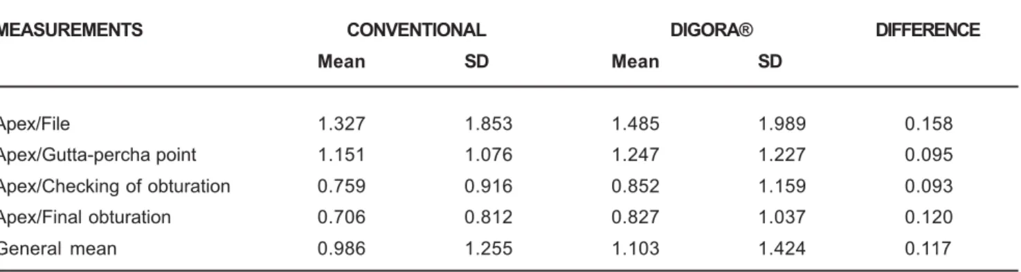
The measurements recorded with the two methods are presented in Table 1. The measurements of the first and second stage were compared by two-way analysis of variance with repeated measurements (Table 2).
There was statistically significant difference between the measurements obtained with the conventional method and the Digora® software. On average, Digora®
measurements were 0.117 mm larger than those recorded
MEASUREMENTS CONVENTIONAL DIGORA® DIFFERENCE
Mean SD Mean SD
Apex/File 1.327 1.853 1.485 1.989 0.158
Apex/Gutta-percha point 1.151 1.076 1.247 1.227 0.095
Apex/Checking of obturation 0.759 0.916 0.852 1.159 0.093
Apex/Final obturation 0.706 0.812 0.827 1.037 0.120
General mean 0.986 1.255 1.103 1.424 0.117
TABLE 1- Means and standard deviations of the measurements (in mm) obtained on the conventional and indirect digitized
radiographs
EFFECT DF MS DF MS F P
effect effect error error
Measurement 3 14.246 219 2.403 5.92774 0.000662*
Method 1 2.0269 73 0.185 10.92811 0.001471*
Interaction 3 0.03345 219 0.134 0.24896 0.862020
* Statistically significant difference (p<0.01)
TABLE 2- Two-way analysis of variance (measurement and method) with repetition
168
COMPARISON OF RADIOGRAPHIC MEASUREMENTS OBTAINED WITH CONVENTIONAL AND INDIRECT DIGITAL IMAGING DURINGon the conventional radiographs (Table 1).
With regard to comparison of image quality, in the conventional method 64.5% of the images were scored as good, 31.5% as acceptable and only 4% as poor; whereas in the Digora® system, there was 100% of good images, as
they had been improved by adjustment of brightness and contrast.
DISCUSSION
The use of digital radiographic resources has demonstrated several advantages over conventional radiographic film1,4,6,11,12. The sensitivity of the conventional
radiographic method is not the problem, but rather the ability of clinicians to interpret the images. In this context, the digital method has several advantages, due to its versatility and possibility of image manipulation1,10,12. The
digitized images may enhance the conditions for diagnosis, treatment planning and follow-up compared to conventional radiographs, due to the technological possibilities available through digital softwares3,11,13.
This study compared the quality of radiographic images and indirect digitization. The examiners scored 64.5% of images as good in conventional radiographs and 100% in indirectly digitized images. The factor that most contributed to the improvement in image quality was the adjustment of brightness and contrast, performed by the examiners according to their own judgment.
The difference between the filling material and apex measurements was 0.117 mm larger when the measurements recorded with the millimeter ruler on the film viewer were compared to those obtained on Digora®. Despite the
statistically significant difference, the clinical significance of a measurement of one tenth of millimeter in endodontic treatment is not relevant. The observation of larger measurements by Digora® may be related to the highest
measuring accuracy of the software and increased image size when analyzed on the computer screen. The accuracy of measurements achieved on indirect digital radiographs by the Digora® system was tested and did not reveal
statistically significant difference between the actual and digital measurements, yet the low sample size may lead to loss of diagnostic information.
Comparison between intraoral digital sensors and conventional radiographic film for root canal length determination and measurement of endodontic files of different sizes has been previously performed2,5,9,13. These
studies revealed the superiority of conventional radiographic film for detection of smaller files, whereas there was no statistically significant difference for size 15 files13. However, the authors recommend the use of the
digital system because of the possibility of reducing the patient’s exposure to ionizing radiation and time reduction in obtaining and processing digital images.
The quality of images obtained on the Digora® system
was higher because brightness and contrast could be adjusted. This is an advantage of this software over
conventional radiographs because the adjustment of images of lower quality avoids repetitions and consequently reduces the patient exposure to radiation.
On the basis of these results it may be concluded that the quality of indirectly digitized images was superior to that of conventional radiographs. The images of the filling material on the digitized images were 0.117mm larger than on the conventional image.
ACKNOWLEDGMENTS
The authors thank FAPESP for the financial grant for this study and to Professor José Roberto Lauris for his assistance with the statistical analysis.
REFERENCES
1-Borg E, Attaelmanan A, Grondahl HG. Subjective image quality of solid-state and photostimulable phosphor systems for digital intra-oral radiography. Dentomaxillofac Radiol. 2000;29:70-5.
2-Cederberg RA, Tydwell E, Frederiksen NL, Benson BW. Endodontic working length assessment: comparinson of storage phosphor digital imaging and radiographic film. Oral Surg Oral Med Oral Pathol Oral Radiol Endod. 1998;85:325-8.
3-Conover GL, Hildebolt CF, Yokoyama-Crothers N. Comparison of linear measurements made from storage phosphor and dental radiographs. Dentomaxillofac Radiol.1996;25:268-73.
4-Emmott LF. The digital revolution, images and X-rays. N Y State Dent J. 2005;71:40-2.
5-Garcia AA, Navarro LF, Castelló VU, Laliga RM. Evaluation of a digital radiography to estimate working length. J Endod. 1997;23:363-5.
6-Huda W, Rill LN, Benn DK, Pettigrew JC. Comparison of a photostimulable phosphor system with film for dental radiology. Oral Surg Oral Med Oral Pathol Oral Radiol Endod. 1997;83:725-31.
7-Kaeppler G, Vogel A, Axmann-Krcmar D. Intra-oral storage phosphor and conventional radiography in the assessment of alveolar bone structures. Dentomaxillofac Radiol. 2000;29:362-7.
8-Lavelle CLB, Wu CJ. Digital radiographic images will benefit endodontic services. Endod Dent Traumatol. 1995;11:253-60.
9-Loushine RJ, Weller RN, Kimbrough WF, Potter BJ. Measurement of endodontic file lengths: calibrated versus uncalibrated digital images. J Endod. 2001;27:779-81.
10-Miles DA. The deal on digital: the status of radiographic imaging. Compendium. 2001;22:1057-64.
11-Nair KM, Nair UP. Digital and advanced imaging in endodontics: a review. J Endod. 2007;33:1-6.
12-Parissis N, Kondylidou-Sidira A, Tsirlis A, Patias P. Conventional radiographs vs digitized radiographs: image quality assessment. Dentomaxillofac Radiol. 2005;34:353-6.
169
13-Sanderink GCH, Huiskens R, Van der Stelt PF, Welander US, Stheeman E. Image quality of direct intraoral x-ray sensors in assessing root canal length: the Radio VisioGraphy, Visualix/VIXA, Sens-A-Ray, and Flash Dent Systems compared with Ektaspeed films. Oral Surg Oral Med Oral Pathol. 1994;78:125-32.
14-Shearer AC, Mullane E, Macfarlane TV, Grondahl HG, Horner K. Three phosphor plate systems and film compared for imaging root canals. Int Endod J. 2001;34:275-9.
15-Versteeg CH, Sanderink GCH, Lobach SR, van der Stelt PF Reduction in size of digital images: does it lead to less detectability or less of diagnostic information? Dentomaxillofac Radiol. 1998;27:93-6.
16-Westphalen VPD, Moraes IG, Westphalen FH, Martins WD, Couto Souza PH. Conventional and digital radiographic methods in the detection of simulated external root resorptions: a comparative study. Dentomaxillofac Radiol. 2004;33:233-5.