R
ABSTRACT
REGIONAL ODONTODYSPLASIA: CASE REPORT
Ana Carolina MAGALHÃES1, Juliano Pelim PESSAN1, Robson Frederico CUNHA2, Alberto Carlos Botazzo DELBEM2
1-DDS, MSc, PhD Student, School of Dentistry of Araçatuba, São Paulo State University, UNESP, São Paulo, SP, Brazil.
2- DDS, MSc, PhD, Associate Professor, School of Dentistry of Araçatuba, São Paulo State University, UNESP, São Paulo, SP, Brazil.
Corresponding address: Prof. Dr. Alberto Carlos Botazzo Delbem - Departamento de Odontologia Infantil e Social - Faculdade de Odontologia de Araçatuba, UNESP - Rua José Bonifácio 1193, 16015-050 Araçatuba - SP - Brasil - Tel: +55 18 3636 3235 - Fax: +55 18 3636 3200 e-mail: [email protected]
Received: September 13, 2007 - Modification: September 29, 2007 - Accepted: October 1, 2007
egional odontodysplasia (RO) is a rare developmental anomaly involving both mesodermal and ectodermal dental components in a group of contiguous teeth. It affects the primary and permanent dentitions in the maxilla and mandible or both jaws. Generally it is localized in only one arch. The etiology of this dental anomaly is uncertain. Clinically, affected teeth have an abnormal morphology, are soft on probing and typically discolored, yellow or yellowish-brown. Radiographically, the affected teeth show a “ghostlike” appearance. This paper reports the case of a 5-year-old girl presenting this rare anomaly on the left side of the maxillary arch, which crossed the midline. The primary maxillary left teeth (except for the canine) and the primary maxillary right central incisor were missing due to previous extractions. The permanent teeth had a “ghostlike” appearance radiographically. The treatment performed was rehabilitation with temporary partial acrylic denture and periodic controls. In the future, the extraction of affected permanent teeth and rehabilitation with dental implants will be evaluated. The presentation of this case adds valuable information to pediatric dentists to review special clinical and radiographic features of RO, which will facilitate the diagnosis and treatment of patients with this condition.
Uniterms:Regional odontodysplasia; Dental dysplasia; Dentition, primary; Dentition, permanent.
INTRODUCTION
Regional odontodysplasia (RO) is a rare developmental anomaly involving both mesodermal and ectodermal dental components in a group of contiguous teeth 8. This condition
was probably first described by Hitchin12 (1934). Many
authors however, credit to McCall and Wald14 (1947) the
first report of this condition, showing only radiographic features.
The word “odontodysplasia” was coined by Zegarelli, et al.30 (1963). Because this abnormality has a tendency to
affect only one quadrant, “regional odontodysplasia” became the most accepted term to define it. The same condition has been described under other denominations, such as “odontogenic dysplasia”21, “localized arrest tooth
development”24, “ghost teeth”19, “odontogenisis
imperfecta”4, “unilateral dental malformation”1 and “familial
amelodentinal dysplasia”15.
RO affects both primary and permanent dentitions. Generally, it is limited to only one arch and sometimes crosses the midline. The maxilla is affected twice as often as the mandible5, the maxillary left quadrant being most commonly
involved5,16. Regarding the teeth, the central and lateral
incisors are more frequently affected than the posterior teeth18. It is also common that in the same quadrant the
teeth are affected in the different degrees10. It has been
suggested that this condition is more common in girls than in boys5. There has been no report of tendency towards a
particular ethnic group9.
The etiology of this dental anomaly is uncertain, although several factors, such as local trauma or infection5,19,
teratogenic drugs, local circulatory disorders27, Rh
incompatibility, irradiation17, neural damage17,
hyperpyrexia17,27, metabolic and nutritional disorders19 and
vitamin deficiency17,27, have been discussed. It seems to be
non-inherited condition because no cases with affection of family members have been described10,16. RO has been
reported in association with vascular nevi27, hemangioma27,
epidermal nevus syndrome22, orbital coloboma28, hypoplasia
of the affected side of the face29, hypophosphatasia20,
ectodermal dysplasia and hydrocephalus6.
The criteria for diagnosis of RO are primarily clinical and radiographic13. Clinically, affected teeth have an abnormal
morphology and an irregular surface contour, with pitting and groves surface7. The teeth appear to be discolored,
hypoplastic and hypocalcified. The thin enamel is soft on probing and teeth are typically discolored, yellow or yellowish-brown7. It is possible to find some teeth without
any alterations in the affected quadrant10. Affected teeth
fracturing at the slightest trauma10. Tooth eruption is delayed
or does not occur. The most frequent clinical symptoms after eruption of teeth with RO are gingival swelling, periapical infection and abscess formation in the absence of caries 10,13. There is a range of severity with this condition.
In some mild cases, the roots could develop quite well without having any infection or complications25.
Radiographically, the affected teeth show a “ghostlike” appearance due to reduced thickness and radiodensity of enamel and dentin19. A demarcation between
hypomineralized dentin and hypomineralized enamel is not visible27. The teeth tend to be shorter, have short roots with
wide open apices and abnormally wide pulp chambers and canals10.
The clinical and radiographic characteristics of RO will be discussed in this article, describing this anomaly with certain unique findings and treatment options that need to be considered with specific reference to pediatric patients.
CASE REPORT
A 5-year-old healthy Brazilian girl was referred to the Pediatric Dentistry Clinic at the Dental School of Araçatuba, São Paulo State University (UNESP), for detailed evaluation of her oral condition. According to the report of her mother, the primary maxillary left teeth had erupted differently from the other child’s teeth. The mother also reported that her daughter’s affected teeth had a yellowish color and were rapidly destroyed by caries and fractured often accompanied by pain and gingival swelling.
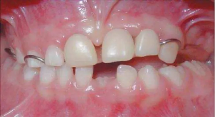
FIGURE 1- Extraoral aspect of the child with RO FIGURES 2, 3, 4 and 5- Intraoral view of the child with RO
FIGURE 3
FIGURE 5 FIGURE 2
The patient’s mother also reported that during pregnancy she had taken medication for high blood pressure control (Enalapril®). Except for this fact, the pregnancy and the birth
occurred uneventfully. There was no history of tooth or genetic anomalies in the family. The child’s general health was good and no congenital or acquired disease was
reported. However, the girl had a urinary infection in the first three days of life, which was treated with Amoxicillin®.
Extraoral examination revealed no facial asymmetry, but a vascular birthmark (haemangioma) on the right side of her face (Figure 1). Intraoral clinical examination revealed a relatively caries-free mouth with normal occlusion, soft tissues and developing dentition except for the maxillary left quadrant (Figures 2-6).
On the left side of the maxilla, the central incisor and the first and second molars were missing as well as the primary right central incisor. According to her mother, the teeth were extracted by a dentist 6 months before the girl was referred to the clinic. The residual root of the left lateral incisor was present and was indicated for extraction. The left canine was previously restored with composite resin and it was less affected compared to the other teeth. It is likely that part of the permanent left first molar had an altered aspect because the primary second molar had been previously extracted. This clinical finding was not presented at the first appointment and it was observed at the third appointment. The panoramic radiograph, which was taken 6 months before the girl was referred to our Clinic at the Dental School of Araçatuba, revealed the presence of primary teeth and the germs of permanent teeth, including the permanent second molars, except in the maxillary left quadrant. In the affected area, it was possible to observe the primary right central incisor, which was extracted straight away. The root of primary left lateral incisor was also present. In addition, the primary left canine presented reduced radiodensity and showed wide open apex and abnormally wide pulp chambers and canals in comparison to unaffected teeth. Germs of permanent teeth from the maxillary left quadrant and also the germ of the permanent right central incisor presented a “ghost like” appearance, showing that this condition had crossed the midline. Dental development appeared
age-FIGURE 8- Rehabilitation of the child with temporary
maxillary partial acrylic denture
FIGURE 6- Intraoral view of the child with RO
FIGURE 7- Panoramic radiograph showing the teeth with “ghostlike” appearance in maxillary left quadrant and the right
appropriate. Normal thickness of enamel and dentin in primary and permanent dentitions was observed in the other quadrants, except for the permanent mandibular left first molar, which showed some alterations in its crown, probably a developmental anomaly (Figure 7). This radiographic finding has to be investigated in the future controls.
After extraction of the root of the maxillary left lateral incisor, the affected edentulous quadrant was rehabilitated with a temporary maxillary partial acrylic denture (Figure 8). Prosthetic rehabilitation of patients is important to maintain mastication and phonation, to improve esthetics and to prevent overeruption of opposite teeth. The girl has been followed up periodically (once a month) to observe if there will be eruption of the “affected teeth” and to monitor the growth and development of the maxillary and mandibular arches.
DISCUSSION
Regional odontodysplasia is a rare developmental anomaly of dental hard tissues. According to a previous literature review25, 138 cases of RO had been published from
1934 up to the end of the year 2002. After their publication in 2004, other 6 cases were retrieved from a Pubmed database search until the present moment. The patient of this case report exhibited many of the common clinical and radiographic features consistent with the diagnosis of RO. The clinical and radiographic characteristics involving the permanent dentition in the maxillary left quadrant (and also including the right permanent central incisor) strongly supported the diagnosis of this condition.
In this case, RO affected only one quadrant in the maxilla, but it crossed the midline, as previously reported11,28. RO
seems to be more prevalent in females and to occur in both dentitions25, as found in this case. In addition, the finding
of possible taurodontism in an unaffected quadrant (mandibular left first molar) suggests that RO might occur in association with other disorders26.
The etiology of RO remains unknown10. Several factors
have been associated to its occurrence11. In this case, the
history of maternal use of medication during pregnancy, as well as the urinary infection in the first three days of the child’s life may have not been related to the condition. Otherwise, the prevalence of this anomaly would be higher. Some authors have suggested a strong correlation between vascular nevi and the pathogenesis of RO27. In this case,
the haemangioma was observed upon examination, but on the right side of her face (not affected by RO) in accordance to Fearne, et al.7 (1986). Although the haemangioma is
localized in the opposite site to that affected by odontodysplasia, it could be an indication of abnormal vasculature, probably due to local ischemia or to morphological defects in blood vessels, perhaps following somatic mutation7. Unfortunately the cause of the present
case remains unknown. In order to confirm possible etiological factors, further detailed investigation of this anomaly through the histological analysis must be reported13. Being RO a rare disease, it seems difficult to get
a significant number of patients to study this anomaly in a more detailed manner, for clinical, microradiographic and histopathological analyses. Microradiography is indicated to evaluate the mineral content of affected teeth because in conventional radiography the density of enamel is not evident due to the thinness of enamel layer in these teeth3.
Several factors must be considered to determine the best treatment option for a child with RO, such as age of the patient, any relevant medical history, previous dental experience, child’s and parental attitude regarding dental treatment and number of affected teeth2,11. There has been
much debate as to whether affected teeth (with or without abscesses) should be extracted or saved5,10. In this case,
the residual root of the left lateral incisor was indicated for extraction because it could not be restored. In contrast, the left canine had been previously restored with composite resin because it was less affected by the anomaly. Other teeth probably were in same condition of that the left lateral incisor and had probably been extracted by another dentist. The partial denture was fabricated to provide function, phonation and esthetics in this critical period. It was not possible to place posterior teeth in this denture yet because there is not space available. The early primary molars loss during the growth phase leads to reduction of vertical occlusal dimension. Because of this, it has to be considered the use of an appliance with screw to compensate maxillary growth. Another treatment option for patients with RO was described by Cahuana, et al.2 (2005), who consider
autotransplantation as an alternative during this phase. However, it is limited by the availability of suitable donor teeth3,13.
Although the child and her mother are satisfied with treatment, the rehabilitation in this case cannot be considered ideal because the patient’s oral esthetic and function were not totally restored. For the time being, the patient is under control visits to observe the growth and development of the maxilla and mandible. During the appointments, the appliance has been adjusted or changed for a new one. The permanent teeth will probably not erupt or will have altered eruption pattern. The noninfected permanent teeth will be not extracted before eruption because these teeth help maintaining the alveolar bone3.
The presence of teeth is important during the skeletal growth phase. However, the prognosis of the affected permanent teeth is poor. Because of this, in the future, the extraction of permanent teeth and the rehabilitation with dental implants must be evaluated3,23.
CONCLUSION
REFERENCES
1- Bergmen G, Lysell L, Pindborg JJ. Unilateral dental malformation: report of two cases. Oral Surg. 1963;16:48-60.
2- Cahuana A, Gonzalez Y, Palma C. Clinical management of regional odontodysplasia. Pediatr Dent. 2005;27:34-9.
3- Cho SY. Conservative management of regional odontodysplasia: case report. J Can Dent Assoc. 2006;72:735-8.
4- Claudhry AP, Wittich HC, Stickel FR, Holland MR. Odontogenesis imperfecta. Oral Surg Oral Med Oral Pathol. 1961;14:1099-103.
5- Crawford PJM, Aldred MJ. Regional odontodysplasia: a bibliography. J Oral Pathol Med. 1989;18:251-63.
6- Dahllöf G, Lindskog S, Theorell K, Ussisoo R. Concomitant regional odontodysplasia and hydrocephalus. Oral Surg Oral Med Oral Pathol. 1987; 63:354-7.
7- Fearne J, Williams DM, Brook AH. Regional odontodysplasia: a clinical and histological evaluation. J Int Assoc Dent Child. 1986;17:21-5.
8- Gardner DG, Sapp JP. Regional Odontodysplasia. Oral Surg. 1973;35:351-65.
9- Gerlach RF, Jorge J Jr, de Almeida OP, Coletta RD, Zaia A. Regional Odontodysplasia. Report of two cases. Oral Surg Oral Med Oral Pathol. 1998;85:308-13.
10- Gomes MP, Modesto A, Cardoso ASC, Hespanhol W. Regional odontodyspalsia: report of a case involving two separate affected areas. ASCD J Dent Child. 1999;66:203-7.
11- Hanks JA, Williams B. Odontodysplasia: report of two cases. Pediat Dent. 1998;20:199-203.
12- Hitchin AD. Unerupted deciduos teeth in a youth aged 15 1/2. Br Dent J. 1934; 56:631-3.
13- Kinirons MJ, O’Brien FV, Gregg TA. Regional odontodysplasia: an evaluation of three cases based on clinical, microradiographic and histopathological findings. Br Dent J. 1998;20:136-9
14- Mc Call JO, Wald SS. In clinical dental roentgenography. Philadelphia: W.B. Saunders Co; 1947. p.169-70.
15- Mock D, Aidelbaum MR, Chapmick P. Familial amelodentinal dysplasia. Oral Surg Oral Med Oral Pathol. 1986;61:485-91.
16- Özer L, Cetiner S, Ersoy E. Regional odontodysplasia: report of a case. J Clin Pediat Dent. 2004;29:45-8.
17- Pandis N, Polido C, Bell WH. Regional odontodysplasia. A case associated with asymmetric maxillary and mandibular development. Oral Surg Oral Med Oral Pathol. 1991;72:492-6.
18- Rosa MC, Marcelino GA, Belchior RS, Souza AP, Parizotto SC. Regional Odontodisplasia: reporto f case. J Clin Pediatr Dent. 2006;30:333-6.
19- Rushton MA. Odontodysplasia: ghost teeth. Br Dent J 1965;119:109-13.
20- Russel K, Yacobi R. Generalized odontodysplasia concomitant with mild hypophophatasia: a case report. J Can Dent Assoc. 1993;59:187-90.
21- Sibley LC, Zimmerman ER. Odontogenic dysplasia: report of a case. Oral Surg. 1962;15:1370-3.
22- Slootweg PJ, Meuwissen PR. Regional odontodysplasia in epidermal nevus syndrome. J Oral Pathol. 1985;14:256-62.
23- Spini TH, Sargenti-Neto S, Cardoso SV, Souza KC, de Souza SO, de Faria PR et al. Progressive dental development in regional odontodysplasia. Oral Surg Oral Med Oral Pathol Oral Rad Endo. 2007;104:40-5.
24- Suher T, Jump EB, Landis RL. Localized arrested tooth development. Oral Surg. 1970;6:1305-14.
25- Tervoten SA, Stratmann U, Mokrys K, Reichart PA. Regional Odontodysplasia: a review of the literature and report of four cases. Clin Oral Investig. 2004;8:45-51.
26- Vaikuntam J, Tatum NB, McGuff HS. Regional odontodysplasia: review of the literature and the report of a case. J Clin Pediatr Dent. 1996;21:35-40.
27- Walton JL, Witkop CJ, Walker PO. Odontodysplasia. Report of three cases with vascular nevi overlying the adjacent skin of the face. Oral Surg. 1978;46:676-84.
28- Williams SA, High AS. Odontodysplasia associated with orbital coloboma. Br Dent J. 1988;164:390-4.
29- Ylipaavalniemi P, Ranta R, Lamberg M. Odontodysplasia associated with unilateral malformations. Proc Finn Dent Soc. 1982;78:134-40.