T
ABSTRACT
SEGMENTAL LEFORT I OSTEOTOMY FOR TREATMENT
OF A CLASS III MALOCCLUSION WITH
TEMPOROMANDIBULAR DISORDER
Marcos JANSON1, Guilherme JANSON2, Eduardo SANT’ANA3, Alexandre NAKAMURA4, Marcos Roberto de FREITAS5
1- DDS, MSc, Private Practice, Bauru, SP, Brazil.
2- DDS, MSc, PhD, MRCDC (Member of the Royal College of Dentists of Canada). Professor, Department of Pediatric Dentistry, Orthodontics and Community Health, Bauru School of Dentistry, University of São Paulo, Bauru, SP, Brazil.
3- DDS, MSc, PhD, Associate Professor, Department of Stomatology, Bauru School of Dentistry, University of São Paulo, Bauru, SP, Brazil. 4- DDS, MSc, Orthodontics Graduate Student, Department of Pediatric Dentistry, Orthodontics and Community Health, Bauru School of Dentistry, University of São Paulo, Bauru, SP, Brazil.
5- DDS, MSc, PhD, Professor, Department of Pediatric Dentistry, Orthodontics and Community Health, Bauru School of Dentistry, University of São Paulo, Bauru, SP, Brazil.
Corresponding address: Dr. Guilherme Janson - Disciplina de Ortodontia - Faculdade de Odontologia de Bauru/ USP - Alameda Octávio Pinheiro Brisolla 9-75 - 17012-901 Bauru, SP, Brazil - Phone/Fax: 55-14-32342650 - e-mail: jansong@travelnet.com.br
Received: February 29, 2008 - Accepted: April 02, 2008
his article reports the case of a 19-year-old young man with Class III malocclusion and posterior crossbite with concerns about temporomandibular disorder (TMD), esthetics and functional problems. Surgical-orthodontic treatment was carried out by decompensation of the mandibular incisors and segmentation of the maxilla in 4 pieces, which allowed expansion and advancement. Remission of the signs and symptoms occurred after surgical-orthodontic intervention. The maxillary dental arch presented normal transverse dimension. Satisfactory static and functional occlusion and esthetic results were achieved and remained stable. Three years after the surgical-orthodontic treatment, no TMD sign or symptom was observed and the occlusal results had not changed. When vertical or horizontal movements of the maxilla in the presence of moderate maxillary constriction are necessary, segmental LeFort I osteotomy can be an important part of treatment planning.
Key words: Malocclusion. Angle Class II. Osteotomy, LeFort. Temporomandibular joint disorders.
INTRODUCTION
Posterior crossbites or transverse maxillary deficiencies are relatively common dentofacial deformities that can be found alone or in association with other maxillary problems21.
Class III malocclusion caused by maxillary retrognathism is often accompanied by posterior crossbite10. If they are
detected before the adolescent growth spurt, maxillary expansion and face-mask therapy provide well-controlled results18,19. Unfortunately, these techniques are of limited
use in adult patients because the maxillary sutures are already fused. Surgical intervention, comprehending expansion and advancement of the maxilla, can be performed in adult subjects to achieve satisfactory esthetic and functional outcomes.
In adult cases of constricted maxilla, expansion of the arch can be performed by surgically assisted rapid palatal expansion (SARPE), or by segmenting the maxilla during the osteotomy. The former is carried out as a first stage of a two-stage surgical treatment. Subtotal LeFort I osteotomy
with midline osteotomy is conducted with an osteotome and a mallet, and thereafter expansion is accomplished with a standard banded hyrax appliance3. Because the expansion
is gradually performed, between 7 to 15 days, allowing the palatal mucosa to adapt to the stretching, practically 7 to 14 mm of expansion can be achieved. Thereafter, 1-piece LeFort I osteotomy is performed to advance the maxilla.
When the decision is to correct the maxillary constriction concomitantly with the osteotomy, the maxilla is segmented into pieces to allow appropriate expansion and advancement in the Class III patient. Traditionally, segmental LeFort I osteotomy have been indicated where a transverse deficiency is associated with other maxillary problems11.
Given this assumption, the present case report demonstrates a segmental LeFort I osteotomy for expansion and advancement of the maxilla in the treatment of a Class III patient with TMD.
CASE REPORT
Diagnosis and Etiology
The patient was a 20-year-old man, who sought treatment in the private orthodontic office of Dr. MJ, due to TMD and esthetic-functional problems. The patient complained of suffering from headache and muscle symptoms for over 3 years in addition to pain on the temporomandibular joints (TMJs) and masticatory muscles, and muscle tenderness to palpation. A history of bruxism and clenching was also reported. Clinical examination showed maximum mouth opening and lateral movement limitations. No clicking, popping or crepitus sound evaluated by auscultation were detected in either TMJ. No mandibular shift during opening or closing movements was noticed.
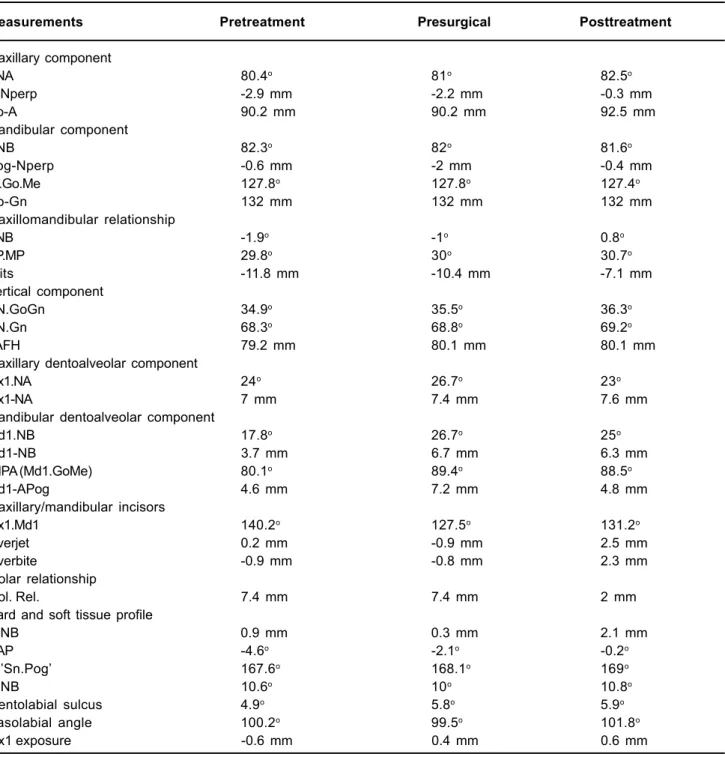
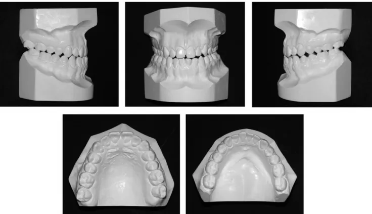
Facial esthetics and occlusal function were also concerns associated to TMD. Cephalometric analysis showed a retrusive maxilla, and a proportionally large mandible, disguised by an increased lower anterior facial height (Tables 1 and 2). Facial examination showed a horizontal deficiency of the midface with flattening of the malar bone and the cheeks, and retrusion of the upper lip (Figure 1). The lower facial third showed a satisfactory horizontal relationship with the entire profile. The face was symmetrical in the frontal aspect. The intraoral examination showed ¾ molar Class III relationship on the right and ¼ Class III relationship on the left side1,15. In centric relation,
the posterior teeth and the incisors occluded in an edge to edge relationship (Figure 2). Satisfactory alignment of the teeth and a mild curve of Spee could be seen, and both midlines were 2 mm deviated to the right of the midsagittal
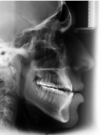
plane. The mandibular left central incisor was treated endodontically, had a composite resin restoration and was darkened, but did not present clinical signs of ankylosis. Cephalometrically, the maxillary incisors were well positioned on the basal bone, and the mandibular incisors were lingually tipped (Table 2 and Figure 3).
Treatment Objectives
The primary treatment goal was to eliminate or alleviate the TMD signs and symptoms. Satisfactory facial esthetics and masticatory function were also objectives to be attained. Proper bilateral Class I molar occlusion and normal overjet and overbite could be established by correcting the compensating tooth positions, and expanding and advancing the maxilla. Attainment of ideal functional occlusion with canine and incisal guidance was an important goal. Also, maxillary advancement and correction of tooth interdigitation would improve the retrognathic aspect of the midface and the intraoral appearance.
Treatment Alternatives
Three treatment options were considered. The first treatment alternative was an orthodontic approach with fixed appliances only, by means of dentoalveolar compensation. Wider maxillary archwires would expand the constricted dental arch, and Class III elastics could be used to correct the posterior occlusion and the anterior crossbite. The maxillary incisors would be labially tipped and the mandibular incisors would be lingually tipped.
The second option involved a surgical orthodontic approach. In this way, the overall treatment goals could be attained, in spite of the risks inherent to the procedure. The maxillary surgical expansion and advancement could help in achieving correct static and functional occlusion and considerable improvement in facial esthetics. In order to perform the surgical expansion of the maxillary arch, two options were presented: it could be done in a first stage,
Dental cephalometric variables
Md1-APog distance between incisal of mandibular incisor to line APog Mx1.Md1 angle formed by the long axes of maxillary and mandibular incisors
Skeletal cephalometric variables
A-Nperp distance between A point to nasion-perpendicular Pog-Nperp distance between Pog point to nasion-perpendicular PP.MP angle formed by palatal and mandibular planes
SN.Gn angle formed by SN and NGn lines
Soft tissue cephalometric variables
Gl’Sn.Pog’ angle formed by soft tissue glabella, subnasale and pogonion H.NB angle formed by Holdaway esthetic and NB lines
Mentolabial sulcus angle formed by the greatest concavity in the midline between the lower lip and chin Mx1 exposure vertical distance between incisal of the maxillary incisor to upper lip stomion
with a subtotal LeFort I osteotomy, and thereafter a 1-piece osteotomy would be performed for advancement; or, concomitantly with the advancement, segmentation of the maxilla in four pieces would provide expansion of the arch. The treatment options were presented to the patient and discussed. Because esthetic appearance was a major concern, the first option was refused and the third was chosen because it would be performed in only one surgical intervention. For the mandibular arch, the choice was to treat with fixed appliances only, by means of decompensation of the incisors. For the maxillary arch, the choice was a segmental LeFort I osteotomy to permit both expansion and advancement.
Treatment Progress
Malocclusion was treated with conventional 0.022-in slot preadjusted edgewise appliances. Leveling and aligning were performed with round nickel-titanium and steel archwires until rectangular 0.018 x 0.025-inch stainless-steel archwires were placed. Class II elastics were used to retract the maxillary incisors and reciprocally mesialize the mandibular molars. After 10 months of presurgical orthodontic treatment, the maxillary archwire was segmented mesially to the canines, in order to avoid postoperative orthodontic relapse13. Conventional orthodontic mechanics
continued for 3 additional months.
A LeFort I osteotomy was performed with segmentation
Measurements Pretreatment Presurgical Posttreatment
Maxillary component
SNA 80.4o 81o 82.5o
A-Nperp -2.9 mm -2.2 mm -0.3 mm
Co-A 90.2 mm 90.2 mm 92.5 mm
Mandibular component
SNB 82.3o 82o 81.6o
Pog-Nperp -0.6 mm -2 mm -0.4 mm
Ar.Go.Me 127.8o 127.8o 127.4o
Co-Gn 132 mm 132 mm 132 mm
Maxillomandibular relationship
ANB -1.9o -1o 0.8o
PP.MP 29.8o 30o 30.7o
Wits -11.8 mm -10.4 mm -7.1 mm
Vertical component
SN.GoGn 34.9o 35.5o 36.3o
SN.Gn 68.3o 68.8o 69.2o
LAFH 79.2 mm 80.1 mm 80.1 mm
Maxillary dentoalveolar component
Mx1.NA 24o 26.7o 23o
Mx1-NA 7 mm 7.4 mm 7.6 mm
Mandibular dentoalveolar component
Md1.NB 17.8o 26.7o 25o
Md1-NB 3.7 mm 6.7 mm 6.3 mm
IMPA (Md1.GoMe) 80.1o 89.4o 88.5o
Md1-APog 4.6 mm 7.2 mm 4.8 mm
Maxillary/mandibular incisors
Mx1.Md1 140.2o 127.5o 131.2o
Overjet 0.2 mm -0.9 mm 2.5 mm
Overbite -0.9 mm -0.8 mm 2.3 mm
Molar relationship
Mol. Rel. 7.4 mm 7.4 mm 2 mm
Hard and soft tissue profile
P-NB 0.9 mm 0.3 mm 2.1 mm
NAP -4.6o -2.1o -0.2o
Gl’Sn.Pog’ 167.6o 168.1o 169o
H.NB 10.6o 10o 10.8o
Mentolabial sulcus 4.9o 5.8o 5.9o
Nasolabial angle 100.2o 99.5o 101.8o
Mx1 exposure -0.6 mm 0.4 mm 0.6 mm
FIGURE 2- Pretreatment study models
of the maxilla in four mobile segments. Vertical interdental osteotomies were implemented between the maxillary lateral incisors and the canines. Two horizontal osteotomies, parallel with the septum were performed to expand the maxilla transversally. Following the osteotomy, the maxillary segments were anteriorly repositioned and connected to the mandible in the correct occlusal relationship. The mandibular and maxillary arches were wired together and acted as a unit, rotating around the condylar heads. Due to the absence of condylar displacement, efforts were made to preserve the preoperative temporomandibular relationship while seating the condyles in the most superior and anterior part of the mandibular fossa. Rigid fixation with miniplates and miniscrews fixed the maxillary segments in the final position. No interocclusal splint or postoperative maxillomandibular fixation was used. The patient was instructed to wear ¼ inch intermaxillary elastics for 20 h/day during 45 days and then gradually reduce the wear time.
Thereafter, post-surgical edgewise treatment continued for 14 months. After debonding, a fixed canine-to-canine retainer was placed in the mandibular anterior teeth and a removable Hawley retainer in the maxillary arch. The overall active treatment period was 2 years and 3 months.
Treatment Outcomes
After surgical orthodontic treatment, headache, pain on the TMJ and jaw muscle tenderness upon palpation had
FIGURE 3- Pretreatment lateral radiograph
FIGURE 5- Posttreatment study models
ceased. Functional analysis showed normal mandibular opening and excursive movements. The patient reported discontinuation of bruxism and clenching.
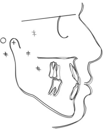
The posttreatment facial photographs show satisfactory changes in frontal and profile views by increasing the cheek support and protrusion of the upper lip (Figure 4). After advancement, the final position of the maxilla showed an improved reciprocal balance with the mandible and the lower anterior facial height. Bilateral Class I molar relationship and positive overjet and overbite were achieved with maxillary advancement. Segmentation of the maxilla allowed transverse expansion and avoided molar buccal inclination (Figure 5). The cephalometric superimposition shows that the maxillary incisors were protruded without inclination changes (Figures 6 and 7). On the other hand, the mandibular incisors had mild labial tipping. Three years after the surgical-orthodontic treatment, no TMD sign or symptom was observed and the occlusal results had not changed.
DISCUSSION
Correction of maxillary constriction is an important part of the surgical-orthodontic treatment plan. When horizontal or vertical movements of the maxilla are also required, segmental LeFort I osteotomy is considered an effective procedure to correct transverse deficiencies. While SARPE is accomplished as a first step of a 2-step approach, segmental LeFort I is performed concomitantly with the osteotomy. Because time is required for expansion and a postoperative healing period is necessary after SARPE, the entire surgical orthodontic treatment time can be prolonged2.
During treatment planning, some factors between SARPE and segmental LeFort I should be considered: presence of other maxillary problem, magnitude of width deficiency and stability. According to Bailey, et al.2 (1997), if other surgery
in the maxilla is necessary after arch expansion, there is little reason to perform surgery twice. One exception is the magnitude of the maxillary constriction. Because of the inelasticity of the palatal mucosa, there is limitation in the amount of expansion with segmental LeFort I5. In the present
case, which required moderate expansion of the arch and advancement of the maxilla, a single surgical approach reduced the clinical steps of the entire treatment. The last point, stability of the expansion, should be seen with some caution. Studies have demonstrated better stability for lateral expansion with SARPE compared to segmental LeFort I osteotomy20,24. An anticipated relapse of about 50% could
be expected with segmentation of the maxilla. However, this amount of skeletal relapse can be controlled by means of dentoalveolar compensation, with the insertion of wide heavy archwires in the maxillary posterior teeth.
Some complications associated with segmental LeFort I have been described and that is the reason the procedure is sometimes avoided. Large spaced transversal “gaps” between the segments can cause lacerations in the mucosa, and dehiscence and resorption of the trabecular bone. Therefore, a correct clinical diagnosis is important. Risks
for root or vascular damage, and difficulty in segment management can compromise the surgical outcome17. Clinical
expertise is mandatory in all types of surgical intervention. Skeletal modifications should not be expected after treatment because the patient was an adult. Nevertheless, this Class III patient could be orthodontically compensated without surgery. Cases with greater skeletal discrepancies can be solved with fixed appliances alone14,16. The result
would be a Class I posterior occlusion and dentoalveolar compensation to achieve normal overjet and overbite. For patients with muscular pain, however, an accurate final functional occlusion must be accomplished, and precaution in this topic is mandatory. Accordingly, because of the indirect retrusive force on the mandible by the use of Class III elastics, care was taken to avoid distal pressure on the TMJ25,28.
The surgical procedures undertaken in this case were limited to segmental expansion and advancement of the maxilla. In a first moment, the increased lower anterior facial height was supposed to be an indication for maxillary impaction. The subsequent counterclockwise rotation of the mandible would produce a prognathic appearance and, therefore, would require a sagittal split osteotomy. Additionally, the maxillary incisors were completely covered by the lips at rest, and the upper lip smile line was located at the level of the gingival margin of the maxillary incisors (Figure 3). In addition, there was an acceptable functional balance in this vertical dimension of occlusion, suggesting maintenance of the original face height.
Generally, orthognathic surgery offers beneficial outcome in the management of TMD cases8, with a success
rate highly dependent on the diagnosis and treatment modalities26. Among patients who receive orthognathic
surgery, those with Class III relationships experiment greater improvement than those with Class II27. With respect to
surgical procedures, favorable outcomes are smaller in cases of bimaxillary or mandibular surgery, while isolated maxillary surgery offers greater chances of success6,12. This is because
mandibular osteotomy techniques require rotation of the condylar axis, sometimes affecting TMJ function. Moreover, changes in the position of the condyle are normally expected to happen after bimaxillary surgery9. Therefore, LeFort I
osteotomy for maxillary advancement can be a worthwhile alternative therapy for TMD patients with Class III malocclusions.
Orthodontic finishing plays an important role in patients with muscular dysfunction. All efforts were focused in reaching the functional treatment goals23. That is why the
duration of post-surgical orthodontics was relatively longer than usual4,7. In addition, because most surgical relapse
occurs during the first year22, continuation of orthodontic
CONCLUSION
Segmental LeFort I osteotomy requires clinical expertise in the management of the maxillary pieces. In surgical cases presenting moderate maxillary constriction associated with other maxillary problems, it may be an important part of the treatment plan. The major advantage refers to the single surgical intervention, reducing the period of convalescence, the psychological impact and the treatment costs. After the orthodontic-surgical intervention, no TMD signs or symptoms were observed.
REFERENCES
1- Andrews LF. The straight wire appliance: syllabus of philosophy and techniques. 2nd ed. San Diego: Larry F. Andrews Foundation of Orthodontic Education and Research; 1975.
2- Bailey LJ, White RP Jr, Proffit WR, Turvey TA. Segmental LeFort I osteotomy for management of transverse maxillary deficiency. J Oral Maxillofac Surg. 1997;55(7):728-31.
3- Bays RA, Greco JM. Surgically assisted rapid palatal expansion: an outpatient technique with long-term stability. J Oral Maxillofac Surg. 1992;50(2):110-3.
4- Bell WH, Jacobs JD. Three-dimensional planning for surgical/ orthodontic treatment of mandibular excess. Am J Orthod. 1981;80(3):263-88.
5- Betts NJ, Sturtz DH, Aldrich DA. Treatment of transverse (width) discrepancies in patients who require isolated mandibular surgery: the case for maxillary expansion. J Oral Maxillofac Surg. 2004;62(3):361-4.
6- Dervis E, Tuncer E. Long-term evaluations of temporomandibular disorders in patients undergoing orthognathic surgery compared with a control group. Oral Surg Oral Med Oral Pathol Oral Radiol Endod. 2002;94(5):554-60.
7- Dowling PA, Espeland L, Sandvik L, Mobarak KA, Hogevold HE. LeFort I maxillary advancement: 3-year stability and risk factors for relapse. Am J Orthod Dentofacial Orthop. 2005;128(5):560-7. 8- Egermark I, Blomqvist JE, Cromvik U, Isaksson S. Temporomandibular dysfunction in patients treated with orthodontics in combination with orthognathic surgery. Eur J Orthod. 2000;22(5):537-44.
9- Fernandez Sanroman J, Gomez Gonzalez JM, Alonso Del Hoyo J, Monje Gil F. Morphometric and morphological changes in the temporomandibular joint after orthognathic surgery: a magnetic resonance imaging and computed tomography prospective study. J Craniomaxillofac Surg. 1997;25(3):139-48.
10- Guyer EC, Ellis EE 3rd, McNamara JA Jr, Behrents RG. Components of class III malocclusion in juveniles and adolescents. Angle Orthod. 1986;56(1):7-30.
11- Hall HD, West RA. Combined anterior and posterior maxillary osteotomy. J Oral Surg. 1976;34(2):126-41.
12- Harper RP. Analysis of temporomandibular joint function after orthognathic surgery using condylar path tracings. Am J Orthod Dentofacial Orthop. 1990;97(6):480-8.
13- Jacobs JD, Sinclair PM. Principles of orthodontic mechanics in orthognathic surgery cases. Am J Orthod. 1983;84(5):399-407.
14- Janson G, Souza JE, Alves FA, Andrade P Jr, Nakamura A, Freitas MR, et al. Extreme dentoalveolar compensation in the treatment of Class III malocclusion. Am J Orthod Dentofacial Orthop. 2005;128(6):787-94.
15- Keeling SD, Wheeler TT, King GJ, Garvan CW, Cohen DA, Cabassa S, et al. Anteroposterior skeletal and dental changes after early Class II treatment with bionators and headgear. Am J Orthod Dentofacial Orthop. 1998;113(1):40-50.
16- Kondo E, Ohno T, Aoba TJ. Nonsurgical and nonextraction treatment of a skeletal Class III patient with severe prognathic mandible: long-term stability. World J Orthod. 2001;2:115-26. 17- Lanigan DT, Hey JH, West RA. Major vascular complications of orthognathic surgery: hemorrhage associated with Le Fort I osteotomies. J Oral Maxillofac Surg. 1990;48(6):561-73.
18- Nartallo-Turley PE, Turley PK. Cephalometric effects of combined palatal expansion and facemask therapy on Class III malocclusion. Angle Orthod. 1998;68(3):217-24.
19- Ngan P, Hagg U, Yiu C, Merwin D, Wei SH. Treatment response to maxillary expansion and protraction. Eur J Orthod. 1996;18(2):151-68.
20- Pogrel MA, Kaban LB, Vargervik K, Baumrind S. Surgically assisted rapid maxillary expansion in adults. Int J Adult Orthodon Orthognath Surg. 1992;7(1):37-41.
21- Proffit WR, Phillips C, Dann C 4th. Who seeks surgical-orthodontic treatment? Int J Adult Orthodon Orthognath Surg. 1990;5(3):153-60.
22- Proffit WR, Phillips C, Prewitt JW, Turvey TA. Stability after surgical-orthodontic correction of skeletal Class III malocclusion. 2. Maxillary advancement. Int J Adult Orthodon Orthognath Surg. 1991;6(2):71-80.
23- Roth RH. Functional occlusion for the orthodontist. J Clin Orthod. 1981;15(1):32-40,44-51.
24- Silverstein K, Quinn PD. Surgically-assisted rapid palatal expansion for management of transverse maxillary deficiency. J Oral Maxillofac Surg. 1997;55(7):725-7.
25- Tucker MR, Thomas PM. Temporomandibular pain and dysfunction in the orthodontic surgical patient: rationale for evaluation and treatment sequencing. Int J Adult Orthodon Orthognath Surg. 1986;1(1):11-22.
26- Westermark A, Shayeghi F, Thor A. Temporomandibular dysfunction in 1,516 patients before and after orthognathic surgery. Int J Adult Orthodon Orthognath Surg. 2001;16(2):145-51. 27- White CS, Dolwick MF. Prevalence and variance of temporomandibular dysfunction in orthognathic surgery patients. Int J Adult Orthodon Orthognath Surg. 1992;7(1):7-14.


