Complete mole in a dichorionic twin
pregnancy after intracytoplasmic
sperm injection
Mola hidatiforme completa em gravidez bicoriônica após injecção
intracitoplasmática de espermatozoides
Carla Maria alMeida raMalho2 Filipa aBreu goMesde Carvalho3 BerTa CeCília CaMpos liMade Carvalho3
oTília gonçalves BenTo Cavaleiro Brandão2
nuno aires MoTa Mendonça MonTenegro2
Abstract
A dichorionic twin pregnancy with complete hydatidiform mole and coexistent fetus is a rare and challenging situation, whose pathogenesis has not been yet fully understood. We present a case of a 39-year-old woman who underwent intracytoplasmic sperm injection with two embryos transfer. The 12-week gestation ultrasound examination revealed normal fetus and placenta with features of hydatidiform mole, leading to pregnancy termination. Autopsy and histological examinations diagnosed a complete mole coexisting with a normal fetus, and the genetic analysis showed a diploid fetus with biparental genome and molar tissue with paternal diploidy. This case highlighted that complete molar pregnancies may still occur even though pregnancy is achieved after intracytoplasmic sperm injection. A review of the literature was performed by collecting data from the few similar reported cases and by commenting on the pathogenesis of this rare condition.
Resumo
Uma gravidez bicoriônica com mola hidatiforme completa e feto normal é uma situação rara e desaiadora, cuja patogênese não foi ainda totalmente compreendida. Apresenta-se o caso de uma mulher de 39 anos submetida à injeção intracitoplasmática de espermatozoides com transferência de dois embriões. Na ecograia pré-natal realizada na 12ª semana de gestação, foi identiicado um embrião morfologicamente normal e uma placenta com características molares. Esta situação resultou na terminação eletiva da gravidez. A autópsia e o estudo histológico permitiram o diagnóstico deinitivo de uma mola hidatiforme completa coexistindo com feto normal. A análise genética mostrou feto diploide com genoma biparental e tecido molar com diploidia paterna. Este caso ressaltou que as gestações com mola hidatiforme completa poderão ainda ocorrer, mesmo que a gravidez seja realizada após uma injeção intracitoplasmática de espermatozoides. Foram realizadas uma revisão dos raros casos descritos na literatura e uma explicação da patogenia desta condição rara.
1Serviço de Ginecologia e Obstetrícia, Centro Hospitalar de São João do Porto – Porto, Portugal. 2Serviço de Anatomia Patológica, Centro Hospitalar de São João do Porto – Porto, Portugal. 3Departamento de Genética da Faculdade de Medicina, Universidade do Porto – Porto, Portugal.
Conlict of interest: none.
Keywords
Hydatidiform mole Sperm injections, intracytoplasmic Pregnancy, twin Case reports
Palavras-chave
Mola hidatiforme Injeções de esperma intracitoplásmicas
Gravidez de gêmeos Relatos de casos
Correspondence
Tiago Matos Ferraz Serviço de Ginecologia e Obstetrícia, Centro Hospitalar de São João Alameda Professor Hernâni Monteiro, 4200 319 CEP: 4202-451 Porto, Portugal
Received
10/02/2012
Accepted with modiications
17/09/2012
Introduction
Estimates from studies conducted in North America, Australia, New Zealand, and Europe have shown that the inci-dence of hydatidiform mole ranges from 0.57 to 1.1 per 1000 pregnancies, whereas others in Southeast Asia and Japan have described an incidence as high as 2.0 per 1,000 pregnancies1. Hydatidiform mole coexisting with live fetuses is a rare event with a reported incidence of 1 in 20,000 to
100,000 pregnancies2,3. In many instances, these
preg-nancies are associated with signiicant maternal and fetal complications including preeclampsia, thromboembolic disease, hyperemesis, hemorrhage, and intrauterine de-mise. Partial hydatidiform mole or twin pregnancy, with complete hydatidiform mole coexisting with a normal fetus (CHMCF), falls into this category. The fetus of a triploid partial mole tends to die in the irst trimester, while a complete mole coexisting with a normal twin
fetus allows for expectant management2,4,5.
With the introduction of assisted reproductive tech-niques (ART), a signiicant rise in the incidence of multiple pregnancies has been reported, which is associated with increased maternal and perinatal complications. Complete hydatidiform mole is possibly associated with advanced maternal age and use of ART, and this relects how dificult the decision of termination is for such couples. A few cases of CHMCF after intracytoplasmic sperm injection (ICSI)
have been reported, mostly in the Asian population6-15.
This case reported an achieved pregnancy after ICSI of a CHMCF with comprehensive genetic analysis.
Case Report
The patient was a 39-year-old Caucasian woman,
gravida 2, para 0with one previous spontaneous abortion.
She had a history of secondary infertility three years pre-ceding this pregnancy and had sought medical advice. Male factor infertility based on oligoastenospermy was diagnosed by a single semen analysis, and the couple was informed. ICSI cycle treatment was started and pregnancy was achieved after two embryos were transferred.
Routine ultrasound examination at the 12th gestational week showed multiple cystic placenta, that resembled hydatidiform mole and a normal developing fetus, sug-gesting a twin pregnancy.
After referral to our department, a more precise diagnosis was attempted by offering genetic analysis of chorionic villous sampling from the hydroptic pa-renchyma and amniocentesis, which was refused by the couple, who decided to terminate the pregnancy. This was performed with vaginal misoprostol followed by
aspiration curettage. The pre-evacuation serum β-hCG
level was 1,402,565 mUI/mL, and the TSH one was 0.006 ng/mL. Based on these results, propylthiouracil as well as hydrocortisone were started in order to prevent thyrotoxic storm. The patient was discharged two days later, asymptomatic.
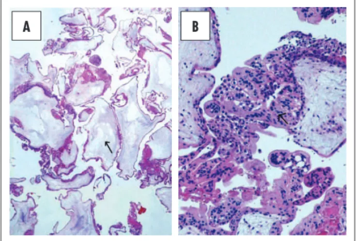
Pathological examination presented a product of conception that macroscopically comprised: abundant fragments of placental tissue with marked cystic swelling of the villi; fragments of grossly normal placenta, and an intact normal female fetus with biometric and growth parameters that were consistent of 14 weeks of gestational age. Microscopic examination (Figure 1) conirmed these macroscopic aspects showing frequent cistern formation and areas of hyperplastic trophoblast, which are typical of a complete hydatidiform mole (Figure 1). The other placental fragments corresponded to normal placental parenchyma (Figure 2). These morphological features allowed diagnosing a twin pregnancy consisting of a
Figure 2. Normal placenta. Histology shows normal placental parenchyma composed of immature intermediate and mesenchymal villi (arrow) adequate to 14 weeks of gestational age.
Figure 1. Complete hydatidiform mole. (A) Histological low power view show-ing numerous edematous villi with frequent cistern formation (arrow); (B) his-tological high power view showing hyperplastic trophoblast (arrow).
complete hydatidiform mole and a normal 14-gestational-week-old placenta and fetus.
Genetic analysis, using polymorphic markers (D13S258; D13S631; D18S51; D18S535; D21S1414; and D21S1437), conirmed normal diploid fetus (46,XX) with biparental genome and a molar tissue with paternal diploidy.
The initial serum β-hCG declined appropriately
during eight weeks reaching 28.74 mUI/mL and then a plateau. Hysteroscopy was performed, and the diagnosis of persistent gestational trophoblastic disease was found. There were no evidences of persistent or metastatic disease during the three-year follow-up.
Discussion
This case presents the dificultness of a differential diagnosis, in which a detailed morphological ultrasound of the fetus is a decisive procedure. Few cases of CHMCF have been reported over the last two decades. These pregnancies are often associated with severe maternal complications, such as persistent vaginal bleeding, thromboembolic disease, severe preeclampsia, and persistent gestational
trophoblastic neoplasia (GTN)2,5,16. Moreover, this case
represents one of the rarest ones occurring after reproduc-tive medicine techniques, namely ICSI.
The challenge was to differentiate a singleton preg-nancy consisting of a partial mole and a live triploid fetus and a twin pregnancy with one placenta exhibiting a
complete mole and the other sustaining a normal fetus4.
Unlike partial hydatidiform mole that is commonly as-sociated with multiple fetal anomalies and is managed by immediate termination of pregnancy, reported cases of twin pregnancy with complete hydatidiform mole (the present case) are not associated with fetal anomalies in the coexisting fetus. Although rare, twin pregnancies com-prising a mole and a healthy fetus are a complex clinical condition. The correct diagnosis is extremely important based on the high frequency of spontaneous abortion and intrauterine death and also for severe maternal compli-cations, mainly preeclampsia, which frequently prompt pregnancy termination. Accordingly, prenatal diagnosis should always be based on fetal karyotype using chorionic villous sampling, amniocentesis, or fetal blood sample2,4.
Many series and several case reports have been published regarding a complete mole and a coexisting
normal twin fetus (CHMCF)2-10,13-15,17,18. Twin
pregnan-cies including a mole and a healthy fetus give rise to complex clinical considerations, especially in a strongly desired pregnancy. Sebire et al.2 reported the largest se-ries so far comprising 77 CHMCF, with approximately 27% of the pregnancies achieving live birth and 19% developed persistent gestational trophoblastic disease (pGTD), without signiicant differences between those
who chose to electively terminate pregnancy and those
who did not. Recently, Massardier et al.5 published a
series of 14 cases with similar live birth percentage and a
50% pGTD. Single case reports were also published13-15,
which prompts for understanding in medical community the importance of this diagnosis.
Pregnancies complicated by CHMCF may result in a viable live-born infant in approximately 40% of the time. Continuation of such pregnancy may be an option, granted the mother has been appropriately counseled on the numerous risks.
The potential risk for pGTD (from 19.0 to 62.5%)3
is the most problematic factor when counseling these couples. Based on small series and case reports, most of these pregnancies were electively terminated in light of this potential risk. A study, undertaken by Niemann
et al.3, in which 270 histological and cytogenetically
conirmed complete hydatidiform moles were analyzed to evaluate the risk of pGTD and other obstetrics com-plications, showed no differences between singleton and twin molar pregnancies. Expectant management in these
cases may be permitted13, and the risk does not change
with advanced gestational age3.
In partial hydatidiform moles, there are fetal or embryonic tissue and chorionic villi with focal edema. Most partial moles have a triploid karyotype (usually 69, XXY), resulting from the fertilization of an apparently normal ovum by two sperms. Complete moles (CM) are usually diploid androgenic conceptus due to loss of the maternal nuclear genome with either fertilization by a single haploid sperm cell that duplicates to produce a 46,XX (monospermic) CM or by two sperms resulting
in a 46,XX or 46,XY (dispermic) CM19. This results in
excessive trophoblastic growth. No embryo develop-ment is observed due to lack of genes transcribed from maternally derived genes.
To our best knowledge, only 17 CHMCF were
re-ported9-12,15,19-22 as a result of ART since 1977, when a
morphological classiication of molar pregnancies was established, with seven of them resulting from the ICSI technique6-12 (Table 1).
1. Bracken MB. Incidence and aetiology of hydatidiform mole: an epidemiological review. Br J Obstet Gynaecol. 1987;94(12):1123-35.
2. Sebire NJ, Foskett M, Paradinas FJ, Fisher RA, Francis RJ, Short D, et al. Outcome of twin pregnancies with complete hydatidiform mole and healthy co-twin. Lancet. 2002;359(9324):2165-6. 3. Niemann I, Sunde L, Petersen LK. Evaluation of the risk of
persistent trophoblastic disease after twin pregnancy with diploid hydatidiform mole and coexisting normal fetus. Am J Obstet Gynecol. 2007;197(1):45.e1-5.
4. Vaisbuch E, Ben-Arie A, Dgani R, Perlman S, Sokolovsky N, Hagay Z. Twin pregnancy consisting of a complete hydatidiform mole and co-existent fetus: report of two cases and review of literature. Gynecol Oncol. 2005;98(1):19-23.
5. Massardier J, Golier F, Journet D, Frappart L, Zalaquett M, Schott AM, et al. Twin pregnancy with complete hydatidiform mole and coexistent fetus: obstetrical and oncological outcomes in a series of 14 cases. Eur J Obstet Gynecol Reprod Biol. 2009;143(2):84-7. 6. Hamanoue H, Umezu N, Okuda M, Harada N, Ohata T, Sakai H,
et al. Complete hydatidiform mole and normal live birth following intracytoplasmic sperm injection. J Hum Genet. 2006;51(5):477-9. 7. Yamada T, Matsuda T, Kudo M, Yamada T, Moriwaki M, Nishi S, et
al. Complete hydatidiform mole with coexisting dichorionic diamniotic
twins following testicular sperm extraction and intracytoplasmic sperm injection. J Obstet Gynaecol Res. 2008;34(1):121-4. 8. Dedes I, Christodoulou E, Ziogas V. Complete hydatidiform
mole coexisting with a viable pregnancy as twins after intracytoplasmic sperm injection: a case report. J Reprod Med. 2008;53(3):227-30.
9. Dolapcioglu K, Gungoren A, Hakverdi S, Hakverdi AU, Egilmez E. Twin pregnancy with a complete hydatidiform mole and co-existent live fetus: two case reports and review of the literature. Arch Gynecol Obstet. 2009;279(3):431-6.
10. Petignat P, Senn A, Hohlfeld P, Blant SA, Laurini R, Germond M. Molar pregnancy with a coexistent fetus after intracytoplasmic sperm injection. A case report. J Reprod Med. 2001;46(3):270-4. 11. Vandenhove M, Amant F, van Schoubroeck D, Cannie M,
Dymarkowski S, Hanssens M. Complete hydatidiform mole with co-existing healthy fetus: a case report. J Matern Fetal Neonatal Med. 2008;21(5):341-4.
12. Kashani EBP, Roshandel G, Roshandel D. Molar pregnancy and co-existent foetus: a report of two cases. J Clin Diagn Res. 2009;3(1):1334-7.
13. Shazly SA, Ali MK, Abdel Badee AY, Alsokkary AB, Khodary MM, Mostafa NA. Twin pregnancy with complete hydatidiform mole and coexisting fetus following ovulation induction with a
References
fertilization process nor guarantees paternal and maternal origins for each pronucleus.
Recently, using polymorphic DNA markers, Niemann et al. investigated the origin of twin pregnancies that comprised androgenetic diploid mole and a normal fetus. Accordingly, duplication of the paternal chromosomes before pronuclear fusion and development of a triploid pronuclear stage with one haploid maternal pronucleus
and two haploid paternal pronuclei was described23.
However, this model cannot be explained according to the normal process for early steps of fertilization. Another author described endoreduplication in the maternal pro-nucleus as a possible cause of digynic triploidy, stating
that there was evidence only for maternal contribution for abnormal oocytes24.
Post-zygotic events may also be a possibility, giv-ing rise to speculations regardgiv-ing the earliest stages of fertilization and more speciically a complete mole and
a normal pregnancy dichorionic pregnancy after ICSI24.
The ability to diagnose a molar placenta in an ongo-ing pregnancy is clinically challengongo-ing. This case dem-onstrated two main features: irst, ultrasound alone may not be suficient for an accurate management although it is a good diagnostic tool in molar pregnancies; secondly, complete molar pregnancies may still occur, despite the pregnancy being achieved by ICSI.
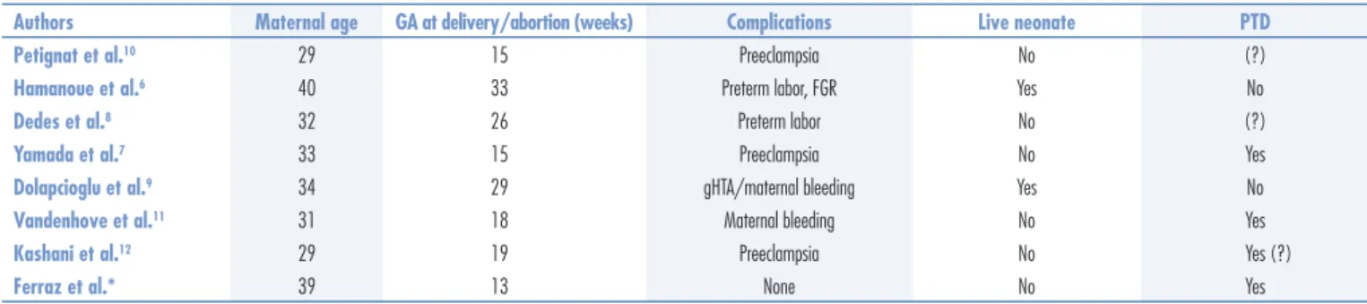
Table 1. Literature reported cases of complete hydatidiform mole coexisting with normal fetus after intracytoplasmic sperm injection
Authors Maternal age GA at delivery/abortion (weeks) Complications Live neonate PTD
Petignat et al.10 29 15 Preeclampsia No (?)
Hamanoue et al.6 40 33 Preterm labor, FGR Yes No
Dedes et al.8 32 26 Preterm labor No (?)
Yamada et al.7 33 15 Preeclampsia No Yes
Dolapcioglu et al.9 34 29 gHTA/maternal bleeding Yes No
Vandenhove et al.11 31 18 Maternal bleeding No Yes
Kashani et al.12 29 19 Preeclampsia No Yes (?)
Ferraz et al.* 39 13 None No Yes
non-prescribed clomiphene citrate regimen: a case report. J Med Case Rep. 2012;6:95.
14. Singh M, Shaltoot N, Emovon E. Twin pregnancy with complete hydatidiform mole and co-existent viable fetus. J Obstet Gynaecol. 2011;31(8):767-8.
15. Chesnais AL, Le Breton F, Devouassoux-Shisheboran M, Huissoud C, Massardier J, Quilichini B, et al. Twin pregnancy with both complete hydatiform mole and coexistent alive fetus: report of a non-antenatal diagnosed case. Ann Pathol. 2011;31(4):299-302. 16. Sánchez-Ferrer ML, Machado-Linde F, Martínez-Espejo Cerezo A,
Peñalver Parres C, Ferri B, López-Expósito I, et al. Management of a Dichorionic twin pregnancy with a normal fetus and an androgenetic diploid complete hydatidiform mole. Fetal Diagn Ther. 2012 [Epub ahead of print].
17. Aguilera M, Rauk P, Ghebre R, Ramin K. Complete hydatidiform mole presenting as a placenta accreta in a twin pregnancy with a coexisting normal fetus: case report. Case Rep Obstet Gynecol. 2012;2012:405085.
18. Sasaki Y, Ogawa K, Takahashi J, Okai T. Complete hydatidiform mole coexisting with a normal fetus delivered at 33 weeks of gestation and involving maternal lung metastasis: a case report. J Reprod Med. 2012;57(7-8):301-4.
19. Fluker MR, Yuzpe AA. Partial hydatidiform mole following transfer of a cryopreserved-thawed blastocyst. Fertil Steril. 2000;74(4):828-9.
20. Guzman González E, Gaviño Gaviño F, Valero Origel A, Deschamps Díaz H, Ramírez Fernández MA, Miranda Lamadrid M. Twin pregnancy with complete mole and coexisting fetus after in vitro fertilization and embryo transfer complicated with placenta previa accreta. A case report. Ginecol Obstet Mex. 2009;77(3):151-5.
21. Montes-de-Oca-Valero F, Macara L, Shaker A. Twin pregnancy with a complete hydatidiform mole and co-existing fetus following in-vitro fertilization: case report. Hum Reprod. 1999;14(11):2905-7. 22. Hsu CC, Lee IW, Su MT, Lin YC, Hsieh C, Chen PY, et al. Triple
genetic identities for the complete hydatidiform mole, placenta and co-existing fetus after transfer of a single in vitro fertilized oocyte: case report and possible mechanisms. Hum Reprod. 2008;23(12):2686-91.
23. Niemann I, Bolund L, Sunde L. Twin pregnancies with diploid hydatidiform mole and co-existing normal fetus may originate from one oocyte. Hum Reprod. 2008;23(9):2031-5.