parameters between the 11
thand 14
thpregnancy weeks in a population sample
from Northeast Brazil
Valores de referência para parâmetros doplervelocimétricos das
artérias uterinas entre a 11
ae 14
asemanas de gestação em uma
amostra populacional do Nordeste do Brasil
BrunA YAngdA CostA silvA2PAulo CesAr PrACiAnode sousA1 sAmmYA BezerrA mAiA1,3 FABríCiodA silvA CostA1,4
Abstract
PURPOSE: To establish reference values for the irst trimester uterine artery resistance index (UtA-RI) and pulsatility index (UtA-PI) in healthy singleton pregnant women from Northeast Brazil. METHODS: A prospective observational cohort study including 409 consecutive singleton pregnancies undergoing routine early ultrasound screening at 11–14 weeks of gestation was performed. The patients responded to a questionnaire to assess maternal epidemiological characteristics. The left and right UtA-PI and UtA-RI were examined by color and pulsed Doppler by transabdominal technique and the mean UtA-PI, mean UtA-RI and the presence of bilateral protodiastolic notching were recorded. Quartile regression was used to estimate reference values. RESULTS: The mean±standard deviation UtA-RI and UtA-PI were 0.7±0.1 and 1.5±0.5, respectively. When segregated for gestation age, mean UtA-PI was 1.6±0.5 at 11 weeks, 1.5±0.6 at 12 weeks, 1.4±0.4 at 13 weeks and 1.3±0.4 at 14 weeks’ gestation and mean UtA-RI was 0.7±0.1 at 11 weeks, 0.7±0.1 at 12 weeks, 0.6±0.1 at 13 weeks and 0.6±0.1 at 14 weeks’ gestation. Uterine artery bilateral notch was present in 261 (63.8%) patients. We observed that the 5th and 95th percentiles of the UtA-PI and UtA-RI uterine arteries were 0.7 and 2.3 and, 0.5 and 0.8, respectively. CONCLUSION: Normal reference range of uterine artery Doppler in healthy singleton pregnancies from Northeast Brazil was established. The 95th percentile of UtA-PI and UtA-RI values may serve as a cut-off for future prediction of pregnancy complications studies (i.e., pre-eclampsia) in Northeast Brazil.
Resumo
OBJETIVO: Estabelecer valores de referência para os índices de resistência (UtA-IR) e de pulsatilidade (UtA-IP) das artérias uterinas em mulheres com gravidezes saudáveis do nordeste do Brasil. MÉTODOS: Um estudo de coorte observacional prospectivo, incluindo 409 gestações únicas consecutivas submetidas a exame de ultrassonograia de rotina entre 11 e 14 semanas de gestação, foi realizado. As pacientes responderam a um questionário para avaliar características epidemiológicas maternas. Os índices UtA-IR e UtA-IP das artérias uterinas esquerda e direita foram examinadas através de Doppler colorido e pulsátil por técnica transabdominal. A média UtA-IP, a média UtA-IR e a presença de incisura protodiastólica bilateral foram registradas. Regressão quartil foi utilizada para estimar os valores de referência.
RESULTADOS: A média±desvio-padrão de UtA-IR e UtA-IP foram de 0,7±0,1 e 1,5±0,5, respectivamente. Quando separadas por idade gestacional, a média de UtA-IP foi de 1,6±0,5 com 11 semanas, 1,5±0,6 com 12 semanas, 1,4±0,4 com 13 semanas e 1,3±0,4 em uma gestação de 14 semanas e a média de UtA-IR foi de 0,7±0,1 com 11 semanas, 0,7±0,1 com 12 semanas, 0,6±0,1 com 13 semanas e 0,6±0,1 com 14 semanas de gestação. Incisura bilateral das artérias uterinas estava presente em 261 (63,8%) pacientes. Observou-se que os percentis 5 e 95 de UtA-IP e UtA-IR foram 0,7 e 2,3 e 0,5 e 0,8, respectivamente. CONCLUSÃO: A curva de valores de referência dos índices de dopplervelocimetria das artérias uterinas no primeiro trimestre foi estabelecida para gestações únicas e saudáveis do nordeste do Brasil. Os valores do percentil 95 para os índices UtA-IP e UtA-IR podem servir como ponto de corte para estudos de predição de complicações em gravidezes (por exemplo, pré-eclampsia) no nordeste do Brasil.
Study carried out at the Service of Obstetrics and Gynaecology, General Hospital of Fortaleza, Universidade Estadual do Ceará – UECE − Fortaleza (CE), Brazil.
1Department of Public Health, Universidade Estadual do Ceará – UECE − Fortaleza (CE), Brazil. 2Instituto Federal de Educação, Ciência e Tecnologia do Ceará – IFCE – Sobral (CE), Brasil. 3Universidade de Fortaleza − UNIFOR − Fortaleza (CE), Brazil.
4University of Melbourne Department of Obstetrics and Gynaecology and Department of Perinatal Medicine, Pregnancy Research Centre, Royal Women’s Hospital – Melbourne, Victoria, Australia.
Conlict of interests: nothing to declare. Keywords
Ultrasonography, Doppler Pregnancy trimester, irst Pre-eclampsia Fetal growth retardation Placental circulation
Palavras-chave
Ultrassonograia Doppler Primeiro trimestre de gravidez Pré-eclâmpsia Retardo do crescimento fetal Circulação placentária
Correspondence
Júlio Augusto Gurgel Alves Av. Dedé Brasil, 1700 – Itaperi Zip code: 60740-000 Fortaleza (CE), Brazil
Received
05/6/2013
Accepted with modiications
08/15/2013
Introduction
Pre-eclampsia (PE) remains the leading cause of ma-ternal and perinatal mortality and morbidity worldwide; in particular, it has been estimated that 10–15% of the 500 000 maternal deaths that occur each year are caused by hypertensive diseases of pregnancy. The ability to predict which women will be at high risk for PE has been a focus of recent research. At present, the irst trimester appears to be the preferred gestational period for PE screening; this preference for early screening has been reinforced by recent evidence suggesting that the prophylactic use of low-dose aspirin beginning in early pregnancy (prior to 16 weeks) can reduce the prevalence of PE by as much as 50% and signiicantly decrease rates of perinatal death1,2.
Although no single screening procedure for predicting PE has been widely adopted in clinical practice, uterine artery Doppler is certainly the most widely studied clinical test available for this purpose. Uterine artery Doppler ultrasound has become a useful method for the indirect assessment of uteroplacental circulation in early pregnancy (11–14 weeks). If combined with examination of maternal history, mean arterial pressure (MAP) and certain biochemical markers (pregnancy-associated plasma protein A and placenta growth factor), uterine artery Doppler may be regarded as an adjunct screening tool for predicting PE and intrauterine growth res-triction. Abnormal uterine artery Doppler results are strongly correlated with adverse maternal and perinatal outcomes1-8.
The clinical use of uterine artery Doppler ultrasound requires the existence of reference values. Reference values for mean uterine artery Doppler indices between 11 and 14 weeks of gestation have not previously been reported for our study population. The objective of this study was to determine reference values for the uterine artery resistance index (UtA-RI) and uterine artery pulsatility index (UtA-PI) between weeks 11 and 14 of gestation among singleton pregnant women from Fortaleza, located in the Northeastern region of Brazil.
Methods
This prospective observational cohort study recruited patients who visited the Maternal-Fetal Medicine Service of Fortaleza General Hospital (Hospital Geral de Fortaleza, HGF) in the Ceará state in Northeastern Brazil for routine irst trimester Down syndrome screening between August 2009 and February 2011. The women were followed up until late postpartum. Information regarding the evolution of these patients’ pregnancies and maternal and perinatal outcomes was obtained from the patients’ hospital records.
The patients were invited to undergo an ultrasound scan to measure fetal crown-rump length and to conirm gestational age (GA). Nuchal translucency and uterine artery Doppler index measurements were also obtained during
this ultrasound. GA was established using menstrual dates and conirmed by irst trimester ultrasound; the ultrasoun-d-based GA estimate was utilised if the GA estimates from menstrual and ultrasound data differed by more than 7 days. This study was approved by the Research Ethics Committee of Fortaleza General Hospital.
The inclusion criteria for this study were singleton pregnant women recruited in the irst trimester who deli-vered a phenotypically normal stillborn or live-born infant at or after 24 weeks of gestation without experiencing hypertensive disease.
The exclusion criteria were pregnancies with major fetal abnormalities, pregnancies ending in miscarriage or fetal death prior to 24 weeks, twin pregnancies, cases lost of fol-low-up and patients with fetal growth restriction, PE and/ or gestational hypertension (GH).
Patients responded to a questionnaire that included the following items: age, race (white, non-white or others), method of conception (spontaneous conception or assisted conception requiring the use of ovulation drugs), the smoking of any number of cigarettes per day during pregnancy (yes or no), the intake of any volume of alcohol during pregnancy (yes or no), drug use during pregnancy (yes or no), medical history, including not only whether the patient has been diagnosed with chronic hypertension or diabetes mellitus but also whether there exists a familial history of PE in the patient’s mother or sister (Fam-PE; scored as yes or no) and obstetric history, including parity and the occurrence of PE in a previous pregnancy (Previous PE; scored as yes or no).
As quantitative variables, UtA-RI and UtA-PI were mea-sured. Transabdominal ultrasound examinations were per-formed using a Voluson 730 Pro (General Electric, USA) ultrasound system. This system, which was equipped with a 3.5-MHz convex transducer, has been described in previously published studies5-10. Pulsed Doppler ultrasound was used to
obtain low velocity waveforms from the ascending branch of the uterine artery at the point closest to the internal os. When three similar consecutive waveforms were obtained, UtA-RI and UtA-PI were measured, and mean index values were calculated using the values obtained for the left and right arteries. The presence or absence of a protodiastolic notch was also recorded5,8-12.
Values of quantitative variables were analysed for each categorical variable. The χ2 test and Fisher’s exact test were
Results
The examined cohort initially consisted of 550 con-secutive, singleton pregnancies with a live fetus between gestational weeks 11 and 14. However, 45 of these cases (8.1%) were excluded due to loss of follow-up. Among the remaining 505 cases, women who developed PE or GH (n=78), women who did not develop PE or GH but delivered newborns who were small for their GA (n=12) and women with pregnancies resulting in fetal death or miscarriage prior to gestational week 24 (n=6) were all excluded from the study.
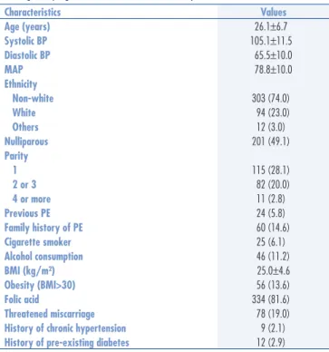
This study included 409 women whose pregnancies progressed to term without the occurrence of hypertensive disorders. The mean age of the participants was 26.1 years (range: 15–43 years). With respect to race, the women were predominantly non-white. The majority of study participants had low parity as the subjects included 201 nulliparous women (49.1%), 115 primiparous women (28.1%) and 93 multiparous women (22.8%). Only 25 study subjects (6.1%) smoked during their pregnancies, and 41 study subjects (11.2%) had consumed alcohol during their pregnancies. Nine participants presented a history of chronic hypertension, and 12 participants were diabetic (type I or II). The mean body mass index (BMI) of the study subjects was 25.0±4.9 (range: 15–38), and 56 subjects (13.6%) had a BMI greater than 30. The mean systolic and diastolic pressures of the study participants were 105.1 mmHg and 65.5 mmHg, respectively. The average MAP was 78±10 mmHg (range: 53–117 mmHg) (Table 1).
The presence of risk factors for PE, such as chronic hypertension, diabetes, a family history of PE, a prior his-tory of PE and obesity, was not associated with signiicant changes in mean UtA-RI and mean UtA-PI values during the irst trimester of pregnancy. The intake of folic acid, smoking, alcohol consumption and miscarriage risk also did not appear to signiicantly affect the values of these Doppler indices (Table 2).
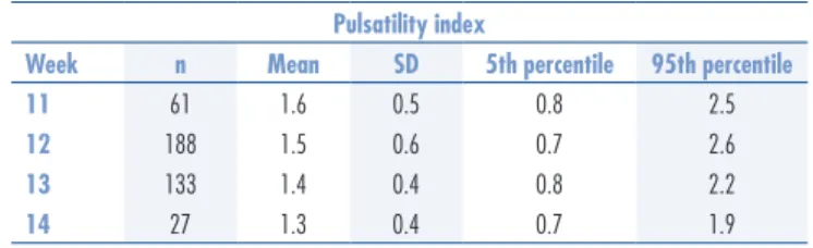
This study examined 61 women at 11 weeks, 188 women at 12 weeks, 133 women at 13 weeks and 27 women at 14 weeks. Deliveries occurred at approximately 39 weeks, and the mean birth weight of the infants was 3272 g.
Overall, the means±standard deviation for the UtA-RI and UtA-PI values measured in this study were 0.7±0.1 and 1.5±0.5, respectively. After breaking down the results by GA, the mean UtA-PI values ranged from 1.6±0.5 at 11 weeks to 1.3±0.4 at 14 weeks (Table 3). As pregnancies progressed from 11 to 14 weeks, a decrease in the presence of a bilateral notch and the mean values of the impedance indices (UtA-RI and UtA-PI) were observed (Tables 3 and 4).
Table 1. Maternal epidemiologic characteristics between weeks 11 and 14 of gestation in 409 singleton pregnancies that evolved satisfactorily
Characteristics Values
Age (years) 26.1±6.7
Systolic BP 105.1±11.5
Diastolic BP 65.5±10.0
MAP 78.8±10.0
Ethnicity
Non-white 303 (74.0)
White 94 (23.0)
Others 12 (3.0)
Nulliparous 201 (49.1)
Parity
1 115 (28.1)
2 or 3 82 (20.0)
4 or more 11 (2.8)
Previous PE 24 (5.8)
Family history of PE 60 (14.6)
Cigarette smoker 25 (6.1)
Alcohol consumption 46 (11.2)
BMI (kg/m²) 25.0±4.6
Obesity (BMI>30) 56 (13.6)
Folic acid 334 (81.6)
Threatened miscarriage 78 (19.0)
History of chronic hypertension 9 (2.1)
History of pre-existing diabetes 12 (2.9)
Data are shown as mean±standard deviation or n (%) of sample.
BP: blood pressure; MAP: mean arterial pressure; PE: pre-eclampsia; BMI: body mass index.
Table 2. The mean values of resistance index and pulsatility index of the uterine arteries between weeks 11 and 14 of gestation among 409 singleton pregnancies that evolved satisfactorily, according to maternal epidemiological characteristics
Variables Present Absent p-value Present Absent p-value
Resistance index Pulsatility index
Cigarette smoker 0.7 0.7 0.7 1.5 1.5 0.8
Alcohol consumption 0.7 0.7 0.8 1.5 1.5 0.9
Obesity (BMI>30) 0.6 0.7 0.4 1.4 1.5 0.2
Diabetes 0.7 0.6 0.6 1.6 1.5 0.6
Folic acid 0.7 0.7 0.8 1.5 1.4 0.1
Chronic hypertension 0.7 0.7 0.6 1.5 1.5 0.2
Threatened miscarriage 0.7 0.7 0.3 1.5 1.4 0.0
Nulliparous 0.6 0.7 0.4 1.5 1.5 0.3
Family history of PE 0.7 0.7 0.8 1.5 1.5 0.5
The difference in UtA-PI values between 11 and 14 weeks was statistically signiicant (p=0.02), whereas the corresponding difference in UtA-RI values was nearly statistically signiicant (p=0.08) (Table 4).
The 95th percentile values for mean UtA-PI and mean UtA-RI were 2.4 (range: 1.9–2.5) and 0.8 (ran-ge: 0.8–0.9), respectively. A bilateral notch occurred in 261 patients (63.8%).
Discussion
Bilateral uterine artery Doppler waveforms were successfully obtained in all included cases. Analyses of the study results demonstrated the feasibility of assessing uteroplacental circulation by transabdominal ultrasono-graphy in the irst trimester of pregnancy (11–14 weeks) and conirmed the possibility of incorporating uterine artery Doppler into routine screening performed during this trimester. The application of this method, in com-bination with other markers, such as maternal history, MAP and biochemical markers, appears to be useful for identifying pregnancies at high risk for complications, and the increased monitoring of these pregnancies could improve maternal and perinatal outcomes5,10,13-16.
We observed that UtA-RI and UtA-PI values decrea-sed as GA increadecrea-sed from 11 to 14 weeks. This inding suggested that trophoblastic invasion may be responsible for the observed decrease in impedance in uterine vessels during this time period and that this decrease is indicative of a good prognosis for a pregnancy16-20.
The mean UtA-PI value was 1.5 (range: 1.3–1.6). This mean UtA-PI value was similar to values obtai-ned for the unaffected group in other studies that utilised
transabdominal uterine artery Doppler21-25. However,
our observed mean UtA-PI value was lower than the mean UtA-PI value of 1.6 reported by another study conducted in Brazil that utilised transvaginal uterine artery Doppler to examine 344 pregnancies with normal pregnancy outcomes26.
We observed that mean UtA-RI and UtA-PI values were lower among women with previous pregnancies of over 24 weeks that resulted in normal outcomes than among nulliparous women. This inding may suggest that the occurrence of a prior healthy pregnancy is associated with improved placental adaptation19,20,27,28.
It is known that PE in a prior pregnancy, obesity, a family history of PE and nulliparity are risk factors for PE8-11,28. The presence of these risk factors did not
signi-icantly alter the mean UtA-RI and UtA-PI values in the irst trimester of pregnancies that evolved satisfactorily. High UtA-PI values have been associated with adverse pregnancy outcomes. An abnormal uterine artery Doppler pattern has been defined as a mean UtA-PI higher than the 95th percentile value, and this cut-off may be useful in clinical practice for PE screening among high-risk populations22,25,29-31. In our study, the 95th percentile
value for mean UtA-PI was 2.3 (range: 1.9–2.5). The 95th percentile value for mean UtA-PI was lower in our study than in other published studies6,14,25.
One of these previously published investigations was a prospective study of 1091 patients that reported 95th percentile values for mean UtA-PI that ranged from a minimum of 2.3 (at gestational week 14) to a maximum of 3.1 (at gestational week 11). Another study by the same research group evaluated 620 patients and repor-ted 95th percentile values for mean UtA-PI that ranged from 2.2 (at gestational week 14) to 2.7 (at gestational week 11)6,25. Less variation in the 95th percentile values
of mean UtA-PI over time was observed in our investi-gation than in other studies; this discrepancy remains even if the comparison is restricted to only include other studies that also used the transabdominal uterine artery Doppler measurement technique. Differences among various investigations with respect to the characteristics of study population may explain this inding.
We observed that an average of 63.8% of preg-nant women presented with a bilateral notch during the first trimester. We can therefore consider this finding normal for this gestational period. Martin et al.3 performed transabdominal scanning of women
in the first trimester of pregnancy and reported that bilateral notches were present in 55% of the 3045 examined cases.
Another recent prospective study with 280 pregnant women reported a sensitivity of 66.7% and a speciicity of 96.5% when using the 95th percentile value for mean
Table 3. Mean, standard deviation, 5th and 95th percentiles for mean uterine artery pulsatility index between weeks 11 and 14 of gestation among 409 singleton pregnancies that evolved satisfactorily
Pulsatility index
Week n Mean SD 5th percentile 95th percentile
11 61 1.6 0.5 0.8 2.5
12 188 1.5 0.6 0.7 2.6
13 133 1.4 0.4 0.8 2.2
14 27 1.3 0.4 0.7 1.9
SD: standard deviation.
Table 4. Mean, standard deviation, 5th and 95th percentiles for mean uterine artery resistance index between weeks 11 and 14 of gestation among 409 singleton pregnancies that evolved satisfactorily
Resistance index
Week Mean SD 5th percentile 95th percentile
11 0.7 0.1 0.5 0.8
12 0.7 0.1 0.5 0.8
13 0.6 0.1 0.5 0.8
14 0.6 0.1 0.5 0.8
UtA-PI during the irst trimester as a cut-off for predicting PE32. This prospective study determined that the 95th
percentile value for mean UtA-PI was 2.3, which was comparable to the 95th percentile that was calculated in our study.
Sample size may be regarded as a limiting factor in our study because we examined a few cases of chronic hypertension and diabetes, undermining attempts to comparatively analyse the impedance behaviour of ute-rine arteries in these cases. The examination of a greater number of patients with obesity, chronic hypertension and/or nulliparity may favour the emergence of statis-tically signiicant differences in uterine artery Doppler index values that are associated with these characteristics. The highlight of this study was the analysis of a sample
population consisting of pregnant women in Northeastern Brazil who were treated by the Brazilian National Health Service (Sistema Único de Saúde, SUS). These patients cha-racteristically exhibited high levels of racial miscegenation and belonged to the most underprivileged socioeconomic strata of Brazil.
In summary, we observed that mean UtA-PI decreased as GA progressed from gestational week 11 to week 14; in addition, the mean UtA-PI observed in this study was lower than the corresponding values reported by other investigations. The presence of a family history of PE, diabetes, smoking, alcohol consumption, obesity and/or chronic hypertension at the beginning of a pregnancy did not signiicantly alter the mean UtA-RI and UtA-PI values in the irst trimester of pregnancies that evolved satisfactorily.
1. Bujold E, Roberge S, Lacasse Y, Bureau M, Audibert F, Marcoux S, et al. Prevention of preeclampsia and intrauterine growth restriction with aspirin started in early pregnancy: a meta-analysis. Obstet Gynecol. 2010;116(2 Pt 1):402-14.
2. Roberge S, Nicolaides KH, Demers S, Villa P, Bujold E. Prevention of perinatal death and adverse perinatal outcome using low-dose aspirin: a meta-analysis. Ultrasound Obstet Gynecol. 2013;41(5):491-9.
3. Martin AM, Bindra R, Curcio P, Cicero S, Nicolaides KH. Screening for pre-eclampsia and fetal growth restriction by uterine artery Doppler at 11-14 weeks of gestation. Ultrasound Obstet Gynecol. 2001;18(6):583-6.
4. Papageorghiou AT, Yu CK, Bindra R, Pandis G, Nicolaides KH; Fetal Medicine Foundation Second Trimester Screening Group. Multicenter screening for pre-eclampsia and fetal growth restriction by transvaginal uterine artery Doppler at 23 weeks of gestation. Ultrasound Obstet Gynecol. 2001;18(5):441-9.
5. Plasencia W, Maiz N, Bonino S, Kaihura C, Nicolaides KH. Uterine artery Doppler at 11 + 0 to 13 + 6 weeks in the prediction of pre-eclampsia. Ultrasound Obstet Gynecol. 2007;30(5):742-9. 6. Gómez O, Figueras F, Fernández S, Bennasar M, Martínez JM,
Puerto B, et al. Reference ranges for uterine artery mean pulsatility index at 11-41 weeks of gestation. Ultrasound Obstet Gynecol. 2008;32(2):128-32.
7. Cnossen JS, Morris RK, ter Riet G, Mol BW, van der Post JA, Coomarasamy A, et al. Use of uterine artery Doppler ultrasonography to predict pre-eclampsia and intrauterine growth restriction: a systematic review and bivariable meta-analysis. CMAJ. 2008;178(6):701-11.
8. Akolekar R, Zaragoza E, Poon LC, Pepes S, Nicolaides KH. Maternal serum placental growth factor at 11 + 0 to 13 + 6 weeks of gestation in the prediction of pre-eclampsia. Ultrasound Obstet Gynecol. 2008;32(6):732-9.
9. Akolekar R, Syngelaki A, Sarquis R, Zvanca M, Nicolaides KH. Prediction of early, intermediate and late pre-eclampsia from maternal factors, biophysical and biochemical markers at 11-13 weeks. Prenat Diagn. 2011;31(1):66-74.
10. Poon LC, Akolekar R, Lachmann R, Beta J, Nicolaides KH. Hypertensive disorders in pregnancy: screening by biophysical and biochemical markers at 11-13 weeks. Ultrasound Obstet Gynecol. 2010;35(6):662-70.
11. Pourcelot L. Applications cliniques de l’examen Doppler transcutané. In: Peronneau P, éditeur. Vélocimetrie ultrasonore Doppler. Paris: Inserm; 1974. p. 213-40.
12. Yu CK, Smith GC, Papageorghiou AT, Cacho AM, Nicolaides KH; Fetal Medicine Foundation Second Trimester Screening Group. An integrated model for the prediction of preeclampsia using maternal factors and uterine artery Doppler velocimetry in unselected low-risk women. Am J Obstet Gynecol. 2005;193(2):429-36. 13. Poon LC, Staboulidou I, Maiz N, Plasencia W, Nicolaides KH.
Hypertensive disorders in pregnancy: screening by uterine artery Doppler at 11-13 weeks. Ultrasound Obstet Gynecol. 2009;34(2):142-8.
14. Gómez O, Martínez JM, Figueras F, Del Río M, Borobio V, Puerto B, et al. Uterine artery Doppler at 11-14 weeks of gestation to screen for hypertensive disorders and associated complications in an unselected population. Ultrasound Obstet Gynecol. 2005;26(5):490-4. 15. Hofmeyr GJ, Lawrie TA, Atallah AN, Duley L. Calcium supplementation
during pregnancy for preventing hypertensive disorders and related problems. Cochrane Database Syst Rev. 2010;(8):CD001059. 16. Tavares NM, Ferreira SG, Bennini JR Marussi EF, Barini R,
Peralta CF. [Longitudinal reference intervals of maternal-fetal Doppler parameters]. Rev Bras Ginecol Obstet. 2013;35(1):33-8. Portuguese.
17. Prefumo F, Sebire NJ, Thilaganathan B. Decreased endovascular trophoblast invasion in irst trimester pregnancies with high-resistance uterine artery Doppler indices. Hum Reprod. 2004;19(1):206-9. 18. Urato AC, Norwitz ER. A guide towards pre-pregnancy management of defective implantation and placentation. Best Pract Res Clin Obstet Gynaecol. 2011;25(3):367-87.
19. Venuto RC, Lindheimer MD. Animal models. In: Lindheimer MD, Roberts JM, Cunningham FG, editors. Chesley’s hypertensive disorders in pregnancy 3rd ed. Amsterdam: Elsevier; 2009. p. 169-188.
20. Mustafa R, Ahmed S, Gupta A, Venuto RC. A comprehensive review of hypertension in pregnancy. J Pregnancy. 2012;2012:105918. 21. Farina A, Zucchini C, Sekizawa A, Purwosunu Y, de Sanctis
P, Santarsiero G, et al. Performance of messenger RNAs circulating in maternal blood in the prediction of preeclampsia at 10-14 weeks. Am J Obstet Gynecol. 2010;203(6):575.e1-7.
22. Audibert F, Boucoiran I, An N, Aleksandrov N, Delvin E, Bujold E, et al. Screening for preeclampsia using irst-trimester serum markers and uterine artery Doppler in nulliparous women. Am J Obstet Gynecol. 2010;203(4):383.e1-8.
23. Abdelaziz A, Maher MA, Sayyed TM, Bazeed MF, Mohamed NS. Early pregnancy screening for hypertensive disorders in women without a-priori high risk. Ultrasound Obstet Gynecol. 2012;40(4):398-405. 24. Odibo AO, Zhong Y, Goetzinger KR, Odibo L, Bick JL, Bower
CR, et al. First-trimester placental protein 13, PAPP-A, uterine artery Doppler and maternal characteristics in the prediction of pre-eclampsia. Placenta. 2011;32(8):598-602.
25. Gómez O, Figueras F, Martínez JM, del Río M, Palacio M, Eixarch E, et al. Sequential changes in uterine artery blood low pattern between the irst and second trimesters of gestation in relation to pregnancy outcome. Ultrasound Obstet Gynecol. 2006;28(6):802-8. 26. Liao AW, Toyama J, Costa V, Ramos C, Brizot M, Zugaib M.
Correlation between the Doppler velocimetry indings of the uterine arteries during the irst and second trimesters of pregnancy. Rev Assoc Med Bras. 2009;55(2):197-200.
27. North RA, McCowan LM, Dekker GA, Poston L, Chan EH, Stewart AW, et al. Clinical risk prediction for pre-eclampsia in nulliparous women: development of model in international prospective cohort. BMJ. 2011;342:d1875.
28. Herraiz I, Escribano D, Gómez-Arriaga PI, Herníndez-García JM, Herraiz MA, Galindo A. Predictive value of sequential models of the uterine artery Doppler in pregnancies at high risk for preeclampsia. Ultrasound Obstet Gynecol. 2012;40(1):68-74.
29. Beneventi F, Locatelli E, Ramoni V, Caporali R, Montecucco CM, Simonetta M, et al. Uterine artery Doppler velocimetry and obstetric outcomes in connective tissue diseases diagnosed during the irst trimester of pregnancy. Prenat Diagn. 2012;32(11):1094-101.
30. Robillard PY, Dekker G, Chaouat G, Hulsey TC, Saftlas A. Epidemiological studies on primipaternity and immunology in preeclampsia–a statement after twelve years of workshops. J Reprod Immunol. 2011;89(2):104-17.
31. Gómez-Arriaga PI, Herraiz I, López-Jiménez EA, Gómez-Montes E, Denk B, Galindo A. Uterine artery Doppler and sFIt-1/PIGF ratio: usefulness in diagnosis of pre-eclampsia. Ultrasound Obstet Gynecol. 2013;41(5):530-7.