Hofbauer cells morphology and
density in placentas from normal and
pathological gestations
Morfologia e densidade das células de Hofbauer em
placentas de gestações normais e patológicas
maria CreaTsa1
panaGioTis Bakas1
anGelos liapis1
aGaThi kondi-pafiTi3
GeorGios CreaTsas1
Study carried out at the University of Athens, Medical School, Aretaieion Hospital, Greece
12nd Department of Obstetrics and Gynecology, University of Athens, Medical School, Aretaieion Hospital, Greece. 21st Department of Anesthesiology, University of Athens, Medical School, Aretaieion Hospital, Greece.
3Pathology Laboratory, University of Athens, Medical School, Aretaieion Hospital, Greece. Conlict of interests: none.
Keywords Hofbauer cells Placenta/cytology Immunohistochemistry Fetal growth retardation Diabetes, gestational
Palavras-chave Células de Hofbauer Placenta/citologia Imunohistoquímica Retardo do crescimento fetal
Diabetes gestacional
Correspondence
Charalampos Grigoriadis 2nd Department of Obstetrics and Gynecology, Aretaieion University
Hospital, 76 Vas.Soias Avenue 11528 Athens, Greece
Received
09/02/2013
Accepted with modiications
09/25/2013
Original Article
Abstract
PURPOSE: In placentas from uncomplicated pregnancies, Hofbauer cells either disappear or become scanty after the fourth to ifth month of gestation. Immunohistochemistry though, reveals that a high percentage of stromal cells belong to Hofbauer cells. The aim of this study was to investigate the changes in morphology and density of Hofbauer cells in
placentas from normal and pathological pregnancies. METHODS: Seventy placentas were examined: 16 specimens from
normal term pregnancies, 10 from irst trimester’s miscarriages, 26 from cases diagnosed with chromosomal abnormality of the fetus, and placental tissue specimens complicated with intrauterine growth restriction (eight) or gestational diabetes mellitus (10). A histological study of hematoxylin-eosin (HE) sections was performed and immunohistochemical study was
performed using the markers: CD 68, Lysozyme, A1 Antichymotrypsine, CK-7, vimentin, and Ki-67. RESULTS: In normal
term pregnancies, HE study revealed Hofbauer cells in 37.5% of cases while immunohistochemistry revealed in 87.5% of cases. In irst trimester’s miscarriages and in cases with prenatal diagnosis of fetal chromosomal abnormalities, both basic and immunohistochemical study were positive for Hofbauer cells. In pregnancies complicated with intrauterine growth restriction or gestational diabetes mellitus, a positive immunoreaction was observed in 100 and 70% of cases, respectively. CONCLUSIONS: Hofbauer cells are present in placental villi during pregnancy, but with progressively reducing density. The most speciic marker for their detection seems to be A1 Antichymotrypsine. It is remarkable that no mitotic activity of Hofbauer cells was noticed in our study, as the marker of cellular multiplication Ki-67 was negative in all examined specimens.
Resumo
OBJETIVO: Em placentas de gestações sem complicações, as células de Hofbauer desaparecem ou se tornam raras após o quarto ou quinto mês de gestação. Entretanto, a imunohistoquímica revela que uma alta porcentagem de células estromais pertencem às células de Hofbauer. O objetivo do presente estudo foi investigar as alterações da morfologia e densidade das
células de Hofbauer em placentas de gestações normais e patológicas. MÉTODOS: Foram examinadas 70 placentas: 16
provenientes de gestações normais a termo, 10 de abortos espontâneos no primeiro trimestre, 26 de casos diagnosticados como anormalidade cromossômica do feto, e amostras de tecido placentário com complicações causadas pela restrição de crescimento intrauterino (8) ou pelo diabetes mellitus gestacional (10). Cortes corados com hematoxilina-eosina (HE) foram submetidos a estudo histológico e imunohistoquímico utilizando-se os seguintes marcadores: CD 68, lisozima, antiquimotripsina A1, CK-7, vimentina, e Ki-67. RESULTADOS: Em gestações normais a termo, o estudo HE revelou células de Hofbauer em 37,5% dos casos, enquanto a imunohistoquímica as revelou em 87,5% dos casos. Em abortos do primeiro trimestre e em casos de diagnóstico prenatal de anormalidades cromossômicas fetais, tanto o estudo básico como o estudo imunohistoquímico foram positivos para células de Hofbauer. Em gestações complicadas pela restrição de crescimento intrauterino ou pelo diabetes mellitus gestacional, imunoreação positiva foi observada respectivamente em 100 e 70% dos
casos. CONCLUSÕES: As células de Hofbauer estão presentes nos vilos placentários durante a gestação, embora com
Introduction
From the middle of the 19th century, several studies have reported the presence of large cells in the stroma of chorionic villi of the human placenta. The precise location of these cells in the villous stroma was irst described by Kastschenko in 1885, while Virchow, and later Chaletzky and Neumann irst commented on the association of hydatidiform mole with large isolated cells having clear cytoplasm. This inding led to the term Chaletzky-Neumann cells, used in the past by several investigators. However, Hofbauer1 was the irst who gave a comprehensive morphological and functional description of these cells in normal villi at the beginning of the 20th century, and the term Hofbauer cell (HBCs) was then widely accepted in the literature.
HBCs have been described as frequent, pleomorphic cells of the villous stroma with round, fusiform, or stellate appearance. Their size depends on the length of their processes. The cells vary from 10 to 30 μm in diameter. Early studies had already found that the most striking aspects of HBCs are their highly vacuolated appearance and their granulated cytoplasm2. Later investigations have pointed out that HBCs are characterized by numerous membrane-bound, electron-lucent vacuoles of different sizes, possessing amorphous material of varying density, dense granules (presumably lysosomes), and short proiles of endoplasmic reticulum1,3-7.
Several theories have been reported trying to explain the origin of the HBCs. These include those proposed by Chaletzky who derived them from cells of maternal decidua; those by Neumann who considered them to be derivatives of the syncytium and an expression of malignancy; and other which supported that these cells could be derivatives of endothelial cells1. A very important inding concerning the origin of HBCs was Wynn’s observation, based on sex chromatin staining, that these are fetal cells8. So, most investigators now believe that HBCs are of chorionic mesenchymal origin. They can be recognized in placental villi at a very early stage of development (after the 18th day of gestation). It has been supported that in placentas from uncomplicated pregnancies, HBCs either disappear or become scanty after the fourth to ifth month of gestation. On the other hand, in cases of pathological placentas due to intrauterine growth restriction (IUGR) or gestational diabetes mellitus their density seems to be increased4,6,9. However, electron microscopy studies and immunohisto-chemistry demonstrated their presence throughout normal uncomplicated pregnancy as well, until term, and not only in immature villi of the center of the placentone.
As far as their function is concerned, HBCs are ca-pable of both immune and non-immune phagocytosis, they can trap maternal antibodies crossing over into the
placental tissues and are probably an important source of cytokines, prostaglandins, and thromboxane within the placenta10-12. The high levels of phagocytosis in HBCs conirm their macrophage function13. Other suggested hypothetical functions such as maintenance of placental water balance, involvement in transport mechanisms, a possible endocrine function, and a role in the control of vasculogenesis remain controversial1,14. Most recently it has been reported that HBCs express sprouty proteins and therefore play an important role in placental development by modulating branching of the villous tree15.
The aim of this study was to investigate the differences in morphology and density of HBCs between placentas from normal and pathological pregnancies, as well as be-tween placentas from term gestations and irst trimester miscarriages. Additionally, the function of these cells, their macrophage character and mitotic potential were examined.
Methods
This was a research immunopathological study, appro-ved by the Institutional Research Committee, which was organized in Aretaieion University Hospital in Athens. Multiple sections from 70 specimens of whole placental tissue were examined: 16 specimens were received from normal term pregnancies; 10 from irst trimester’s miscarriages; 26 from pregnancies between the 13th and 23rd week of gestation, diagnosed with chromosomal abnormality of the fetus, that were led to termination; eight from pregnancies between the 32nd and 38th week of gestation, complicated with IUGR; and, inally, 10 specimens were received from pregnancies between the 32nd and 38th week of gestation, associated with gestational diabetes mellitus.
A histological study of hematoxylin-eosin (HE) sections from formalin-ixed and parafin-embedded placental tissues was performed for semi-quantitative determination of HBCs concentration per villus and their basic morphology. At least 50 villi per case were examined under high-power ield (×400) observation. The density of HBCs per villus was determined by two independent observers and subsequently graded as ‘focal’ (+) (1–3 HBCs/villus), ‘intermediate’ (++) (3–6 HBCs/ villus), or ‘diffuse’ (+++) (>6 HBCs/villus).
Results
Group A. Placentas from normal term uncomplicated pregnancies — 16 cases
A focal presence of HBCs in mainly intermediate villi in 6/16 (37.5%) placental specimens of normal term gestations was observed during the basic HE (Table 1). The rest 10 placental specimens were negative for HBCs presence at the basic study. However, additional immuno-histochemical study revealed, via the macrophage marker A1 Antichymotrypsine, focal density of HBCs in 14/16 (87.5%) specimens, while positive immunostain was noticed for the marker CD 68 as well in half of the cases.
Their morphological analysis showed cells with oviform shape, round nuclei, and granulated cytoplasm (Figure 1). No mitotic activity was observed and the marker of cellular proliferation Ki-67 was positive in less than 5% of all examined specimens.
Group B. Placentas from first trimester’s miscarriages — 10 cases
Basic HE study was positive for HBCs presence in all cases (10/10). Diffuse density of HBCs was observed in 10% of the cases, intermediate in 50% of the cases, while focal presence of HBCs was seen in the rest 40% of irst trimester’s placentas during HE study (Figure 2). Immunohistochemistry was positive for presence of HBCs in these placental tissue specimens too, as markers A1 Antichymotrypsine, CD 68, and Lysozyme had positi-ve immunostain reaction in 100%, 80%, and 50% of examined specimens, respectively (Table 2). No posi-tive immunostain reaction was noticed after the use of markers Vimentin, CK-7, or Ki-67. According to their morphology, HBCs in these early weeks of gestation were recognized having a round shape with thin-colored nuclei and coccophile cytoplasm.
Group C. Placentas from pregnancies between 13th and 23rd week of gestation, diagnosed with
chromosomal abnormality of the fetus that were led to termination — 26 cases
Basic HE study was positive for HBCs presence in all cases (26/26). Diffuse density of HBCs was observed in 15.4% of the cases, intermediate in 26.9%, while focal presence of HBCs was seen in 57.7% of the cases. Immunohistochemistry was positive for presence of HBCs in these placental tissue specimens too, as markers A1 Antichymotrypsine showed positive immunostain in all the cases. Positive immunostain reaction was also obser-ved using the markers CD 68 and Lysozyme in 88.5% and 50% of examined specimens, respectively (Figure 3). No positive immunostain reaction was noticed after the use of markers Vimentin, CK-7, or Ki-67 (<5%). As to
their morphology, no remarkable differences were noti-ced between HBCs from placentas with chromosomal abnormalities and HBCs from irst trimester’s placental specimens. HBCs in both categories were recognized with a round shape, thin-colored nuclei without mitoses, and coccophile cytoplasm.
Group D. Placentas from pregnancies between the 32nd and 38th week of gestation, complicated with
IUGR — eight cases
A focal presence of HBCs in 5/8 (62.5%) placental specimens of pregnancies complicated with IUGR was observed during the basic HE study. The rest three placental specimens were negative for HBCs presence at the basic Table 1. Results of basic hematoxylin-eosin study
HBCs Presence Density
No % %
Normal term uncomplicated
pregnancies 6/16 37.5 F100
First trimester’s miscarriages 10/10 100 D10, IN50, F40
Chromosomal abnormality 26/26 100 D15, IN27, F58
IUGR 5/8 62.5 F100
Gestational diabetes mellitus 7/10 70 IN14, F86
D: diffuse density of HBC; F: focal density; IN: intermediate density; HBC: Hofbauer cell.
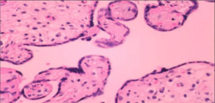
Figure 1. Histological section of mature villi with rare isolated Hofbauer cells located by stromal channels (HE ×120)
study. However, additional immunohistochemical study revealed, via the macrophage marker A1 Antichymotrypsine (Figure 4), focal presence of HBCs in all specimens (8/8) while positive immunostain was noticed for the marker CD 68 as well in 37.5% of the cases. Negative results were noticed in all cases after the use of markers such as Vimentin, CK-7, and Ki-67 (<5%). The morphological characteristics of HBCs from IUGR pregnancies did not present signiicant differences from HBCs of the same gestational age normal placentas.
Group E. Placentas from pregnancies between the 32nd and 38th week of gestation complicated with
gestational diabetes mellitus — 10 cases
Both basic HE study and immunohistochemistry, using the macrophage marker A1 Antichymotrypsine (Figure 5), revealed presence of H.c in 7/10 (70%) pla-cental specimens from pregnancies complicated with gestational diabetes mellitus. Immunohistochemistry failed to show HBCs presence in cases in which basic study was negative. In the vast majority of cases, the density of HBCs was characterized as focal (6/7, 85.7%), while only in one case it was intermediate (1/6, 14.2%). Positive immunostain reaction was observed using the marker CD 68 in 40% of the cases. No positive reaction was noticed after the use of Vimentin, CK-7, or Ki-67 (<5%). The morphological characteristics of HBCs from
pregnancies complicated with maternal diabetes mellitus did not present signiicant differences compared with HBCs of the same gestational age normal placentas.
Discussion
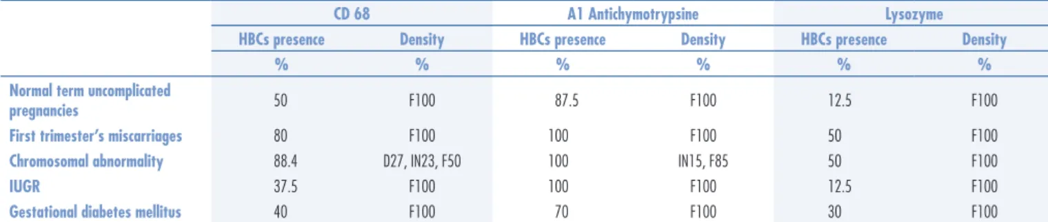
The indings of our report are in agreement with recently published reports which support that HBCs are macrophages of fetal origin and can be found in human placental villi, playing a potential role in villitis of unk-nown etiology13,16. The positive immunostain reaction of these cells after incubation with macrophage markers was characteristic. Antichymotrypsine (A1) was recognized as Table 2. Results of immunohistochemical study
CD 68 A1 Antichymotrypsine Lysozyme
HBCs presence Density HBCs presence Density HBCs presence Density
% % % % % %
Normal term uncomplicated
pregnancies 50 F100 87.5 F100 12.5 F100
First trimester’s miscarriages 80 F100 100 F100 50 F100
Chromosomal abnormality 88.4 D27, IN23, F50 100 IN15, F85 50 F100
IUGR 37.5 F100 100 F100 12.5 F100
Gestational diabetes mellitus 40 F100 70 F100 30 F100
D: diffuse density of HBCs; F: focal density; IN: intermediate density; HBC: Hofbauer cell.
Figure 3. Intermediate villous from placenta with chromosomal abnormal-ity of the fetus. Diffuse concentration of Hofbauer cells, stained with the marker CD 68 (CD 68×120)
Figure 5. Hofbauer cells in placenta from pregnancy complicated with gestational diabetes mellitus (A1 Antichymotrypsine ×120)
the most speciic marker for HBCs determination, as it revealed HBC presence even in cases in which basic HE study was negative. Positive immunostain reaction was also observed in a signiicant percentage of cases, using the monocyte-macrophage marker CD 68, while a poor response was noticed after incubation with Lysozyme. On the other hand, HBCs did not show any reaction after incubation with markers of glandular-epithelial (CK-7) or mesenchymatic differentiation (Vimentin).
Several studies support an important role of A1 Antichymotrypsine in inhibition of natural killing and antibody-dependent cell-mediated cytotoxicity17. If A1 Antichymotrypsine functions in this capacity in the vicinity of the placenta this could provide a mechanism for preventing the two modes of the cellular immune response that would otherwise become engaged during an allogenic recognition reaction18. So, the extensive distribution of A1 Antichymotrypsine within placental HBCs may have particular relevance to the lack of ma-ternal response toward the fetus. Since the location and repertory of HBCs secreted products makes them ideal candidates for a second line of defense against infection, their possible ‘immature macrophage’ status, because of their poor response after incubation with Lysozyme, but with a full phagocytosis function, leads to further investigations concerning their potential role in the pathogenesis of placental infection13-16.
It becomes important to examine whether the poor immunostain reaction for Lysozyme is characteristic of HBCs, or whether its synthesis and secretion can be induced by infection, particularly in bacterial infection where this enzyme would have an effective bactericidal action19. Thus, macrophages that are immature or lack the ability to be activated, such as the HBCs, may play an important role in the transmission of organisms capable to infect the placenta (like bacteria, chlamydia, toxoplasma, cytomegalovirus), as they have full phagocytosis capacity but may lack substances, such as lysozyme and peroxidase, necessary to destroy or inactivate the infectious agent. Additionally, the fact that these specialized placental macrophages express receptors, such as PC-SIGN and L-SIGN, sheds light to HIV-1 vertical transmission20.
The marker of cellular proliferation Ki-67 was nega-tive in all examined placental specimens. This inding, in correlation with the fact that no mitoses were observed during the morphological study of HBCs nuclei, leads to the conclusion that these cells do not have mitotic activity.
According to the literature, HBCs originate from mesenchymal cells during the early stages of pregnancy, before the fetal circulation is established. Later, once fetal circulation is established, HBCs may additionally originate from fetal bone marrow-derived monocytes, as macrophages in other organs do. Finally, it has been
proposed that HBCs may have different origins throughout pregnancy and that may represent a heterogeneous group of cells6. These data are related to in vivo and in vitro observations, showing that HBCs can undergo mitotic division6. Their potential mitotic activity could mean that HBCs may be, in part, an independent self-replicating cellular population, allowing rapid increase in concen-tration when required by the local microenvironment. However, our results, employing the marker Ki-67, do not agree with the previous mentioned research studies, as we found that HBCs do not have proliferative activity.
Our study shows that HBCs are easily detected, with both basic HE study and immunohistochemistry, in the intermediate villi of irst trimester’s gestations. Their density in these cases could be diffuse, associated with a possible role in the control of villous development by remodeling of the villous core by stimulating or inhibiting the proliferation of other mesenchymal cells, controlling villous angiogenesis by secreting angiogenetic cytokines, or controlling trophoblast turnover by inducing tropho-blast apoptosis and syncytial fusion.
In both, irst trimester’s miscarriages as well as in termination of pregnancy because of prenatal diagnosis of fetal chromosomal abnormality between the 13th and 23rd week of gestation, basic HE study and immunohis-tochemistry, via A1 Antichymotrypsine, was positive for HBCs presence in all examined placental specimens. The higher density of HBCs per villous was also observed in these two groups, in comparison with the other three examined categories (normal term placentas, placentas from pregnancies complicated with IUGR or gestational diabetes mellitus).
Immunohistochemistry revealed, by the use of marker A1 Antichymotrypsine, the presence of HBCs in 87.5% of uncomplicated term pregnancies. However, HBCs’ density was characterized focal in all cases, as no more than three cells per villous were found. So, there is a signiicant difference in HBCs’ density between early and term ges-tations. The reduction of HBCs in number as pregnancy progresses possibly occurs because the cells are compressed and masked by the condensation of the villous stroma du-ring placental maturation. Additionally, in placentas from pregnancies complicated with pre-eclampsia, a reduced HBCs’ density has been found via immunohistochemistry, using macrophage markers folate receptor (FR)-β, CD 163, and CD 6821. On the other hand, in cases of miscarriage, chromosomal abnormalities, or maternal diabetes as well as in other pathologic conditions such as Rhesus incom-patibility, these macrophages are more easily identiiable because of the edematous morphology of the villi which can unmask numerous HBCs.
the vast majority of cases, as immunohistochemistry was positive in 87.5% of the examined specimens. So, our study is in agreement with recently published reports which su-pport that HBCs are present in placental villi throughout pregnancy, but with progressively reducing concentration. The signiicant difference in recognition of HBCs presence in normal term placentas between basic HE study (37.5%) and immunohistochemistry (87.5%) could be explained from the dificulty of HE study to morphologically identify HBCs in term placentas due to their compression. So, the role of immunohistochemistry in normal term placentas is important in order to recognize these cells.
Additionally, the characteristics of HBCs were analyzed in cases of pregnancy pathologies, such as in IUGR ges-tations and in cases diagnosed with maternal diabetes mellitus. The marker A1 Antichymotrypsine revealed
HBCs presence in all cases (100%) of IUGR pregnancies and in 70% of placental specimens from pregnancies complicated with gestational diabetes mellitus. Recent literature suggests that diabetes/hyperglycemia affect the anti-inlammatory proile of HBCs by stimulating these cells to acquire an inlammatory proile leading to adverse consequences for the fetal-placental-maternal axis22.
In conclusion, HBCs are present in placental villi of both normal and pathological pregnancies. Remarkable is their presence in all specimens from IUGR pregnancies, but with focal concentration.
There were also signiicant morphological changes observed, with the irst trimester’s round shape of the cells that had coccophile cytoplasm to be replaced, with the progress of gestation, by an oviform cellular shape with granulated cytoplasm in term placentas.
1. Hofbauer J. The function of the Hofbauer cells of the chorionic villus, particularly in relation to acute infection and syphilis. Am J Obstet Gynecol. 1925;10(1):1-14.
2. Enders AC, King BF. The cytology of Hofbauer cells. Anat Rec. 1970;167(2):231-6.
3. Vinnars MT, Rindsjo E, Ghazi S, Sundberg A, Papadogiannakis N. The number of CD68(+) (Hofbauer) cells is decreased in placentas with chorioamnionitis and with advancing gestational age. Pediatr Dev Pathol. 2010;13(4):300-4.
4. Demir R, Erbengi T. Some new indings about Hofbauer cells in the chorionic villi of the human placenta. Acta Anat (Basel). 1984;119(1):18-26.
5. Castellucci M, Zaccheo D, Pescetto G. A three-dimensional study of the normal human placenta villous core. I. The Hofbauer cells. Cell Tissue Res. 1980;210(2):235-47.
6. Kondi-Paiti A, Grigoriadis C, Samiotaki D, Filippidou-Giannopoulou A, Kleanthis C, Hassiakos D. Immunohistochemical study of inhibin A and B expression in placentas from normal and pathological gestations. Clin Exp Obstet Gynecol. 2013;40(1):109-12. 7. King BF. Ultrastructural differentiation of stromal and vascular
components in early macaque placental villi. Am J Anat. 1987;178(1):30-44.
8. Wynn RM. Derivation and ultrastructure of the so-called Hofbauer cell. Am J Obstet Gynecol. 1967;97(2):235-48.
9. Martinoli C, Castellucci M, Zaccheo D, Kaufmann P. Scanning electron microscopy of stromal cells of human placental villi throughout pregnancy. Cell Tissue Res. 1984;235(3):647-55. 10. Hauguel-de Mouzon S, Guerre-Millo M. The placenta cytokine
network and inlammatory signals. Placenta. 2006;27(8):794-8. 11. Wood GW, King GR Jr. Trapping antigen-antibody complexes
within the human placenta. Cell Immunol. 1982;69(2):347-62. 12. Wetzka B, Clark DE, Charnock-Jones DS, Zahradnik HP, Smith
SK. Isolation of macrophages (Hofbauer cells) from human term placenta and their prostaglandin E2 and thromboxane production. Hum Reprod. 1997;12(4):847-52.
References
14. Tang Z, Tadesse S, Norwitz E, Mor G, Abrahams VM, Guller S. Isolation of Hofbauer cells from human term placentas with high yield and purity. Am J Reprod Immunol. 2011;66(4):336-48. 15. Seval Y, Korgun ET, Demir R. Hofbauer cells in early human
placenta: possible implications in vasculogenesis and angiogenesis. Placenta. 2007;28(8-9):841-5.
16. Anteby EY, Natanson-Yaron S, Greenield C, Goldman-Wohl D, Haimov-Kochman R, Holzer H, et al. Human placental Hofbauer cells express sprouty proteins: a possible modulating mechanism of villous branching. Placenta. 2005;26(6):476-83.
17. Tamblyn JA, Lissauer DM, Powell R, Cox P, Kilby MD. The immunological basis of villitis of unknown etiology – Review. Placenta. 2013;34(10):846-55.
18. Gravagna P, Gianazza E, Arnaud P, Neels M, Ades EW. Modulation of the immune response by plasma protease inhibitors. III. Alpha 1-antichymotrypsin inhibits human natural killing and antibody-dependent cell-mediated cytotoxicity. J Reticuloendothel Soc. 1982;32(2):125-30. 19. Braunhut SJ, Blanc WA, Ramanarayanan M, Marboe C, Mesa-Tejada
R. Immunocytochemical localization of lysozyme and alpha-1-antichymotrypsin in the term human placenta: an attempt to characterize the Hofbauer cell. J Histochem Cytochem. 1984;32(11):1204-10. 20. Gajewska E, Woyton J. Lysozyme activity in parturient women
and their newborns in cases of infection. Arch Immunol Ther Exp (Warsz). 1978;26(1-6):665-70.
21. da Silva RC, Segat L, Zanin V, Arraes LC, Crovella S. Polymorphisms in DC-SIGN and L-SIGN genes are associated with HIV-1 vertical transmission in a Northeastern Brazilian population. Hum Immunol. 2012;73(11):1159-65.
22. Tang Z, Buhimschi IA, Buhimschi CS, Tadesse S, Norwitz E, Niven-Fairchild
T, et al. Decreased levels of folate receptor-β and reduced numbers of
fetal macrophages (Hofbauer cells) in placentas from pregnancies with severe pre-eclampsia. Am J Reprod Immunol. 2013;70(2):104-15. 22. Sisino G, Bouckenooghe T, Aurientis S, Fontaine P, Storme L,